
Clenchiella varicosa, Ponder, Winston F., Fukuda, Hiroshi & Hallan, Anders, 2014
|
publication ID |
https://doi.org/10.11646/zootaxa.3872.2.1 |
|
publication LSID |
lsid:zoobank.org:pub:F9F81CC8-E033-46B7-B73B-9FB777DF4116 |
|
DOI |
https://doi.org/10.5281/zenodo.5631013 |
|
persistent identifier |
https://treatment.plazi.org/id/1013A0BA-2333-4A13-80F4-063B911748E9 |
|
taxon LSID |
lsid:zoobank.org:act:1013A0BA-2333-4A13-80F4-063B911748E9 |
|
treatment provided by |
Plazi |
|
scientific name |
Clenchiella varicosa |
| status |
sp. nov. |
Clenchiella varicosa n. sp.
Figures 1 View FIGURE 1 , 3 View FIGURE 3 , 4 View FIGURE 4 , 8 View FIGURE 8 , 10 View FIGURE 10 , 14 View FIGURE 14
Clenchiella sp.; Tong, 1986: 443: 443 (in part?), fig. 2F.
? Clenchiella cf. microscopica ; Ueng & Wang, 2006: 31 –36, figs 1–3 [not of Nevill, 1887].
Etymology. Varicose: strictly meaning a dilated vein (Latin, but here based on varix, a commonly used term for a thickening on the shell, typically behind the aperture).
Types and type locality. Holotype: E Mong Tseng Wai, Hong Kong, in mud and leaves etc. in mangroves, 22°30’ S, 114°00’ E, 16 Apr. 1983. Coll: W.F. Ponder (AMS C.462960). Paratypes: Same data (AMS C.460738, 6 spms).
Material examined. Type material.
Specimens from Taiwan, Tainan City, Wildlife Reserve, 23° N, 120°06’E, 1 Oct. 2006. Coll: Y. Ueng (AMS, C.462993, 6 spms).
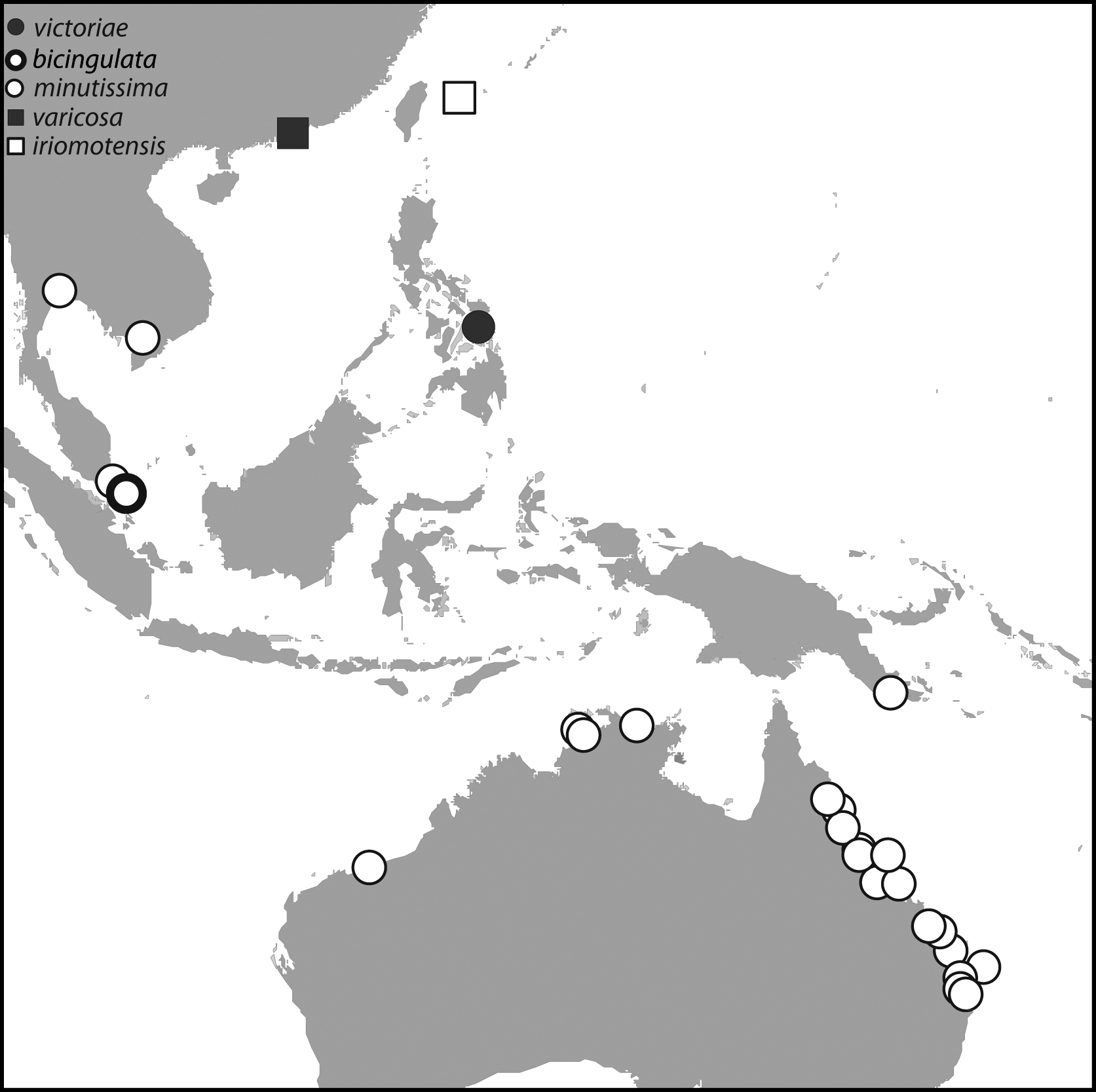
Distribution. Known only from Hong Kong and Taiwan, but is presumably more widespread in China.
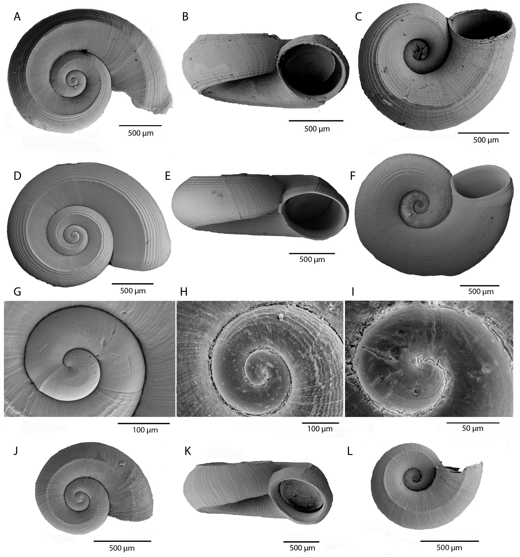
Description. Shell. Small (up to 2 mm in maximum diameter; Table 3), spire mostly flat with apex slightly raised ( Fig. 1 View FIGURE 1 G–I). Protoconch slightly elevated above spire, damaged in available specimens but protoconch I with 4 widely spaced spiral threads, protoconch II smooth. Teleoconch of about 2 whorls, rounded, moderate to rather weak mid-dorsal spiral cord, causing weak subangulation especially in subadults (in some adults not sufficiently strong to cause angulation), with 2–5 slightly weaker spirals on outer side of main spiral; on inner dorsal part of shell spirals very weak to subobsolete or absent; on outer side of dorsal carina spiral lirae have approximately linear interspaces while on periphery lirae with narrow interspaces. Periphery evenly convex. Outer base with 1–2 mid-ventral spiral cords, not sufficiently strong to cause subangulation; several weaker cords on outer and inner side of mid-ventral spiral(s). Base evenly convex; umbilicus wide (more than half width of base), spiral sculpture subobsolete within inner part of umbilicus. Sutures moderately impressed. Aperture near circular, with simple, slightly thickened peristome, external varix weakly to moderately developed, narrow, slightly behind edge of aperture. Colour yellowish (periostracum), shell white.
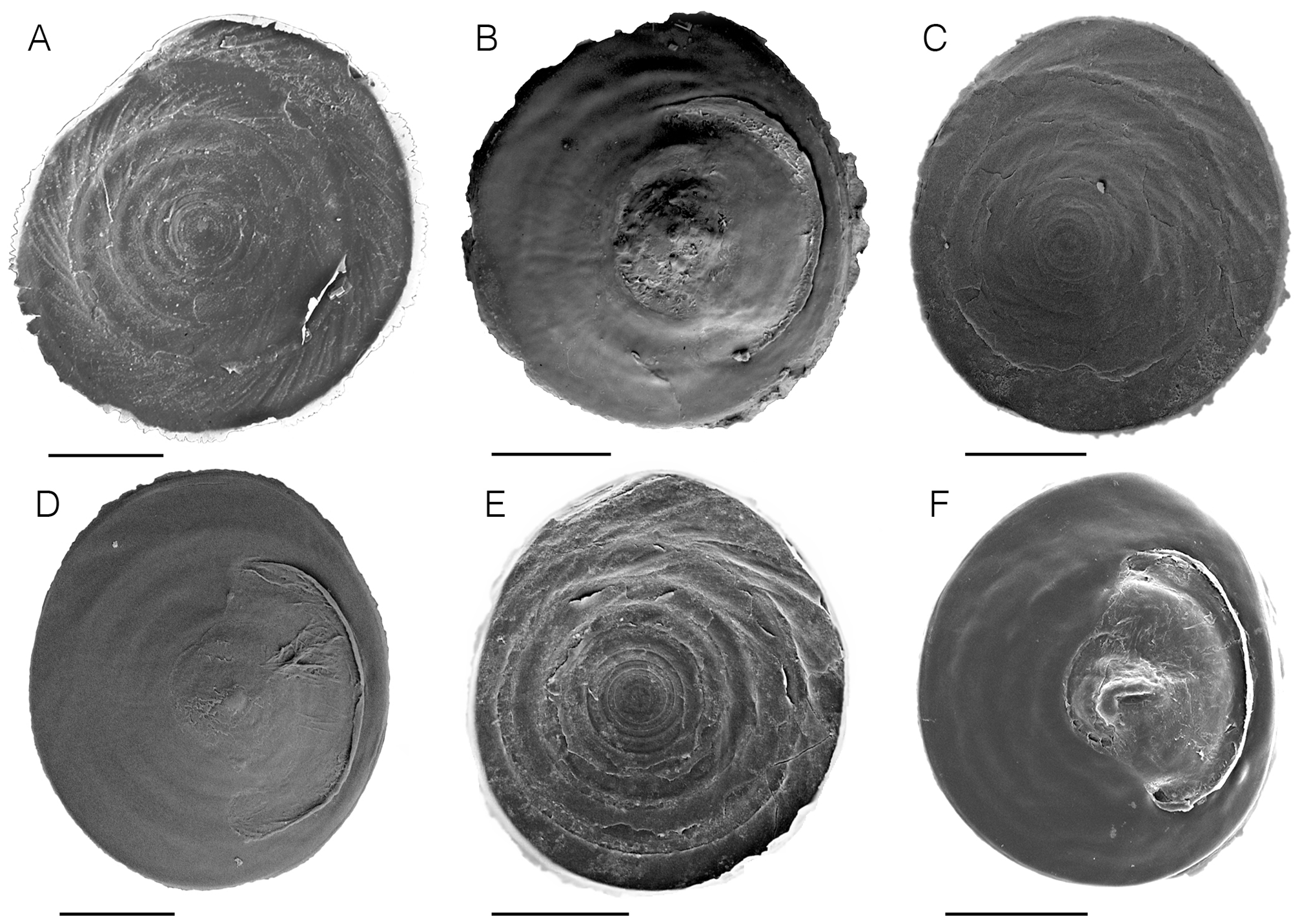
Operculum and radula . Not examined.
Ctenidium . About 30 filaments.
Gut. Anterior oesophagus with two weak folds, rectum with two tight loops.
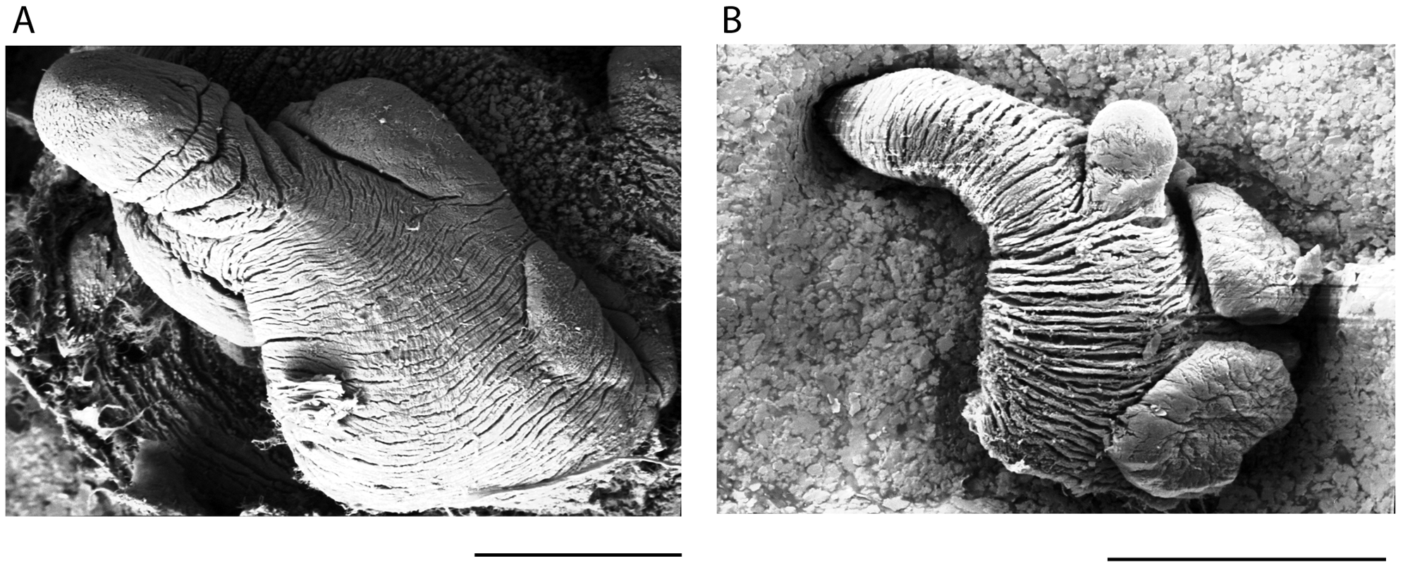
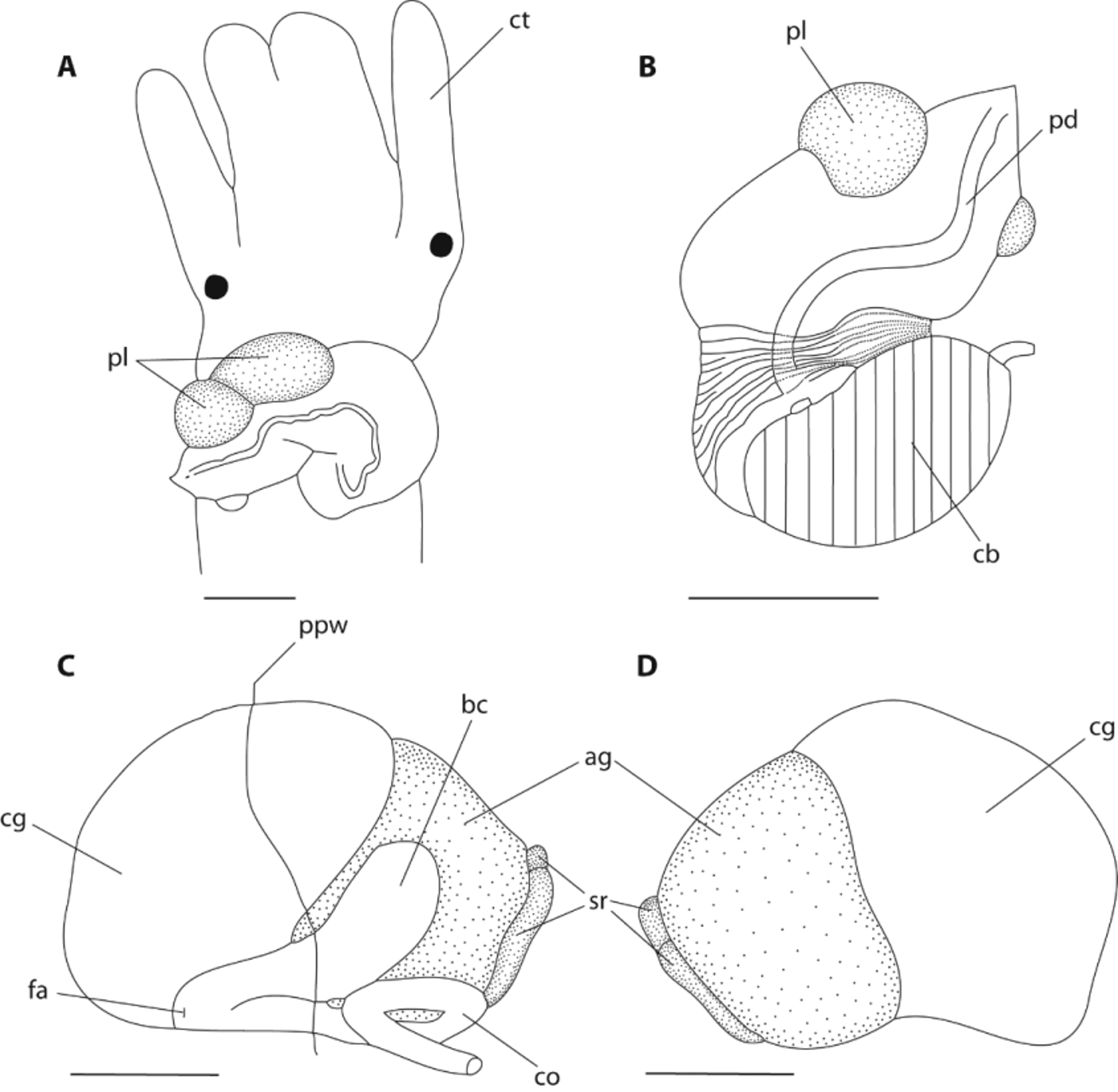
Penis. Similar to Cl. bicingulata , but two lobes on distal portion, one round lobe on right edge, more proximal lobe horizontally elongated; on left edge only one small swelling very close to narrow, pointed tip ( Figs 8 View FIGURE 8 B and 10A, B). Penial duct slightly undulating.
Oviduct. Albumen gland slightly shorter than capsule gland ( Fig. 10 View FIGURE 10 C, D). Coiled oviduct narrow, with single, very large coil. Seminal receptacles short, dorsal (left) longer than ventral (right). Ventral channel short, narrow, straight. Bursa small, lying on left side of oviduct gland, about 4/5 behind posterior wall of mantle cavity; posterior part wide, narrows anteriorly to nearly straight, short bursal duct on left side of capsule gland. Genital opening simple, on anterior end of bursal duct.
Nervous system. Pleural-supraoesophageal connective short. Suboesophageal ganglion shorter and narrower than left pleural ganglion.
Remarks. This species is similar to Cl. bicingulata but has a smaller shell, weaker dorsal and ventral ridges and a weaker varix (shells of both species can be compared in Figures 1 View FIGURE 1 and 3 View FIGURE 3 ) The penial morphology also differs in the arrangement of the glands (see description above), notably in having only two distinct glandular lobes distally instead of three ( Figure 8 View FIGURE 8 ). Unfortunately, the condition of available shell material from Hong Kong was rather poor due to corrosion in the preservative, so SEM figures are not provided.
Shells of the species recorded as Clenchiella cf. microscopica from Shihchu Wildlife Reserve, Tainan City, Southwest Taiwan ( Ueng & Wang 2006) are very similar to this species and we treat them as conspecific. These authors provided photographs of the living animal and SEM figures of the shell and inner side of the operculum. We obtained specimens from the same locality from Dr Y.-T. Ueng, and from our examination of the shells ( Fig. 3 View FIGURE 3 D–F), we regard them as this species pending a more detailed study.

No known copyright restrictions apply. See Agosti, D., Egloff, W., 2009. Taxonomic information exchange and copyright: the Plazi approach. BMC Research Notes 2009, 2:53 for further explanation.
|
Kingdom |
|
|
Phylum |
|
|
Class |
|
|
SubClass |
Caenogastropoda |
|
Order |
|
|
SuperFamily |
Truncatelloidea |
|
Family |
|
|
Genus |
Clenchiella varicosa
| Ponder, Winston F., Fukuda, Hiroshi & Hallan, Anders 2014 |
Clenchiella cf. microscopica
| Ueng 2006: 31 |
Clenchiella
| Tong 1986: 443 |