
Nipponbathynella shigaensis, Park & Cho, 2015
|
publication ID |
https://doi.org/ 10.1080/00222933.2015.1023226 |
|
DOI |
https://doi.org/10.5281/zenodo.4328922 |
|
persistent identifier |
https://treatment.plazi.org/id/03CDB43A-482A-FFF6-FE6B-F4703C2CFB68 |
|
treatment provided by |
Carolina |
|
scientific name |
Nipponbathynella shigaensis |
| status |
sp. nov. |
Nipponbathynella shigaensis sp. nov.
( Figures 15–19 View Figure 15 View Figure 16 View Figure 17 View Figure 18 View Figure 19 )
Etymology
The species name is derived from the province, Shiga-Ken (Ken means prefecture), where the species was collected.
Material examined
Type material
Holotype. Male , dissected on seven slides. Japan, Shiga-Ken (prefecture), Takashima- Shi (city), Imazu-Cho (commune), Hamabun , Imazu Beach, Groundwater 35–02, 4 October 2011 (M.J. Grygier and M. Matsuda) ( LBM1430005567 View Materials ).
Allotype. Female, dissected on six slides, same data as of holotype ( LBM1430005568 View Materials ).
Paratypes. One female and one juvenile each kept as a whole specimen in a slide, same data as holotype ( LBM1430005569 View Materials , LBM1430005570 View Materials ) .
Description of adult male (holotype)
Body ( Figure 15A View Figure 15 ). Elongated and cylindrical, length 1.50 mm, approximately 10 times as long as wide. Head as long as wide, shorter than anterior three thoracic segments combined.
Antennule ( Figure 15B View Figure 15 ) six-segmented. First segment with one seta on inner distal margin, with two simple dorsal setae and each one plumose seta dorso-laterally, laterally and ventro-laterally. Second segment with one group of four plumose setae, with one simple seta on inner distal margin and inner laterally with one ventral seta. Third segment with two lateral setae, one seta on inner distal margin and inner laterally with one ventral seta. Inner flagellum of third segment with three simple setae. Fourth segment with one stub seta and one plumose seta on dorsal margin, and with two stub setae and two plumose setae on outer distal apophysis being slightly protruded. Fifth segment with a medial group of two inner setae, one dorsal aesthetasc and one dorsal simple seta, and distally with three setae, two dorsal aesthetascs, one dorsal seta and one lateral aesthetasc. Sixth segment as large as one half of the fifth segment, with three subterminal aesthetascs and four simple setae.
Antenna ( Figure 15C View Figure 15 ) two-segmented, as long as the length of the first antennular segment. Proximal segment without setae, distal segment with two simple terminal setae and one plumose seta.
Labrum ( Figure 15D View Figure 15 ) flat, with 11 median teeth of more or less similar size flanked by three lateral teeth, lateral-most teeth with tiny terminal spinule. Inner surface concave, with two pairs of nipple-like lateral protrusions and with ctenidia and tiny projection in middle region.
Mandible ( Figure 15E View Figure 15 ) with incisor process of three teeth. Tooth of ventral edge triangular. Spine row consisting of four spines. Palp of one segment, with one apical seta not exceeding incisor process in length.
Maxillule ( Figure 15F View Figure 15 ) two-segmented. Proximal segment with four setae on inner distal margin and with two medial rows of ctenidia. Distal segment with two terminal spines, with four spines and one small spine on inner edge, and with three simple setae on outer distal margin.
Maxilla ( Figure 15G View Figure 15 ) four-segmented, setal formula 2-4-9-7.
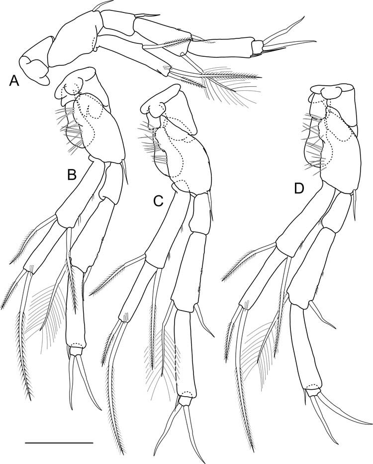
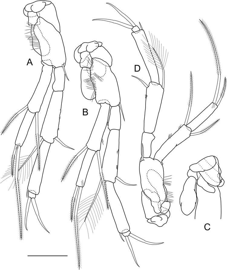
Thoracopods I–VII ( Figures 16A–D View Figure 16 , 17A–D View Figure 17 ) increasing in size up to thoracopod IV, thoracopods IV–VII similar in size. Thoracopods II–VII each bearing one epipod on protopod. Basis of thoracopods I–VII with one seta each. Basis of thoracopods II–VII with long and strong hairs along outer margin. Exopod of thoracopod I one-segmented with one medial seta on ventral margin. Exopod of thoracopods II– VII two-segmented. Endopods of thoracopods I–VII four-segmented, setal formulae:
Thoracopod I 1 + 0/1 + 1/0 + 1/3(1)
Thoracopods II–VII 0 + 0/1 + 1/0 + 1/2(0).
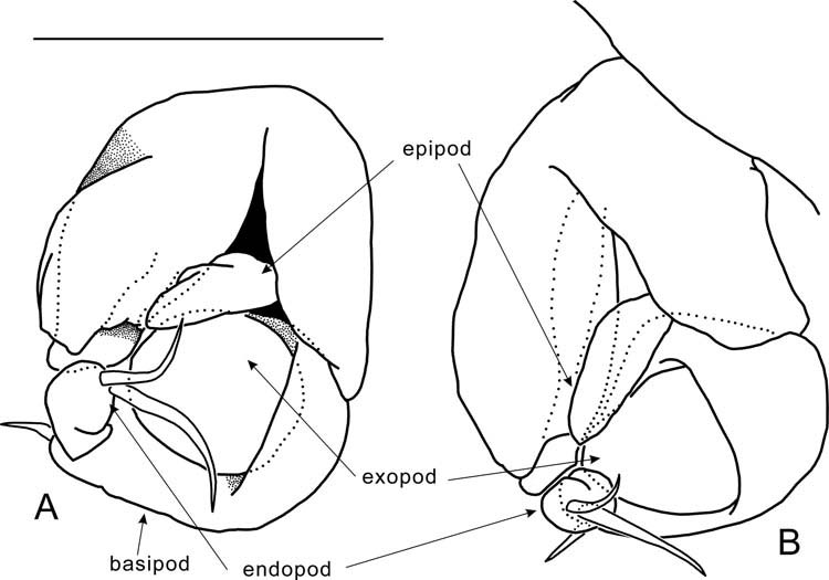
Thoracopod VIII ( Figure 18A, B View Figure 18 ) bell-shaped and tilting backwards in lateral view, 1.2 times longer than wide. Protopod massive, with penial region of two smooth lobes, inner lobe absent. Epipod narrow and elongated, distal end barely reaching penial region. Basis nearly trapezoid, without distal spur, with one seta. Exopod half as long as basis, longer than wide, one-lobed and smooth. Endopod longer than wide, with two distal setae of different sizes.
First pleopod absent ( Figure 15A View Figure 15 ).
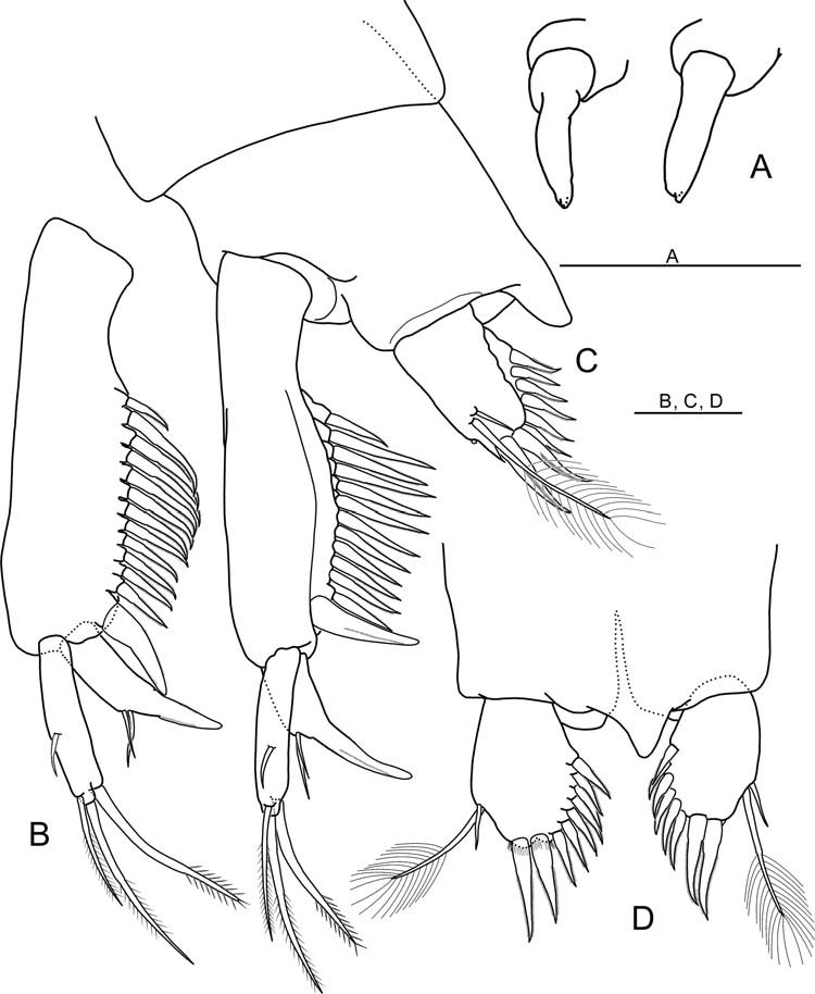
Uropod ( Figure 19B, C View Figure 19 ) with load-shaped sympod bearing 13 spines on inner margin. Proximal-most spine smaller than remaining ones decreasing slightly in size distally. Distal-most spine thicker and longer than others. Endopod 24% as long as sympod length, drawn into spur, with two setae on the outer basis of spur. Exopod longer than endopod, 46% as long as sympod, with one outer seta, two terminal setae and one inner seta. Inner seta nearly subterminal, strong, thicker and slightly shorter than inner terminal seta. Outer terminal seta nearly subterminal. All setae and spines on uropod barbed.
Pleotelson ( Figure 19C, D View Figure 19 ) without seta.
Anal operculum protruded.
Furcal rami ( Figure 19C, D View Figure 19 ) approximately 1.2 times longer than wide, with two distal spines and six (right) or seven (left) additional spines on inner margin, and dorsally with one plumose seta and one simple seta and ventrally with furcal organ.
Description of adult female (allotype)
The female differs from the male in the inner margin of the protopod of the thoracopod VI and in the form of the thoracopod VIII. Body length 1.55 mm (other females: 1.50 mm). Inner margin of the protopod of the thoracopod VI ( Figure 17C View Figure 17 ) undulated and with opening of oviduct. Thoracopod VIII ( Figure 19A View Figure 19 ) in form of two radicles bearing one tiny spine distally

No known copyright restrictions apply. See Agosti, D., Egloff, W., 2009. Taxonomic information exchange and copyright: the Plazi approach. BMC Research Notes 2009, 2:53 for further explanation.