
Nipponbathynella donggangensis, Park & Cho, 2015
|
publication ID |
https://doi.org/ 10.1080/00222933.2015.1023226 |
|
DOI |
https://doi.org/10.5281/zenodo.4328924 |
|
persistent identifier |
https://treatment.plazi.org/id/03CDB43A-4837-FFE3-FE0A-F0EC3CCAFE5A |
|
treatment provided by |
Carolina |
|
scientific name |
Nipponbathynella donggangensis |
| status |
sp. nov. |
Nipponbathynella donggangensis sp. nov.
( Figures 6–10 View Figure 6 View Figure 7 View Figure 8 View Figure 9 View Figure 10 )
Etymology
The species name is derived from a stream (Donggang, a tributary of The Han-River) where the species was collected.
Material examined
Type material
Holotype. Female , dissected on six slides. South Korea, Kangwon-Do (province), Jeongseon-Gun (county), Jeongseon-Eup (town), A gravel bank of Donggang (37° 22'49.3 ''N 128°39'24.9'' E), 24 May 2014 (J.-L. Cho and J.-G. Park) ( NIBR IV0000267099 View Materials ). GoogleMaps
Allotype. Male, dissected on six slides, same data as holotype ( NIBR IV0000267100 View Materials ). No paratype. GoogleMaps
Description of adult female (holotype)
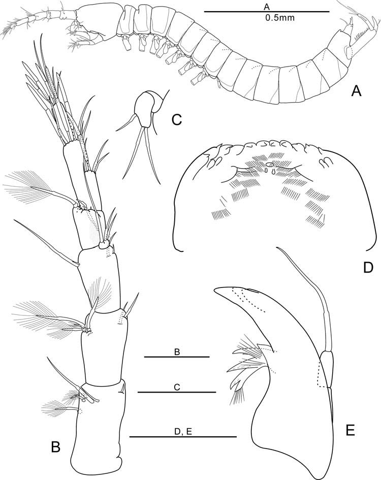
Body ( Figure 6A View Figure 6 ). Elongated and cylindrical, length 1.38 mm, approximately 12 times as long as wide. Head as long as wide, slightly shorter than anterior three thoracic segments combined.
Antennule ( Figure 6B View Figure 6 ) six-segmented. First segment with one seta on inner distal margin, with two simple dorsal setae and each one plumose seta dorso-laterally, laterally and ventro-laterally. Second segment with one group of four plumose setae, with one simple seta on inner distal margin and with one ventral seta inner laterally. Third segment with two lateral setae, one seta on inner distal margin and with one ventral seta inner laterally. Inner flagellum of third segment with three simple setae. Fourth segment with one stub seta and one plumose seta on dorsal margin, and with two stub setae and two plumose setae on outer distal apophysis being slightly protruded. Fifth segment with a medial group of two inner setae, one dorsal aesthetasc and one dorsal simple seta, and distally with three setae, two dorsal aesthetascs, one dorsal seta and one lateral aesthetasc. Sixth segment as large as one half of the fifth segment, with three subterminal aesthetascs and four simple setae.
Antenna ( Figure 6C View Figure 6 ) two-segmented, as long as the length of the first antennular segment. Proximal segment without setae, distal segment with two simple terminal setae and one subterminal seta.
Labrum ( Figure 6D View Figure 6 ) flat, with eight median teeth of more or less similar size flanked by one (left) or two (right) lateral teeth. Inner surface concave, with two pairs of nipple-like lateral protrusions and with ctenidia and three tiny projections in middle region.
Mandible ( Figure 6E View Figure 6 ) with incisor process of three teeth. Tooth of ventral edge triangular. Spine row consisting of four spines. Palp of one segment, with one apical seta not exceeding incisor process in length.
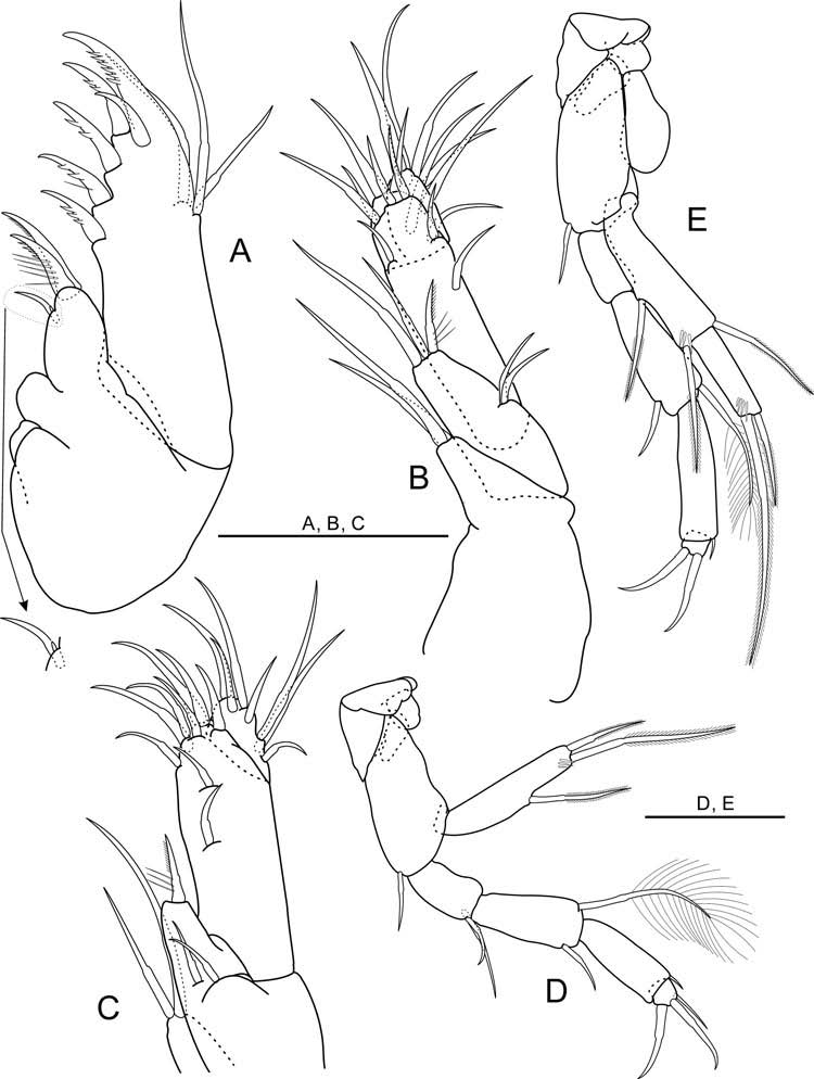
Maxillule ( Figure 7A View Figure 7 ) two-segmented. Proximal segment with three setae and one tiny seta on inner distal margin. Distal segment with two terminal spines, with four spines on inner edge, and with three simple setae on outer distal margin. Most distal spine with six dentils.
Maxilla ( Figure 7B, C View Figure 7 ) four-segmented, setal formula 2-5-10-7.
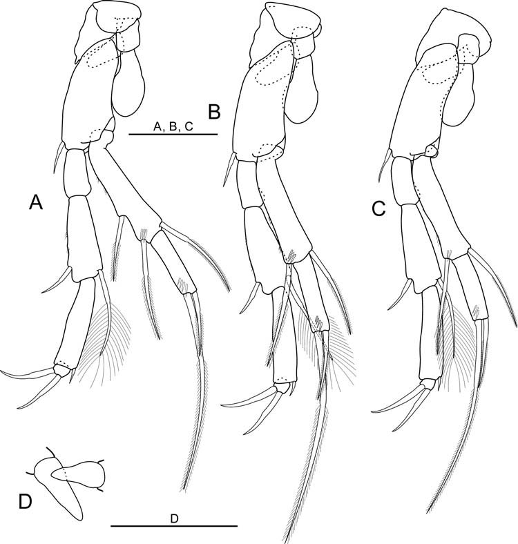
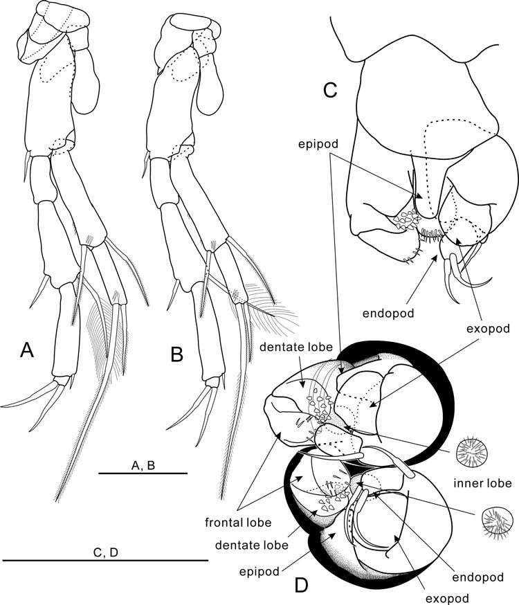
Thoracopods I–VII ( Figures 7D, E View Figure 7 , 8A–C View Figure 8 , 9A, B View Figure 9 ) increasing in size up to thoracopod III, thoracopods III–VII similar in size. Protopod of thoracopod VI protruding and with distal opening. Thoracopods II–VII each bearing one epipod on protopod. Basis of thoracopods I–VII with one seta each. Exopod of thoracopod I one-segmented with one medial seta on ventral margin. Exopod of thoracopods II–VII two-segmented. Exopod of thoracopods II and III with one medial seta on ventral margin of proximal segment. Endopods of thoracopods I–VII foursegmented, setal formulae:
Thoracopod I 2 + 0/1 + 1/0 + 1/3(1)
Thoracopods II–VII 0 + 0/1 + 1/0 + 1/2(0).
Thoracopod VIII ( Figure 8D View Figure 8 ) in form of two radicles.
First pleopod absent ( Figure 6A View Figure 6 ).
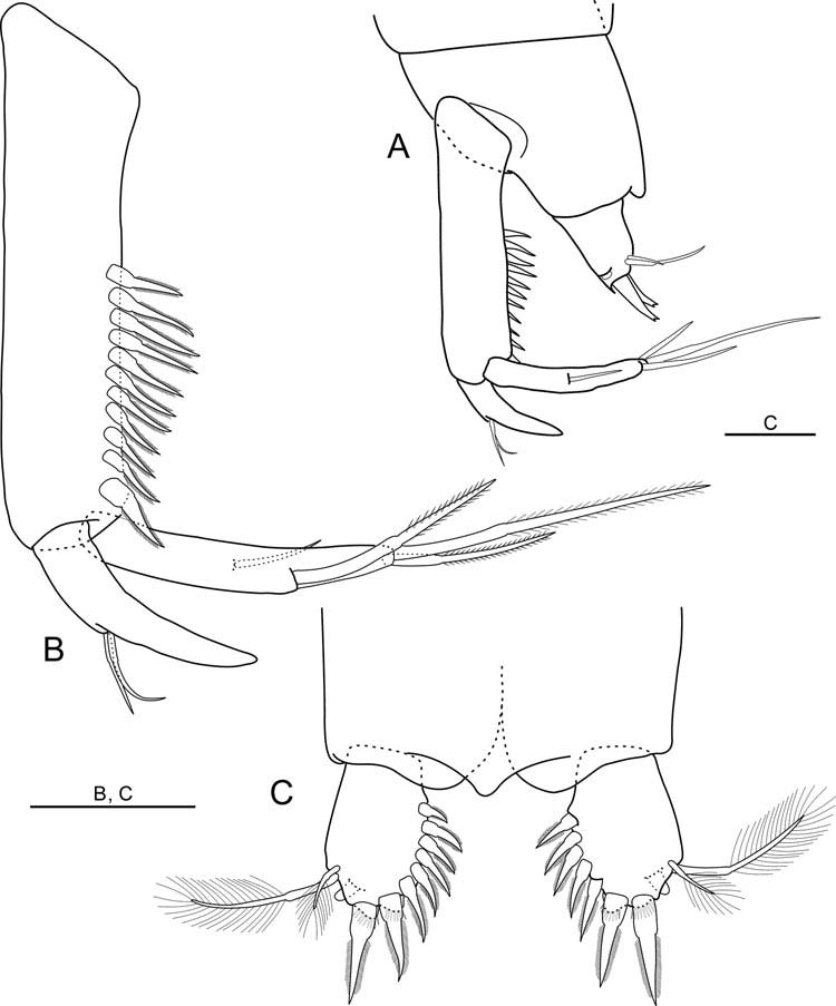
Uropod ( Figure 10A, B View Figure 10 ) with load-shaped sympod bearing 11 spines on inner margin. Most distal spine significantly thicker than others decreasing slightly in size distally. Endopod 21.3% as long as sympod length, drawn into spur, with two setae on the outer basis of spur. Exopod longer than endopod, 55.2% as long as sympod, with one outer seta, two terminal setae and one inner medial seta. Inner seta strong, longer and thicker than outer terminal seta.
Pleotelson ( Figure 10A, C View Figure 10 ) without seta.
Anal operculum protruded.
Furcal rami ( Figure 10C View Figure 10 ) longer than wide, with two distal spines and five (right) or six (left) spines on inner margin, and dorsally with two plumose setae of different length and ventrally with furcal organ.
Description of adult male (allotype)
Male differing from female in smooth inner margin of protopod of thoracopod VI and in form of thoracopod VIII. Body length 1.35 mm. Thoracopod VIII ( Figure 9C, D View Figure 9 ) bell-shaped tilting backwards in lateral view, 1.3 times longer than wide. Protopod massive, with penial region of three lobes: frontal lobe with seven spinules; dentate lobe with 13 teeth; inner lobe spinulated, in form of bur. Epipod small, round distal end not reaching penial region. Basis nearly trapezoid, without distal spur, with one seta. Exopod one-third as long as basis, longer than wide, one-lobed and smooth. Endopod longer than wide, with two distal setae.
| NIBR |
National Institute of Biological Resources |
No known copyright restrictions apply. See Agosti, D., Egloff, W., 2009. Taxonomic information exchange and copyright: the Plazi approach. BMC Research Notes 2009, 2:53 for further explanation.
|
Kingdom |
|
|
Phylum |
|
|
Class |
|
|
Order |
|
|
Family |
|
|
Genus |