
Notocomplana septentrionalis (Kato, 1937)
|
publication ID |
https://doi.org/10.11646/zootaxa.4282.3.6 |
|
publication LSID |
lsid:zoobank.org:pub:182D4546-A38A-49FA-A308-9D82770CA9D6 |
|
DOI |
https://doi.org/10.5281/zenodo.6049800 |
|
persistent identifier |
https://treatment.plazi.org/id/03CE3523-FFD7-FFD7-FF25-FA35FB4BD98B |
|
treatment provided by |
Plazi |
|
scientific name |
Notocomplana septentrionalis (Kato, 1937) |
| status |
|
Notocomplana septentrionalis (Kato, 1937)
( FigS. 4 View FIGURE 4 C, 6, 7)
Notoplana septentrionalis Kato, 1937b : pp. 127–129, text-figs. 5, 6, pl. 8, figs. 4–6 [Muroran, Hokkaido, Japan]. Notocomplana septentrionalis: Faubel (1983) , p. 115 [secondary literature].
Material examined. Fourteen SpecimenS: eleven from OShoro ( 43°12′36″N, 140°51′28″E), Hokkaido, Japan, of which four ( ICHUM 5287 View Materials , Sagittal SectionS, 4 SlideS; ICHUM 5288 View Materials , croSS SectionS, 11 SlideS; ICHUM 5289 View Materials , Sagittal SectionS, 9 SlideS; ICHUM 5290 View Materials , Sagittal SectionS, 6 SlideS) were collected on 25 May 2015, one ( ICHUM 5294 View Materials , Sagittal SectionS, 9 SlideS) on 30 May 2016, and Six ( ICHUM 5295 View Materials , croSS SectionS, 7 SlideS; ICHUM 5296 View Materials , Sagittal SectionS, 8 SlideS; ICHUM 5297 View Materials , Sagittal SectionS, 7 SlideS; ICHUM 5298 View Materials , Sagittal SectionS, 7 SlideS; ICHUM 5299 View Materials , Sagittal SectionS, 9 SlideS; ICHUM 5300 View Materials , Sagittal SectionS, 9 SlideS) on 6 June 2016 GoogleMaps ; and three SpecimenS ( ICHUM 5291 View Materials , Sagittal SectionS, 7 SlideS; ICHUM 5292 View Materials , Sagittal SectionS, 6 SlideS; ICHUM 5293 View Materials , whole Specimen preServed in 70% ethanol) from Zenibako ( 43°09′07″N, 141°11′16″E), Hokkaido, Japan, on 28 May 2016 GoogleMaps .
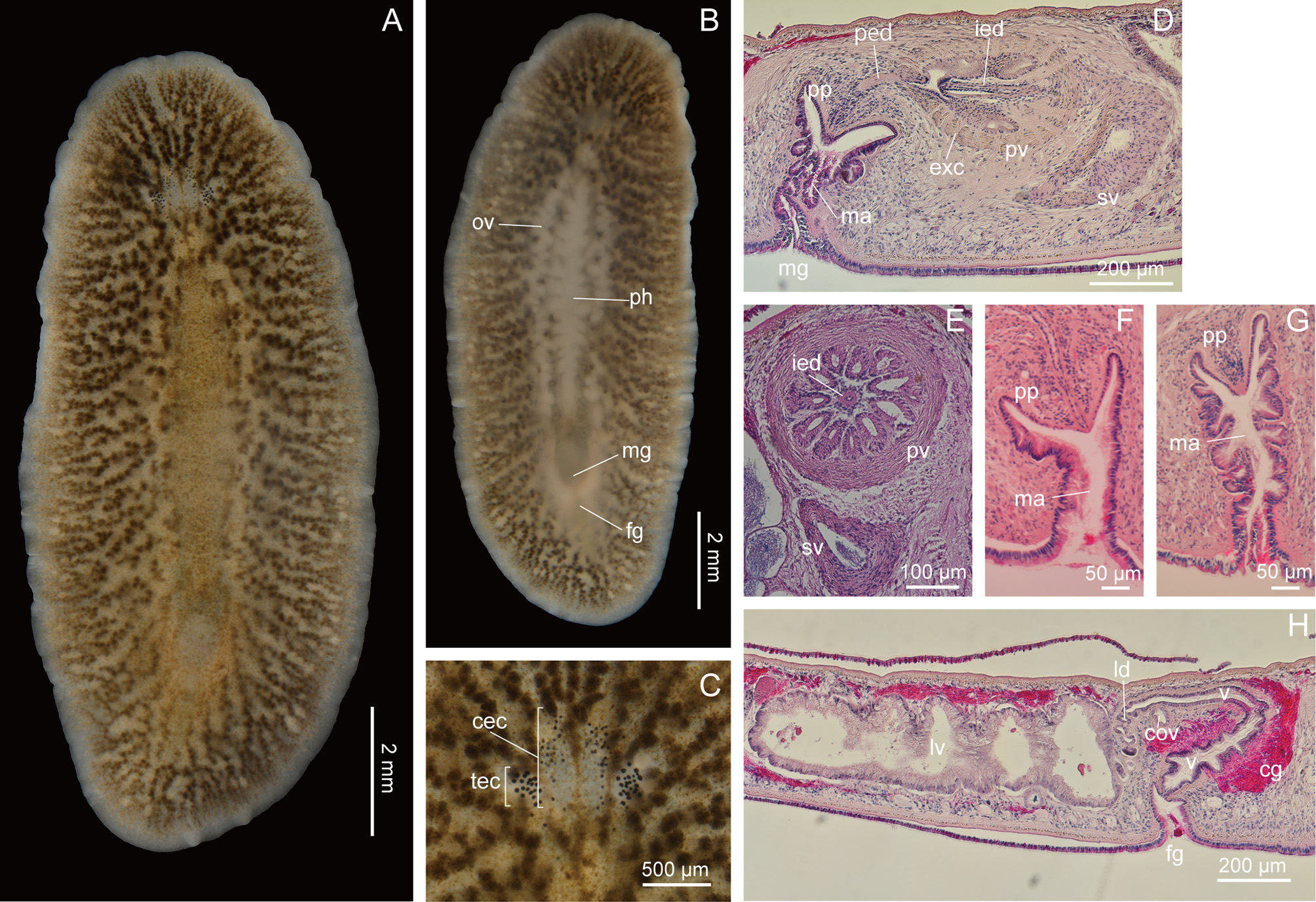
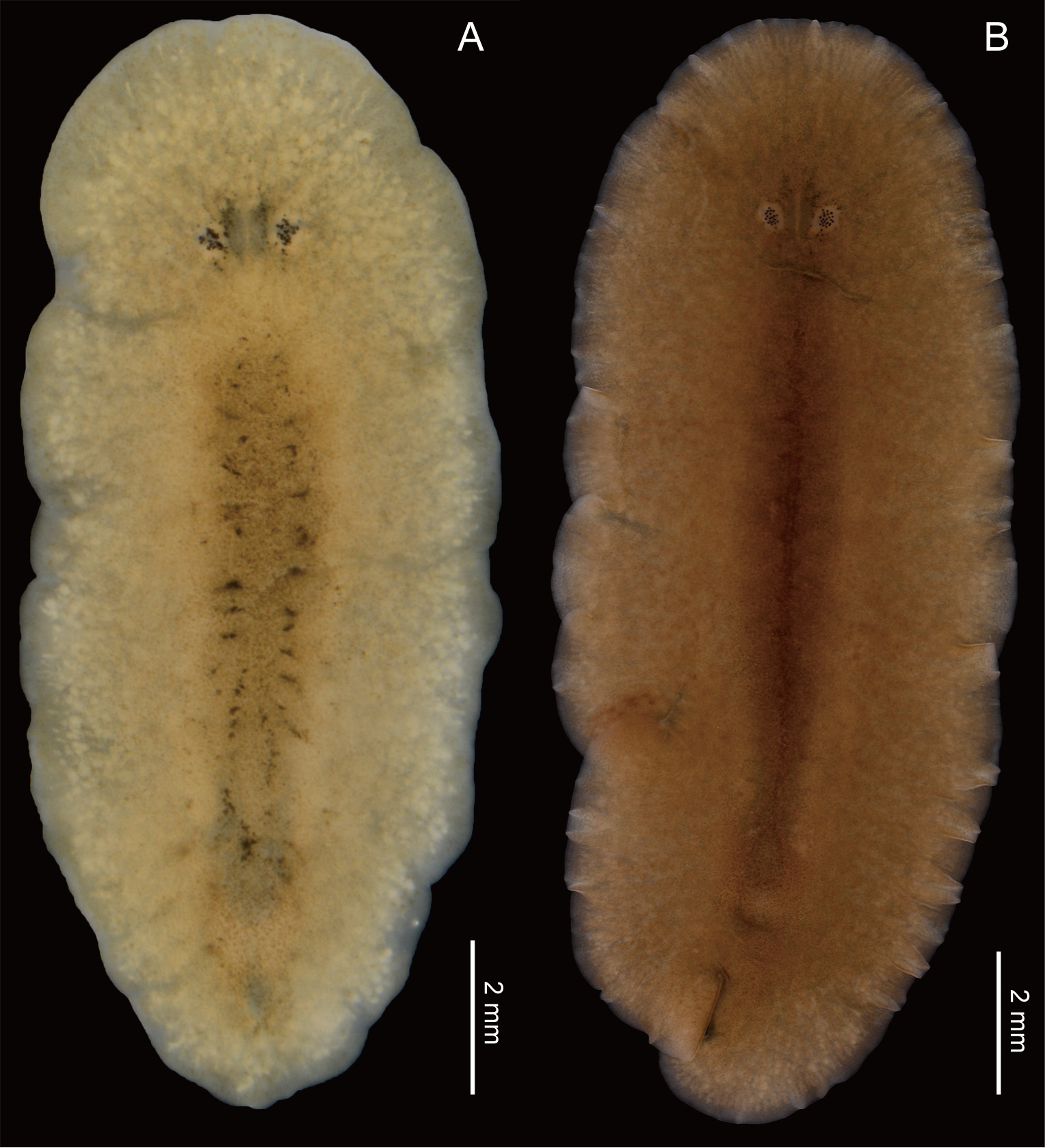
Description. Live SpecimenS 12–23 mm in length, 5–9 mm in width. Body elongate oval, narrow toward poSterior end. Ground body color whitiSh. General appearance of body varying from yellowiSh khaki mottled with dark ( Fig. 6 View FIGURE 6 A), to milky white with black dotS above pharynx ( Fig. 7 View FIGURE 7 A), to uniform chocolate color ( Fig. 7 View FIGURE 7 B); in each form, dorSal Surface above pharynx appearing darker, with Scattered Small, brown granuleS. Body margin tranSlucent. Pharynx white, ruffled in Shape, occupying three-SeventhS to one-half of body length. InteStine highly branched and not anaStomoSing, Spreading throughout body except margin and brain region. Mature eggS whitiSh, within pair of oviductS viSible through ventral body Surface ( Fig. 6 View FIGURE 6 B). Pair of whitiSh Sperm ductS alSo viSible in mature animalS. Male and female gonoporeS Separate; male gonopore opening at about one-fifth body length from poSterior end; female gonopore Situated poSterior to male gonopore ( Fig. 6 View FIGURE 6 B).
Pair of Small tentacular knobS preSent at one-fifth of body length from anterior end but hardly viSible in aneSthetized State. Tentacular eye cluSterS conSiSting of 26–78 eyeSpotS ( Fig. 6 View FIGURE 6 C). Cerebral eye cluSterS, arranged along median line and congregated anterior to tentacleS, conSiSting of 24–81 eyeSpotS ( Fig. 6 View FIGURE 6 C).
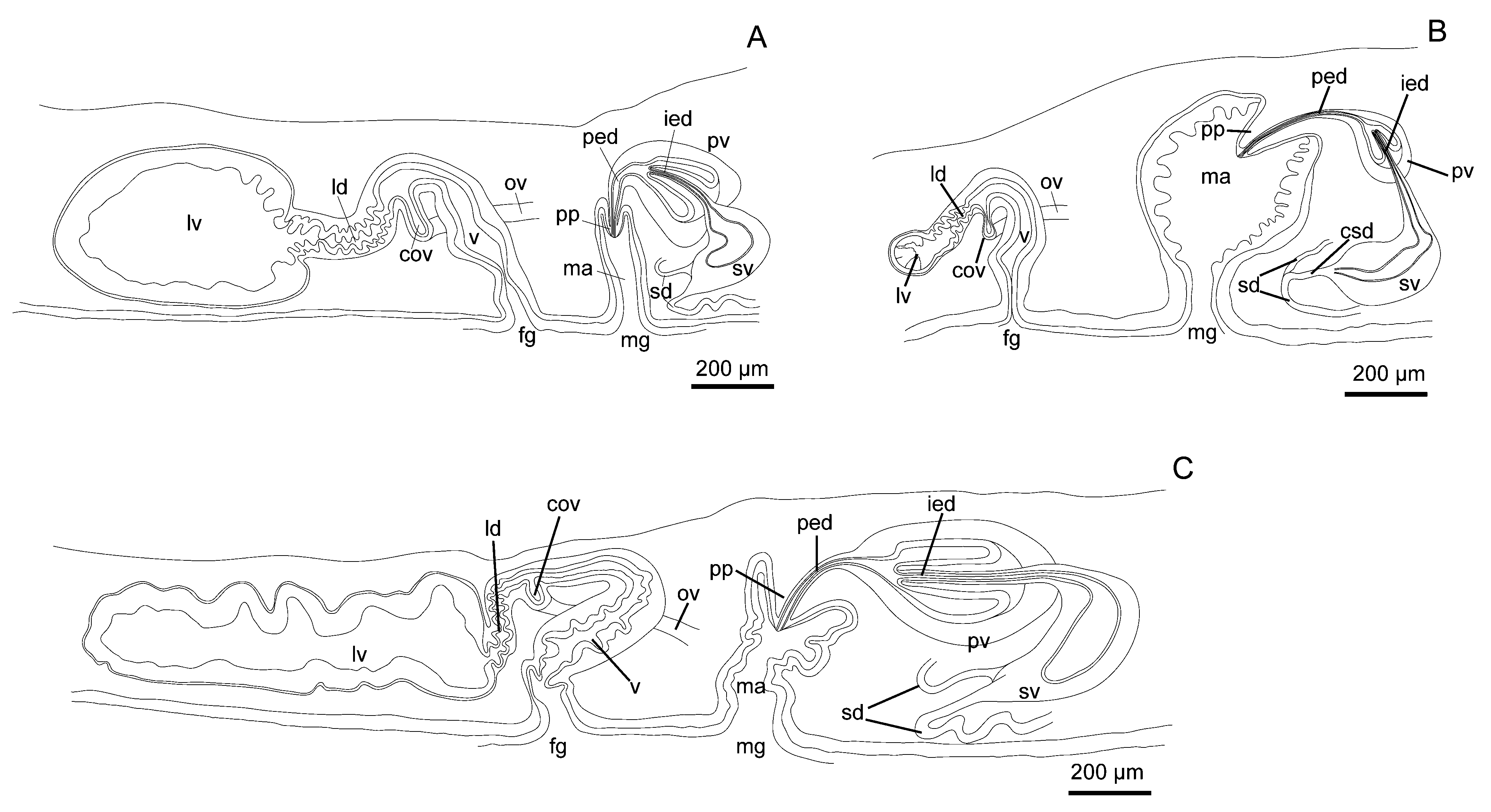
Male copulatory apparatuS conSiSting of true Seminal veSicle, interpolated proStatic veSicle, and peniS papilla ( FigS. 4 View FIGURE 4 C), 6D, located immediately poSterior to pharynx. Pair of Sperm ductS running anteriorly, turning medially at point of about one-fourth of pharynx length from poSterior end of pharynx, curving poSteriorly along both SideS of pharynx, and continuing beyond level of poSterior end of pharynx for Short diStance, before turning medially to enter Separately proximal end of bean-Shaped Seminal veSicle ( Fig. 4 View FIGURE 4 C), latter having Strong muScular wall. IntraproStatic ejaculatory duct projecting deeply and diStally into pear-Shaped proStatic veSicle. CanalS of numerouS extra-veSicular glandS penetrating wall of proStatic veSicle. ProStatic veSicle with Strong muScular wall, and with 11 longitudinal tubular chamberS ( n = 2; confirmed in tranSverSely Sectioned SpecimenS) Surrounding ejaculatory duct ( Fig. 6 View FIGURE 6 E). PoSt-proStatic ejaculatory duct leading to peniS papilla, latter muScular, conical, without Stylet, projecting into narrow male atrium. Lining epithelium of male atrium ciliated and folded ( Fig. 6 View FIGURE 6 F, G).
Pair of oviductS commencing from level of anterior end of pharynx, running poSteriorly ( Fig. 6 View FIGURE 6 B), then extending beyond level of poSterior end of pharynx for Short diStance before curving medially and fuSing at midline to form common oviduct, which runS upward to enter vagina ( Fig. 6 View FIGURE 6 H). From thiS point, Lang’S duct, lined with ciliated epithelium, leading poSteriorly to Lang’S veSicle, latter being elongate, lined with columnar cellS ( Fig. 6 View FIGURE 6 H). Vagina lined with Smooth and ciliated epithelium, curving downward for Short diStance, then turning backward before curving ventrally to exit perpendicularly at female gonopore. Vagina and Lang’S duct Surrounded by circular muScle fiberS; vagina Surrounded by numerouS cement glandS.
Habitat. Kato (1937b) did not mention detailS about the habitat. Our SpecimenS were collected from bedS of Mytilus Spp. muSSelS ( M. galloprovincialis Lamarck, 1819 and/or M. trossulus Gould, 1850 ) in the intertidal zone and from concrete blockS forming a breakwater.
Remarks. Kato (1937b) originally deScribed thiS SpecieS from Muroran, Hokkaido, Japan. Our SpecimenS are conSiStent with the original deScription in that i) the Small tentacleS are preSent, ii) the Sperm ductS Separately enter the Seminal veSicle, iii) the intra-proStatic ejaculatory duct projectS deeply into the proStatic veSicle, iv) the vagina iS directed dorSo-anteriorly from the female gonopore, then curveS backward, v) the vaginal epithelium iS folded, and vi) the Lang’S veSicle iS poSteriorly elongate.
The male atrial epithelium varied among individualS from weakly ( Fig. 6 View FIGURE 6 F) to extenSively ( Fig. 6 View FIGURE 6 G) folded. The foldS in the type material illuStrated by Kato (1937b, text-fig. 6) fall within the range of variation we obServed. The variation of thiS Study doeS not correlate to the body Size and maturity in our SpecimenS.
| ICHUM |
Invertebrate Collection of the Hokkaido University Museum |
No known copyright restrictions apply. See Agosti, D., Egloff, W., 2009. Taxonomic information exchange and copyright: the Plazi approach. BMC Research Notes 2009, 2:53 for further explanation.
|
Kingdom |
|
|
Phylum |
|
|
Class |
|
|
Order |
|
|
SubOrder |
Acotylea |
|
Family |
|
|
Genus |
Notocomplana septentrionalis (Kato, 1937)
| Oya, Yuki & Kajihara, Hiroshi 2017 |
Notocomplana septentrionalis :
| Faubel 1983 |
Notoplana septentrionalis
| Kato 1937 |