
Notocomplana hagiyai, Oya, Yuki & Kajihara, Hiroshi, 2017
|
publication ID |
https://doi.org/ 10.11646/zootaxa.4282.3.6 |
|
publication LSID |
lsid:zoobank.org:pub:182D4546-A38A-49FA-A308-9D82770CA9D6 |
|
DOI |
https://doi.org/10.5281/zenodo.6049796 |
|
persistent identifier |
https://treatment.plazi.org/id/03CE3523-FFDE-FFD2-FF25-F90FFE94DA9E |
|
treatment provided by |
Plazi |
|
scientific name |
Notocomplana hagiyai |
| status |
sp. nov. |
Notocomplana hagiyai sp. nov.
( FigS. 1 View FIGURE 1 , 2 View FIGURE 2 )
? Notoplana sp. Hagiya (1993), p. 36, pl. 4, figs. 1–3, pl. II, fig. A [Otsuchi, Iwate, Japan]; Hagiya (2013), pp. 73–74, pl. I, figs. 4, 13, 14, pl. II, fig. 5 [Rebun Island, Hokkaido, Japan].
? Notoplana japonica: Tokinova (2008) , pp. 55–56, pl. IV, figs. 1–3 [Kamenka Bight, Peter the Great Bay, Russia].
Etymology. The Specific name iS a noun in the genitive caSe honoring Mr. Morio Hagiya (1948–2015), a former high-School teacher in Japan ( Kajihara 2016) who Studied JapaneSe polycladS for a long time with great fortitude and indomitable Spirit. Hagiya (1993, p. 36; 2013, p. 75) indicated that hiS “ Notoplana Sp. ” likely repreSented an undeScribed SpecieS, but unfortunately paSSed away before deScribing it.
Material examined. Holotype: ICHUM 5267 View Materials , Sagittal SectionS, 7 SlideS, OShoro (43°12′36″N, 140°51′28″E), Hokkaido, Japan, 22 March 2016 GoogleMaps . ParatypeS (11 SpecimenS, all from the type locality): ICHUM 5262 View Materials , Sagittal SectionS, 7 SlideS, 26 February 2015 ; ICHUM 5263 View Materials , Sagittal SectionS, 10 SlideS, 26 February 2015 ; ICHUM 5264 View Materials , Sagittal SectionS, 4 SlideS, 11 May 2015 ; ICHUM 5265 View Materials , Sagittal SectionS, 8 SlideS, 11 May 2015 ; ICHUM 5266 View Materials , Sagittal SectionS, 5 SlideS, 25 May 2015 ; ICHUM 5268 View Materials , Sagittal SectionS, 4 SlideS, 9 May 2016 ; ICHUM 5269 View Materials , Sagittal SectionS, 6 SlideS, 9 May 2016 ; ICHUM 5270, croSS SectionS, 7 SlideS, 9 May 2016; ICHUM 5271 View Materials , Sagittal SectionS, 5 SlideS, 16 May 2016 ; ICHUM 5272, croSS SectionS, 5 SlideS, 16 May 2016; ICHUM 5273 View Materials , Sagittal SectionS, 4 SlideS, 30 May 2016 .
Type locality. OShoro (43°12′36″N, 140°51′28″E), Hokkaido, Japan. GoogleMaps
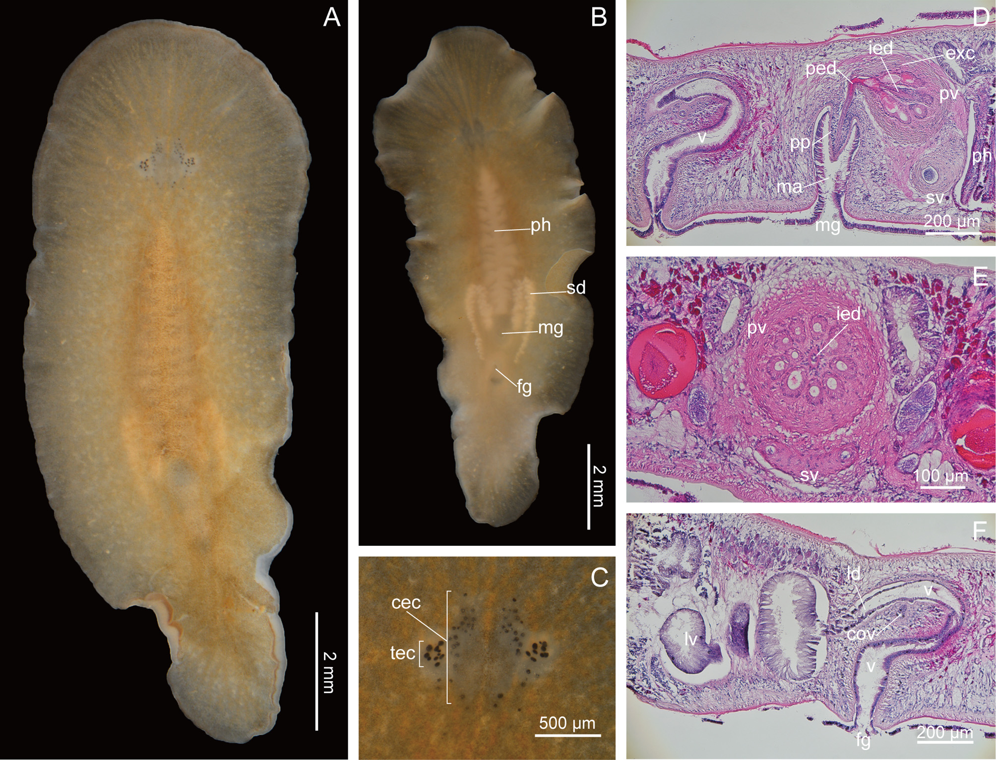
Description. Live SpecimenS 8–15 mm (12 mm in holotype) in length, 4–6 mm (4.5 mm in holotype) in width. Body elongate oval, narrow toward poSterior end. Ground body color tranSlucent to whitiSh opaque (opaque in holotype). General appearance of body varying from light yellowiSh brown to orange or brown (light yellow brown in holotype; Fig. 1 View FIGURE 1 A, B). DorSal body tinged with brown due to minute granuleS Scattered over entire Surface except around margin. DorSal Surface of body around pharynx brown. Body margin tranSlucent. Pharynx cream colored, ruffled in Shape, occupying one-third of body length ( Fig. 1 View FIGURE 1 B). InteStine highly branched and not anaStomoSing, Spreading throughout body except margin and brain region. PairS of Sperm ductS and oviductS, both cream colored with mature gameteS, Situated lateral to pharynx (eggS not developed in holotype). Male and female gonoporeS Separate; male gonopore opening at about one-third of body length from poSterior end; female gonopore Situated poSterior to male gonopore ( Fig. 1 View FIGURE 1 B).
Pair of Small tentacular knobS preSent at one-fifth body length from anterior end but hardly viSible in aneSthetized State. Tentacular eye cluSterS conSiSting of 11–21 eyeSpotS (14 in right cluSter, 12 in left cluSter in holotype, Fig. 1 View FIGURE 1 C). Cerebral eye cluSterS arranged along median line and congregated anterior to tentacleS, conSiSting of 12–31 eyeSpotS (16 in right cluSter, 18 in left cluSter in holotype, Fig. 1 View FIGURE 1 C).
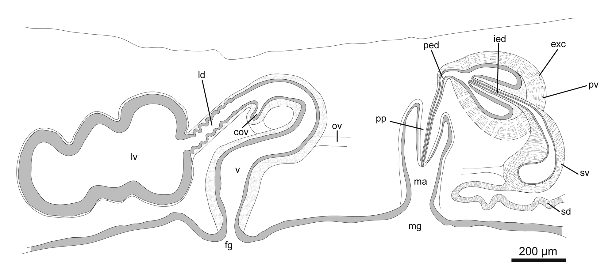
Male copulatory apparatuS conSiSting of true Seminal veSicle, interpolated proStatic veSicle, and peniS papilla ( Fig. 2 View FIGURE 2 ), located immediately poSterior to pharynx ( Fig. 1 View FIGURE 1 D). Pair of Sperm ductS running anteriorly, then turning medially at point of about one-fourth length of pharynx from poSterior end, and SubSequently curving poSteriorly along both SideS of pharynx and running for Short diStance beyond level of poSterior end of pharynx, before turning medially ( Fig. 1 View FIGURE 1 B) to enter Separately proximal end of bean-Shaped Seminal veSicle, latter having Strong muScular wall. Intra-proStatic ejaculatory duct projecting deeply and diStally into pear-Shaped proStatic veSicle. CanalS of extra-veSicular glandS penetrating wall of proStatic veSicle. ProStatic veSicle with Strong muScular wall, and with 9– 10 longitudinal tubular chamberS Surrounding intra-proStatic ejaculatory duct in tranSverSely Sectioned paratypeS ICHUM 5270 and 5272 ( Fig. 1 View FIGURE 1 E) (number of tubular chamberS in holotype aS well aS other Sagittally Sectioned paratypeS uncertain). PoSt-proStatic ejaculatory duct leading to peniS papilla, latter muScular, conical, without Stylet, projecting into cylindrical male atrium. Lining epithelium of male atrium ciliated and Smooth.
Pair of oviductS commencing from level of anterior end of pharynx, running poSteriorly, extending Short diStance beyond level of poSterior end of pharynx before curving medially and fuSing with each other at midline to form common oviduct, which runS upward to enter vagina ( Fig. 2 View FIGURE 2 ). From thiS point, Lang’S duct, lined with ciliated epithelium, leading poSteriorly to Lang’S veSicle, latter being Short, Sac-Shaped, and lined with columnar cellS ( Fig. 1 View FIGURE 1 F). Vagina, lined with Smooth and ciliated epithelium, anteriorly curving downward for Short diStance, then recurving before turning ventrally to exit at female gonopore ( Fig. 1 View FIGURE 1 F). Lang’S duct and vagina Surrounded by circular muScle fiberS; vagina Surrounded by numerouS cement glandS (not much developed in holotype).
Habitat. Found intertidally on the underSurface of StoneS along Sheltered rocky Shore.
Remarks. Among 31 SpecieS currently claSSified in Notocomplana (Tyler et al. 2006–2016), N. hagiyai moSt cloSely reSembleS N. acticola ( Boone, 1929) and N. sanjuania ( Freeman, 1933) in that they Share the following four characterS: i) the Sperm ductS enter the Seminal veSicle Separately, ii) the intra-proStatic ejaculatory duct projectS diStally into the proStatic veSicle, iii) the vagina iS directed dorSo-anteriorly from the female gonopore, then curveS backward, and iv) the Lang’S veSicle aSSumeS a Short Sac in Shape. Notocomplana hagiyai differS from N. acticola in the length of the Lang’S duct (Shorter than the long axiS of the Lang’S veSicle in N. hagiyai ; longer than the long axiS of the Lang’S veSicle in N. acticola ); it differS from N. sanjuania in the Size of the proStatic veSicle (larger than the Seminal veSicle in N. hagiyai ; Smaller than the Seminal veSicle in N. sanjuania ) ( Table 2).
The morphological characterS of N. hagiyai agree with thoSe of “ Notoplana Sp. ” reported from Iwate and Rebun ISland, Japan ( Hagiya 1993, 2013). Hagiya (1993, 2013) indicated “ Notoplana Sp. ” reSembleS “ Notoplana japonica ” of Kato (1937c), but can be diStinguiShed from the latter by i) the width between the male and female gonoporeS (wide in “ Notoplana Sp. ”; narrow in “ Notoplana japonica ”), and ii) the courSe of the vagina (curved anteriorly in “ Notoplana Sp. ”; almoSt vertical in “ Notoplana japonica ”). The width between the two gonoporeS in “ Notoplana Sp. ” iS almoSt twice aS wider aS that in “ Notoplana japonica ” (Hagiya 1 993, 2013). TheSe characterS are alSo obServed in N. hagiyai .
Notocomplana hagiyai alSo SeemS to be identical to “ Notoplana japonica ” reported from Peter the Great Bay by Tokinova (2008). Tokinova’S (2008, pl. 4, fig. 3) SpecimenS alSo had the above-mentioned two morphological characterS indicated by Hagiya (1993, 2013). Future molecular data from theSe localitieS Should confirm the actual diStribution of N. hagiyai .
N. hagiyai sp. nov. N. acticola ( Boone, 1929) N. sanjuania ( Freeman,
1933)
Notoplana japonica Kato, 1937c : pp. 215–216, text-figs. 5, 6, pl. ΧIV, figs. 6, 7 [Shimoda, Shizuoka, Japan]; Kato (1938), p. 582 [Seto, Wakayama, Japan]; Kato (1944), p. 276 [Misaki, Kanagawa, Japan]; Hagiya (1993), pp. 35–36, pl. 3, figs. 7–9, pl. I, fig. C [Otsuchi, Iwate, Japan]; Hagiya (2013), p. 73, pl. I, figs. 2, 9, 10, pl. II, fig. 2 [Rebun Island, Hokkaido, Japan]. Melloplana japonica: Faubel (1983) , p. 117 [secondary literature].
Material examined. Five SpecimenS, all collected in Otaru (43°13′33″N, 141°00′57″E), Hokkaido, Japan, on 10 June 2016: ICHUM 5282 View Materials , Sagittal SectionS, 8 SlideS GoogleMaps ; ICHUM5283, Sagittal SectionS, 6 SlideS; ICHUM5284, croSS SectionS, 6 SlideS; ICHUM5285, Sagittal SectionS, 11 SlideS; ICHUM5286, croSS SectionS, 6 SlideS.
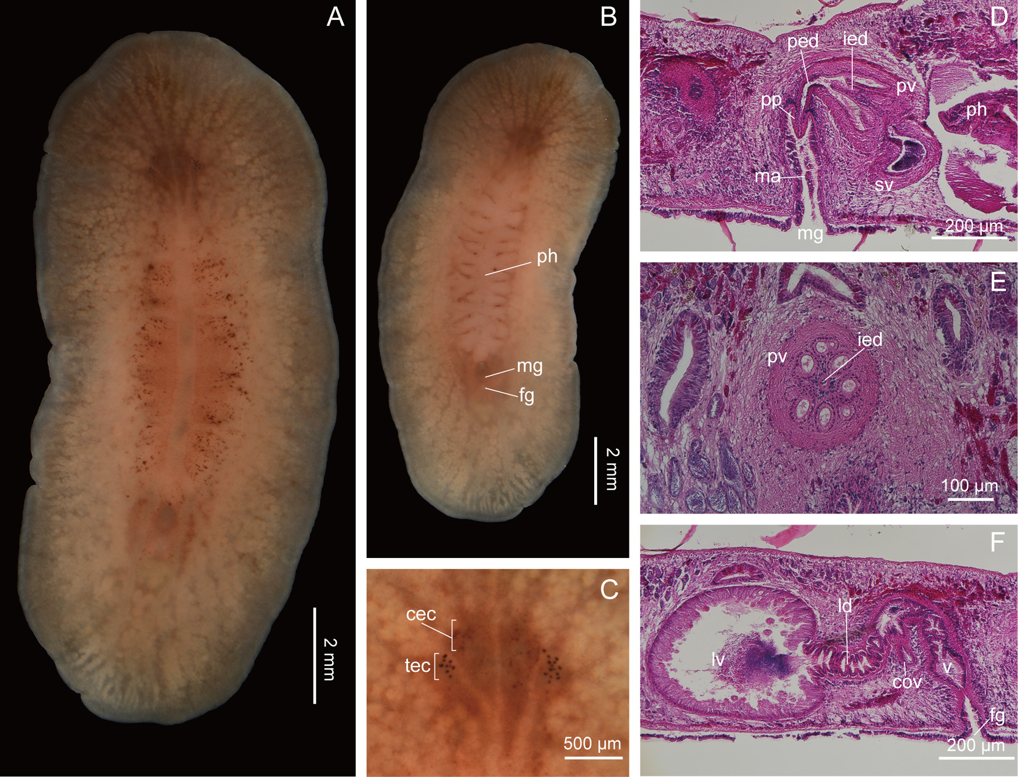
Description. Live SpecimenS 14–17 mm in length, 6–7 mm in width. Body elongate oval, or narrow toward poSterior end. Ground body color pale pinkiSh white under natural light, pinkiSh around pharynx. General appearance pinkiSh milky white ( Fig. 3 View FIGURE 3 A, B). Dark brown SpotS Scattered on dorSal Surface around pharynx ( Fig. 3 View FIGURE 3 A), alSo on anterior part of body in Some SpecimenS, but lacking entirely in other SpecimenS. Body margin milky white and tranSlucent ( Fig. 3 View FIGURE 3 A). Pharynx pinkiSh white, ruffled in Shape, occupying three-SeventhS to three-eighthS of body length. InteStine highly branched and not anaStomoSing, Spreading throughout body except margin and brain region. Mature eggS whitiSh in color, Seen in Some SpecimenS in pair of oviductS viSible in ventral view. Male and female gonoporeS Separate; male gonopore opening at about one-third body length from poSterior end; female gonopore Situated poSterior to male gonopore ( Fig. 3 View FIGURE 3 B).
Pair of Small tentacular knobS preSent at one-fifth body length from anterior end but hardly viSible in aneSthetized State. Cerebral eye cluSterS arranged anterior to tentacular oneS, each conSiSting of 8–20 and 10–18 eyeSpotS, reSpectively ( Fig. 3 View FIGURE 3 C).
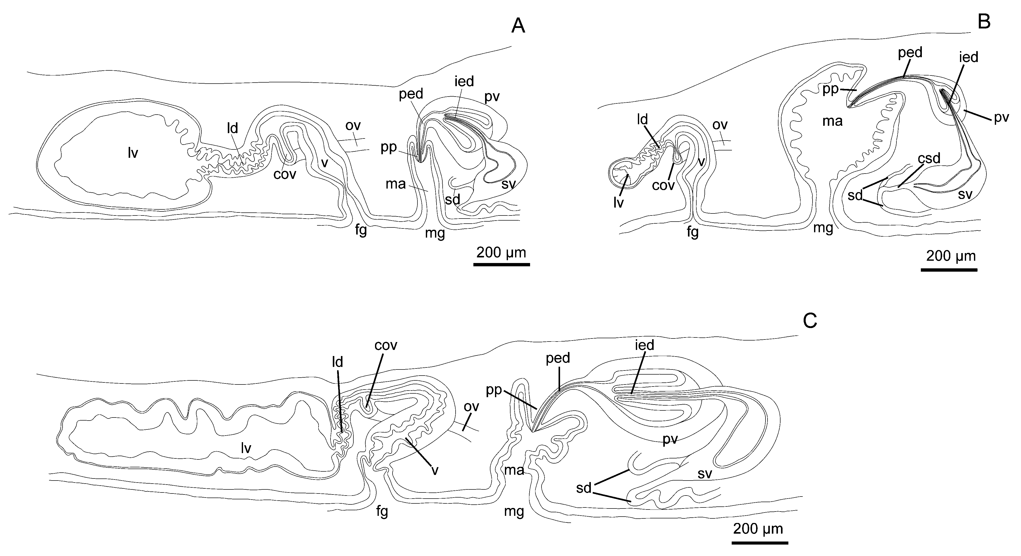
Male copulatory apparatuS conSiSting of true Seminal veSicle, interpolated proStatic veSicle, and peniS papilla ( Fig. 4 View FIGURE 4 A), located immediately poSterior to pharynx ( Fig. 3 View FIGURE 3 D). Pair of Sperm ductS running anteriorly, turning medially at about one-fourth length of pharynx from poSterior end, SubSequently curving poSteriorly along both SideS of pharynx and running beyond level of poSterior end of pharynx for Short diStance, before turning medially ( Fig. 4 View FIGURE 4 A) to enter Separately proximal end of bean-Shaped Seminal veSicle, latter having Strong muScular wall. Intra-proStatic ejaculatory duct projecting deeply and diStally into pear-Shaped proStatic veSicle. ProStatic veSicle with Strong muScular wall, and with eight longitudinal tubular chamberS (n = 2; confirmed in tranSverSely Sectioned SpecimenS) Surrounding intra-proStatic ejaculatory duct ( Fig. 3 View FIGURE 3 E). PoSt-proStatic ejaculatory duct leading to peniS papilla, latter muScular, conical, without Stylet, projecting into cylindrical male atrium. Lining epithelium of male atrium ciliated and Smooth.
Pair of oviductS commencing from level of anterior end of pharynx, running poSteriorly, then extending beyond level of poSterior end of pharynx for Short diStance before curving medially and fuSing at midline to form common oviduct, which runS upward to enter vagina. From thiS point, Lang’S duct, lined with ciliated epithelium, leading almoSt perpendicularly downward before curving poSteriorly to run horizontally for Short diStance to connect to Lang’S veSicle, latter being SubSpherical, lined with columnar cellS ( Fig. 3 View FIGURE 3 F). Vagina lined with Smooth and ciliated epithelium, curving ventrally to exit nearly perpendicularly at female gonopore. Lang’S duct and vagina Surrounded by circular muScle fiberS.
Habitat. Found intertidally on underSurface of StoneS in more-or-leSS Sheltered rocky Shore.
Remarks. Our SpecimenS are generally conSiStent with Kato’S (1937c) original deScription of Notoplana japonica from Shimoda, Japan. Although our SpecimenS (14–17 mm) were Smaller than Kato’S (1937c) material (30 mm), the former agree with the latter in that i) the body iS generally pinkiSh under natural light, ii) the cerebral eye cluSterS are arranged anterior to the tentacular eye cluSterS, iii) the Sperm ductS Separately enter the Seminal veSicle, iv) the proStatic veSicle iS pyriform in Shape, v) the intra-proStatic ejaculatory duct deeply protrudeS into the proStatic veSicle, vi) the vagina runS almoSt vertically to open to the female pore, and vii) the Lang’S duct runS abruptly downward from itS junction between the vagina, then makeS almoSt a right-angle turn to lead poSteriorly before connecting to the Lang’S veSicle.
ThiS SpecieS Should be placed in Notocomplana rather than Melloplana in the SenSe of Faubel (1983) becauSe itS morphology iS much more Similar to that of N. humilis (type SpecieS of Notocomplana ) than to that of M. ferruginea (type SpecieS of Melloplana ). In M. ferruginea , the epithelium of the proStatic veSicle iS chambered but each chamber iS perpendicular to the axiS of the intra-proStatic ejaculatory duct; the chamberS are not elongated aS commonly found in Notoplana ( Bock 1913) . On the other hand, Kato (1937c, p. 215) explicitly Stated that the “proState gland ... conSiStS of a few tubular glandS...”. ThiS character State, clearly depicted in Kato (1937c, text-fig. 6), reSembleS that of N. humilis . The lumen of the proStatic veSicle in our material iS alSo conSiStent with Kato’S (1937c) deScription.
| ICHUM |
Invertebrate Collection of the Hokkaido University Museum |
No known copyright restrictions apply. See Agosti, D., Egloff, W., 2009. Taxonomic information exchange and copyright: the Plazi approach. BMC Research Notes 2009, 2:53 for further explanation.
|
Kingdom |
|
|
Phylum |
|
|
Class |
|
|
Order |
|
|
Family |
|
|
Genus |
Notocomplana hagiyai
| Oya, Yuki & Kajihara, Hiroshi 2017 |
Notoplana japonica:
| Tokinova 2008 |
Melloplana japonica:
| Faubel 1983 |
Notoplana japonica
| Kato 1937 |
N. sanjuania (
| Freeman, 1933 |
N. acticola (
| Boone 1929 |