
Enhydrosoma apimelon, Karanovic, Tomislav, Kim, Kichoon & Lee, Wonchoel, 2015
|
publication ID |
https://doi.org/ 10.11646/zootaxa.3990.4.1 |
|
publication LSID |
lsid:zoobank.org:pub:90BE4EAF-8594-4164-AE26-4F47E04A4A5D |
|
DOI |
https://doi.org/10.5281/zenodo.5616522 |
|
persistent identifier |
https://treatment.plazi.org/id/03CE8781-FFD3-E643-6180-2CC4FD2AE085 |
|
treatment provided by |
Plazi |
|
scientific name |
Enhydrosoma apimelon |
| status |
sp. nov. |
Enhydrosoma apimelon sp. nov.
( Figs. 1–9 View FIGURE 1 View FIGURE 2 View FIGURE 3 View FIGURE 4 View FIGURE 5 View FIGURE 6 View FIGURE 7 View FIGURE 8 View FIGURE 9 )
Type locality. South Korea, South Sea, Gwangyang Bay, sampling station 10 (see Karanovic et al. 2014), muddy sediments, 3455'15.4" N 12747 View Materials '07.9"E.
Specimens examined. Holotype female (NIBRIV 0000287210) preserved in 70% ethanol. Paratypes: two males and three females (NIBRIV 0000287211) preserved in 70% ethanol; one dissected female (NIBRIV 0000287212) mounted on nine slides; one dissected male (NIBRIV 0000287213) mounted on nine slides; one male and 12 females together on one SEM stub (NIBRIV 0000287214); three males and three females together on another SEM stub (NIBRIV 0000287215); collected on 8 December 2012 by K. Kim. One male and two females used for molecular analyses were collected by K. Kim on 8 December 2012 and 26 April 2012, respectively (see Table 1 View TABLE 1 ).
Etymology. The species name derives from the Greek adjective apimelos, meaning “slender”, “without fat”, “lean”, and refers to the slender shape of the caudal rami. It was treated as an adjective, agreeing in gender with the neuter generic name.
Species Cοde Sex Cοuntry Lοcality Statiοn Cοοrdinates Date Bases GenBank Description of female. Based on holotype and several paratypes. Total body length, measured from anterior margin of rostrum to posterior margin of caudal rami, from 291 to 335 µm (mean = 309 µm, n = 10). Colour of preserved specimens yellowish; live specimens not observed. Nauplius eye not visible. Prosome comprising cephalothorax with completely fused first pedigerous somite, and three free pedigerous somites; urosome comprising first urosomite (= fifth pedigerous somite), genital double-somite (fused genital and third urosomites) and three free urosomites (last one being anal somite). No sclerotized joint between prosome and urosome. Habitus ( Figs. 1 View FIGURE 1 A, 2A, 3A) almost cylindrical in dorsal view, widest at posterior end of cephalothorax and tapering posteriorly, boundary between prosome and urosome inconspicuous; prosome/urosome length ratio about 1.3, and prosome only slightly more voluminous than urosome. Body length/width ratio close to 3.5 in dorsal view; cephalothorax only about 1.3 times as wide as genital double-somite. Free pedigerous somites without lateral or dorsal expansions but heavily sculptured; pleurons only partly covering coxae of swimming legs in lateral view. Integument of all somites strongly sclerotized, generally very rough, without discernible cuticular windows or pits, but with characteristic relief of numerous surface ridges and depressions; majority of cuticular depressions and posterior margin of somites covered by dense hair-like spinules, bacterial growth, and detritus, making observation of cuticular pores and sensilla in those regions very difficult. Hyaline fringe of all somites narrow and rough. In addition to hair-like spinules, surface ornamentation of somites and caudal rami consisting of at least two different types of sensilla (slender and bottle-shaped), simple cuticular pores, tubular pores, and few large spinules; exact number of pores and spinules difficult to establish.
Rostrum ( Figs. 1 View FIGURE 1 B, 3B, E) small, fused to cephalic shield, dorsally recurved in lateral view, bifid at tip, bearing two sensilla between apical horn-like projections, pore located on ventral side; space between sensilla greater than width of one apical horn.
Cephalothorax ( Figs. 1 View FIGURE 1 B, C, 2H, 3B) tapering anteriorly in dorsal view, about as long as wide; comprising 30% of total body length. Surface of cephalic shield with at least 19 pairs of large sensilla (not counting those on rostrum), of those four pairs along posterior margin on conical mound-looking protuberances and four pairs along lateral margin. Majority of dorsal sensilla in depressions, majority of lateral sensilla on ridges. Interior surface of lateral margin of cephalic shield with characteristic comb of long setules in anterior part ( Fig. 2 View FIGURE 2 H), touching basis of antenna. Lateral surface of cephalic shield with two large semi-circular depressions, dorsal surface with three central and six paired small and oval depressions; long and strong ridge separating dorsal and lateral depressions.
Pleuron of second pedigerous somite (first free) ( Figs. 1 View FIGURE 1 A, B, 3A, B) with irregular and shallow relief, posterior margin with at least four pairs of sensilla on conical mound-looking protuberances and row of sparse hairlike spinules; ridge between lateral and dorsal side pronounced but not produced posteriorly; posterior lateral depression large, triangular.
Third pedigerous somite ( Figs. 1 View FIGURE 1 A, 3A, C) slightly shorter than second pedigerous somite but of similar width; pleuron relief with two smooth dorsal triangles with sensilla on posterior tip, dorso-lateral ridges more pronounced than in second pedigerous somite and slightly produced posteriorly; low laying surface in between triangles and dorso-lateral ridges covered with dense pattern of small, hair-like spinules; posterior margin with at least three pairs of sensilla on conical mound-looking protuberances (in addition to dorsalmost pair on triangles) and row of sparse but long hair-like spinules.
Fourth pedigerous somite ( Figs. 1 View FIGURE 1 A, 3A, C) similar in size, shape, relief and ornamentation to third pedigerous somite, with slightly more posteriorly produced dorso-lateral ridges and longer dorsal triangles.
First urosomite ( Figs. 1 View FIGURE 1 A, 3A) slightly longer but not narrower than fourth pedigerous somite; pleuron without free lateral margin; relief similar to that of fourth pedigerous somite but with more pronounced dorso-lateral ridges and smaller and less clearly defined dorsal triangles; posterior margin with at least four pairs of sensilla.
Genital double-somite ( Figs. 1 View FIGURE 1 A, 2A, C, 3A, 5A) 1.3 times as wide as long in ventral view; completely fused ventrally but with deep dorso-lateral suture indicating original segmentation between genital and third urosomites, thus dividing double-somite into equally long and similarly wide halves. Ventral surface relatively smooth, flat, with shallow relief but dorso-lateral and ventro-lateral ridges pronounced together with pronounced dorsal triangular structures result in almost hexagonal cross-section; dorso-lateral and ventro-lateral ridges significantly produced posteriorly, each bearing single large sensilla on tip. Two additional ventral and two dorsal sensilla visible on posterior margin, and at least three pairs of sensilla visible in anterior half; both anterior and posterior half with ventro-lateral pair of simple pores; posterior margin with row of hair-like spinules, and one ventral row of strong spinules in anterior half, at base of genital operculum. Female genital complex ( Fig. 5 View FIGURE 5 A) weakly sclerotized and hardly distinguishable from internal sutures and soft tissue, except large copulatory pore in posterior half and wide copulatory duct; genital aperture ventrally in proximal quarter, covered by reduced sixth legs acting as genital operculum.
Fourth urosomite (third free) ( Figs. 1 View FIGURE 1 A, 2A, C, 3A, 5A) similar in shape and ornamentation to posterior part of genital double-somite, except narrower and with tubular ventro-lateral pores; posterior projections of ventro-lateral and dorso-lateral ridges long, each with apical sensilla.
Fifth urosomite (preanal) ( Figs. 1 View FIGURE 1 A, F, G, H, 2A, 5A) shorter and narrower than third urosomite, with shorter posterior projections of dorso-lateral and ventro-lateral ridges, without sensilla, ornamented with pair of ventrolateral tubular pores, posterior row of long hair-like spinules, and several minute spinules on ventro-lateral ridge.
Anal somite ( Figs. 1 View FIGURE 1 F, G, H, 2B, G, 3D, 5A, B, C) clefted medially in posterior fifth, with one pair of large dorsal sensilla at base of anal operculum, one pair of lateral tubular pores, one pair of ventral tubular pores, and several stronger terminal spinules. Dorso-lateral and medial corners produced into small serrated flaps; anal operculum ( Fig. 3 View FIGURE 3 D) semi-circular, long, wide, reaching beyond posterior margin of somite, strongly serrated, representing nearly half of somite's width. Anal sinus ( Fig. 1 View FIGURE 1 G, H) widely open, with one dorsal and two diagonal rows of long hair-like spinules.
Caudal rami ( Figs. 1 View FIGURE 1 F, G, H, 2B, G, 3D, 5A, B, C) long, slender, about twice as long as anal somite, widest at base, gently tapering posteriorly, about 3.2 times as long as wide (ventral view), slightly divergent and nearly cylindrical, separated by distance of about ramus width; armature consisting of seven setae (three lateral, one dorsal, three apical). Ornamentation consisting of lateral tubular pore and numerous hair-like spinules on all sides, those on inner margin anteriorly especially long (see Fig. 2 View FIGURE 2 B), those on outer posterior corner forming dense tuft ( Fig. 1 View FIGURE 1 G); dorsal seta ( Fig. 2 View FIGURE 2 G) smooth, slender, inserted close to inner margin at about first quarter of ramus’ length, approximately half as long as caudal ramus, triarticulate at base (i.e. inserted on two pseudojoints); two proximal lateral setae smooth, of equal length, 0.24 times as long as ramus, inserted very close to each other at about two fifths of ramus length, very close to lateral tubular pore. Ancestral distal lateral seta also smooth, only slightly longer than proximal lateral setae, inserted on outer posterior corner of caudal ramus, flanked by tuft of spinules; inner apical seta smooth and very slender, about as long as proximal lateral setae. Principal apical setae fused basally, both without breaking planes and sparsely pinnate; middle apical seta much stronger and longer, about 3.7 times as long as outer apical one and 1.2 times as long as caudal ramus.
Antennula ( Figs. 1 View FIGURE 1 D, 3E, 6 A–H) short, stout, five-segmented, joined to cephalothorax with small triangular pseudo-segment, approximately half as long as cephalothorax, without cuticular processes, with three rows of strong spinules on first segment. Long aesthetasc on third segment relatively slender, fused basally with adjacent large seta, reaching slightly beyond tip of appendage; even more slender and much shorter apical aesthetasc on fifth segment fused basally with two apical setae, forming apical acrothek. Armature formula: 1.9.7+ae.1.11+ae. Seta on first segment, two setae on second segment, two setae on third segment, and three setae on fifth segment pinnate, all other setae smooth; one pinnate seta on third segment and all pinnate setae on fifth segment recurved, spiniform, with very strong spinules only along convex surface, all other setae slender; six lateral smooth setae on fifth segment biarticulate at base (i.e. inserted on small pseudojoint); dorsal seta on second segment inserted into funnelshaped depression. Length ratio of antennular segments, measured along caudal margin and from proximal end as:, 1: 4: 2: 0.6: 4.5.
Antenna ( Figs. 2 View FIGURE 2 H, 6I) relatively short, composed of coxa, allobasis, one-segmented endopod and onesegmented exopod. Coxa short, with arched row of long posterior spinules. Allobasis longest and most robust segment of antenna, more than three times as long as coxa and about 1.1 times as long as endopod, widest at base and about 2.5 times as long as wide, unarmed, with several long spinules along inner (convex) margin in proximal half. Endopod about as wide as distal part of allobasis, almost cylindrical, about three times as long as wide, with two subdistal surface frills, and two diagonal rows of very large spinules on anterior surface; two strong, pinnate lateral spines flanking minute seta. Apical armature consisting of five elements: two stout but short unipinnate spines, two slender geniculate setae plus massive pectinate spine. Exopod short, forked distally, about as long as coxa but much narrower, with several slender spinules along outer margin, with two apical bipinnate setae; both exopodal setae slender, long, inner seta about 1.3 times as long as outer one, with longer spinules.
Labrum ( Fig. 2 View FIGURE 2 H) large and complex tri-dimensional structure, trapezoidal in anterior view, rigidly sclerotized, with relatively wide, somewhat convex cutting edge, with subapical row of strong spinules and many apical rows of slender spinules and along posterior surface, additional short row of slender long spinules centrally on anterior surface.
Paragnaths ( Fig. 2 View FIGURE 2 H) also forming complex tri-dimensional structure, trilobate, with two ellipsoid anterior lobes and one central posterior lobe, all fused at base, all lobes with numerous rows of slender anterior and apical spinules; posterior surface with single transverse row of extremely long spinules.
Mandibula ( Figs. 2 View FIGURE 2 F, 7H) small, with narrow cutting edge on relatively long coxa, with two strong bicuspidate teeth ventrally, three unicuspidate teeth dorsally, and single dorsalmost slender and smooth seta; dorsalmost tooth longest, ventralmost tooth strongest; dorsal seta slightly shorter than dorsalmost tooth. Palp small, one-segmented, bifurcated distally, proximal half as long and wide as outer branch, inner branch much shorter; proximal part smooth, while each branch with long spinules on anterior surface; inner branch with single bipinnate apical seta, with very strong and long spinules; outer branch with two slender and sparsely bippinate apical setae; all palpal setae slender, subequally long, about 1.5 times as long as palp.
Maxillula ( Figs. 1 View FIGURE 1 E, 2F, H, 8A) relatively small; praecoxa large, with two outer rows of spinules; praecoxal arthrite about as long as wide, with two large tube setae on anterior surface and four distal elements (three naked spines and one naked seta), transverse row of spinules on posterior surface. Coxa short, with two distal setae (one pinnate and one naked) and row of spinules on posterior surface; exopod and endopod fused to basis, as long as praecoxal arthrite but much more slender, with six naked setae and row of spinules on posterior surface.
Maxilla ( Figs. 1 View FIGURE 1 E, 2F, H, 7I) small; syncoxa with three spinular rows on anterior surface and two endites; syncoxal proximal endite with one pinnate stout spine, one slender seta, and one tube seta Syncoxal distal endite similar to proximal endite, except slightly larger and tube seta shorter; basis with short row of spinules and one endite; basal endite with smooth spine distinct at base, tube seta on posterior surface, naked seta and short tubular pore distally.Endopod represented by two tubular setae fused basally.
Maxilliped ( Figs. 1 View FIGURE 1 E, 2F, H, 8B) prehensile, three-segmented, composed of coxa, basis, and one-segmented endopod. Coxa slender, about twice as long as wide, cylindrical, unornamented, armed with single strong bipinnate seta on inner-distal corner. B basis largest and longest segment, about 2.2 times as long as wide and 1.6 times as long as coxa, with longitudinal row of slender inner spinules, unarmed. Endopod minute, subrectangular, apically with two one short and smooth and one smooth recurved claw; endopodal claw fused to endopod at base, 1.2 times as long as coxal seta and about 1.4 times as long as basis.
All swimming legs ( Figs. 1 View FIGURE 1 A, 2A) of similar size, long in comparison to body length, composed of small triangular and unarmed praecoxa, large rectangular and unarmed coxa, shorter and nearly pentagonal basis, slender three-segmented exopod, and even more slender two-segmented endopod; none of exopods or endopods prehensile. Pairs of legs joined by simple intercoxal sclerite.
First swimming leg ( Figs. 3 View FIGURE 3 F, 8C) with smooth, short intercoxal sclerite, distal margin slightly concave. Praecoxa subtriangular, shorter than intercoxal sclerite and about as long as coxa, ornamented with distal row of hair-like spinules on anterior surface. Coxa 2.5 times as wide as long, with longitudinal row of long outer spinules on anterior surface and another inner row of much smaller spinules, both without spinules along distal margin. Basis with one short but strong and finely bipinnate outer spine and one longer and stronger, also bipinnate inner spine, inner spine about 1.4 times as long as outer spine; ornamentation of basis consists of posterior row of very strong spinules, short row of more slender spinules at base of inner spine, and short row of strong spinules at base of outer spine, all on anterior surface. Exopod with all segments of about same length, each about 1.5 times as long as wide and with outer strong and very long spinules and subdistally on anterior surface and more slender spinules along inner margin; first and second exopodal segment each armed with single large outer spine, longer than segment; third exopodal segment with two strong and pinnate outer spines and two slender and very long apical setae; exopodal setae of about same length, 1.6 times as long as entire exopod, and about 2.6 times as long as distalmost exopodal spine, not prehensile, with short outer pinnules and long and sparse inner pinnules. Endopod 0.7 times as long as exopod, also with strong outer and slender inner spinules; first endopodal segment half as long as shortest exopodal segment, about as long as wide, unarmed; second endopodal segment slender, about 5.6 times as long as wide and 4.2 times as long as first endopodal segment, with one strong subapical outer spine, and long apical seta; apical endopodal seta slightly longer than apical exopodal setae, about three times as long as second endopodal segment, and 4.4 times as long as subapical endopodal spine.
Second swimming leg ( Fig. 8 View FIGURE 8 D) similar to first swimming leg, except for shorter and wider intercoxal sclerite, without inner basal spine, first endopodal segment smaller, and second endopodal segment more slender and armed with two long setae.
Third swimming leg ( Fig. 9 View FIGURE 9 A) similar to second swimming leg, except for more slender outer seta of basis, and third exopodal segment with additional inner slender seta.
Fourth swimming leg ( Fig. 9 View FIGURE 9 B) as third swimming leg.
Fifth leg ( Figs. 2 View FIGURE 2 C, D, E, 6J) biramous, comprising conical exopod and baseoendopod. Exopod longer than endopodal lobe and partly covered by it in ventral view, armed with two outer short spines and one longer apical spine, ornamented with several hair-like spinules and two tubular pores on anterior surface and distal frill on posterior surface. Baseoendopod armed with two inner strong spines, and one apical minute seta fused to several basal spinules, ornamented with numerous hair-like spinules on anterior surface and one long tubular pore at base of each inner spine; apical exopodal spine about 1.2 times as long as proximal endopodal spine, 1.4 times as long as distal endopodal spine, 2.4 times as long as either of outer exopodal spines, and nearly 0.6 times as long as entire exopod.
Sixth legs ( Fig. 5 View FIGURE 5 A) fused into simple genital operculum, with row of strong spinules at its base; each leg armed with smooth inner seta and unipinnate outer spine; seta slightly longer than spine, both armature elements shorter than some spinules at base of sixth leg.
Description of male. Based on five paratypes. Body length from 288 to 319 Μm (mean = 307 µm, n = 5). Genital somite and third urosomite not fused. Habitus ( Figs. 3 View FIGURE 3 G, 4A), colour, rostrum ( Figs. 3 View FIGURE 3 H, 4G), shape and ornamentation of cephalothorax ( Figs. 3 View FIGURE 3 G, H), shape and ornamentation of free prosomites ( Fig. 3 View FIGURE 3 G), shape and ornamentation of first urosomite ( Fig. 3 View FIGURE 3 G), general shape in dorsal view and most dorsal ornamentation of other urosomites ( Fig. 3 View FIGURE 3 G), general shape and armature of caudal rami ( Figs. 3 View FIGURE 3 G, 4B, 5D), antenna ( Fig. 4 View FIGURE 4 H), labrum, paragnaths, mandibula, maxillula, maxilla, maxilliped, and all swimming legs ( Fig. 4 View FIGURE 4 A) as in female. Prosome/ urosome ratio about 1.2, greatest width at posterior end of cephalothorax, body length/width ratio about 3.7; cephalothorax 1.5 times as wide as genital somite in dorsal view.
Genital somite ( Figs. 3 View FIGURE 3 G, 4C, 5D) homologous to anterior part of genital double-somite in female but much narrower compared to other parts of urosome, 1.5 times as wide as long in ventral view, with all sensilla and pores homologous to those in female, no additional sensilla and pores observed; ventral surface without ridges or ornamentation, except spinules as base of sixth leg. Right sixth leg developed into genital operculum, while left sixth leg completely reduced, posterior short row of slender spinules as its only remnant; no spermatophore visible inside any examined specimens.
Third urosomite ( Figs. 3 View FIGURE 3 G, 4C, 5D) homologous to posterior part of genital double-somite in female but proportionately much narrower, without ventro-lateral cuticular pores, and with ventral posterior row of very strong spinules.
Fourth urosomite ( Figs. 3 View FIGURE 3 G, 4B, 5D) also narrower than in female, without tube on its ventro-lateral pores, and with medially interrupted ventral posterior row of very strong spinules.
Fifth urosomite ( Figs. 3 View FIGURE 3 G, 4B, 5D) similar to that in female, only proportionately slightly narrower in ventral view.
Anal somite ( Figs. 3 View FIGURE 3 G, 4B, 5D) similar to that in female but posterior-medial cluster of spinules not fused together.
Caudal rami ( Figs. 3 View FIGURE 3 G, 4B, 5D) similar to those in female but longer in proportion to anal somite and generally more elongated; armature and ornamentation as in female.
Antennula ( Figs. 3 View FIGURE 3 H, 4A, D–F, 7A–G) much longer than in female, almost as long as cephalothorax, strongly geniculate, six-segmented, with geniculation between fourth and fifth segments, without strong spiniform setae; segments in geniculation sclerotized, without cuticular ridges along anterior surface; first two and last to segments clearly homologous to those in female, but all except first two segments with numerous differences in shape and/or armature and ornamentation; second segment also with characteristic dorsal seta in funnel-shaped depression ( Fig. 3 View FIGURE 3 H) as in female; third segment small, triangular, without clear homology with segments on female, unornamented, with seven slender setae; fourth segment largest and strongest, with continuous row of strong spinules on dorsal surface directed anteriorly ( Fig. 3 View FIGURE 3 H), with characteristic brush-like organ on anterior surface ( Fig. 4 View FIGURE 4 E), large aesthetasc as in female, and 11 smooth and slender setae, one of them fused basally with aesthetasc ( Fig. 4 View FIGURE 4 D). Penultimate segments with single slender seta as in female ( Fig. 4 View FIGURE 4 E) but significantly more elongated; last segment with distal tip produced into powerful spiniform process ( Fig. 4 View FIGURE 4 F), another smaller process near midlength, acrothek as in female but displaced to about midlength, six lateral slender setae as in female on pseudojoints, no robust spiniform setae.
Fifth leg ( Figs. 4 View FIGURE 4 C, 7J) somewhat smaller than in female but with similar basic structure; exopod with one lateral armature element, longer spinules, longer tubular pore, lacking distal frill. Endopodal spines inserted closer to each other, proximal spine significantly longer than distal spine; endopodal lobe without distal frill of fused spinules and without distal seta, with tubular pore on tip instead. Apical exopodal element about 1.3 times as long as proximal endopodal spine, 2.7 times as long as distal endopodal spine, 1.8 times as long as lateral exopodal element, and 1.3 times as long as exopod.
Sixth legs ( Figs. 4 View FIGURE 4 C, 5D) simple cuticular plates, unarmed, with transverse row of slender, long spinules; left leg completely fused with somite, right leg transformed into functional genital operculum.
Variability. Despite numerous examined specimens and detailed examination using SEM, we are not able to report on any significant morphological variability.
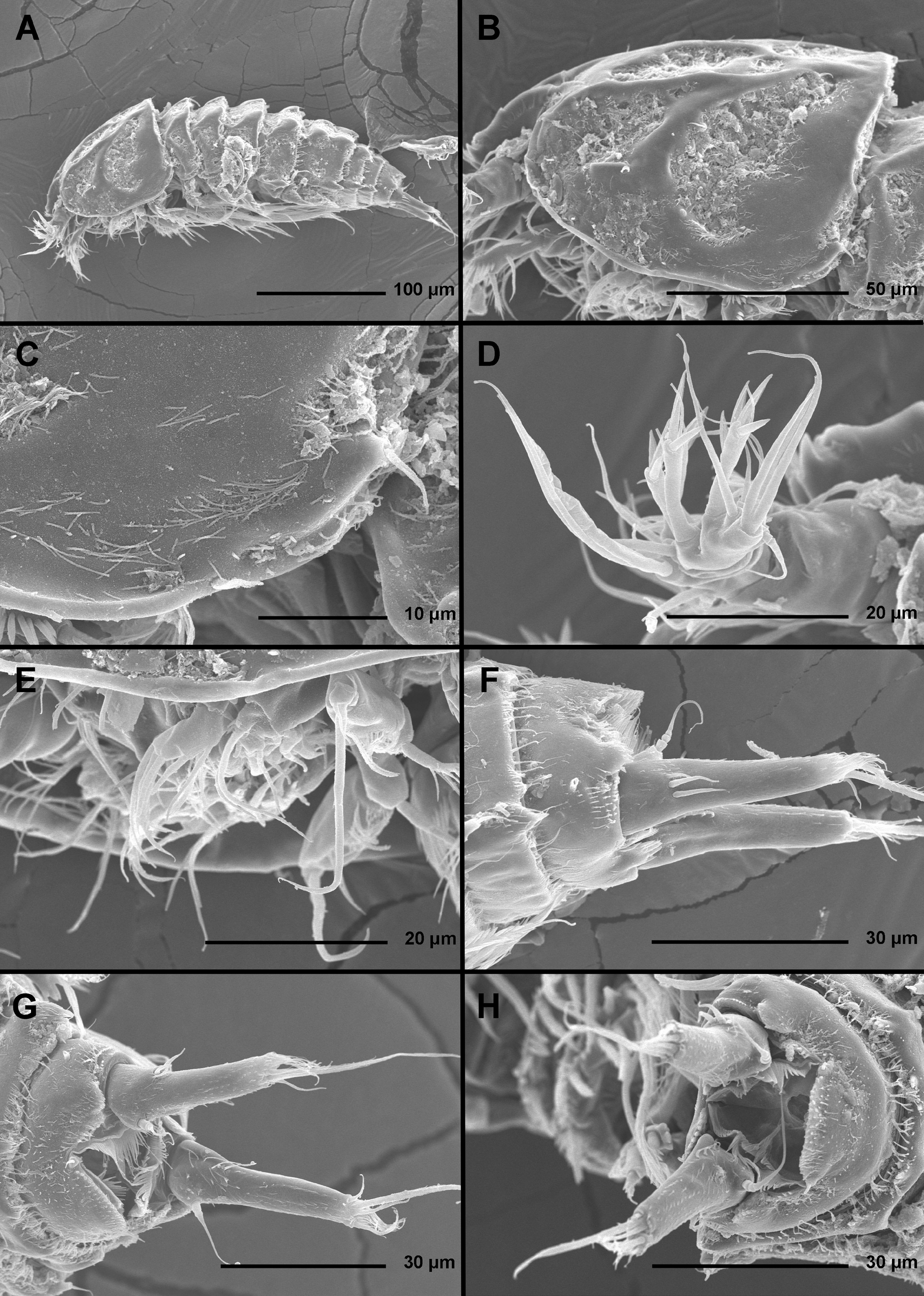
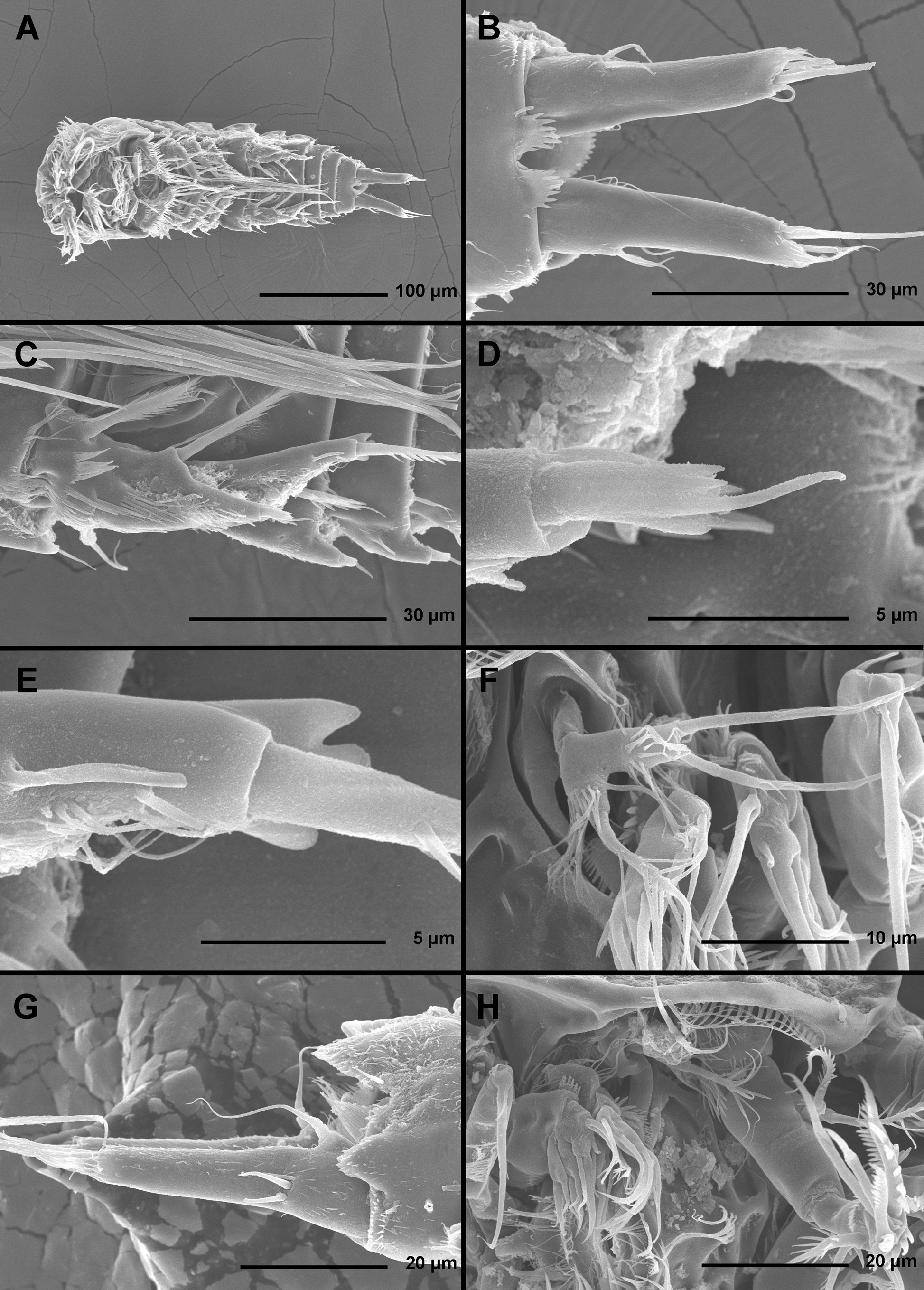
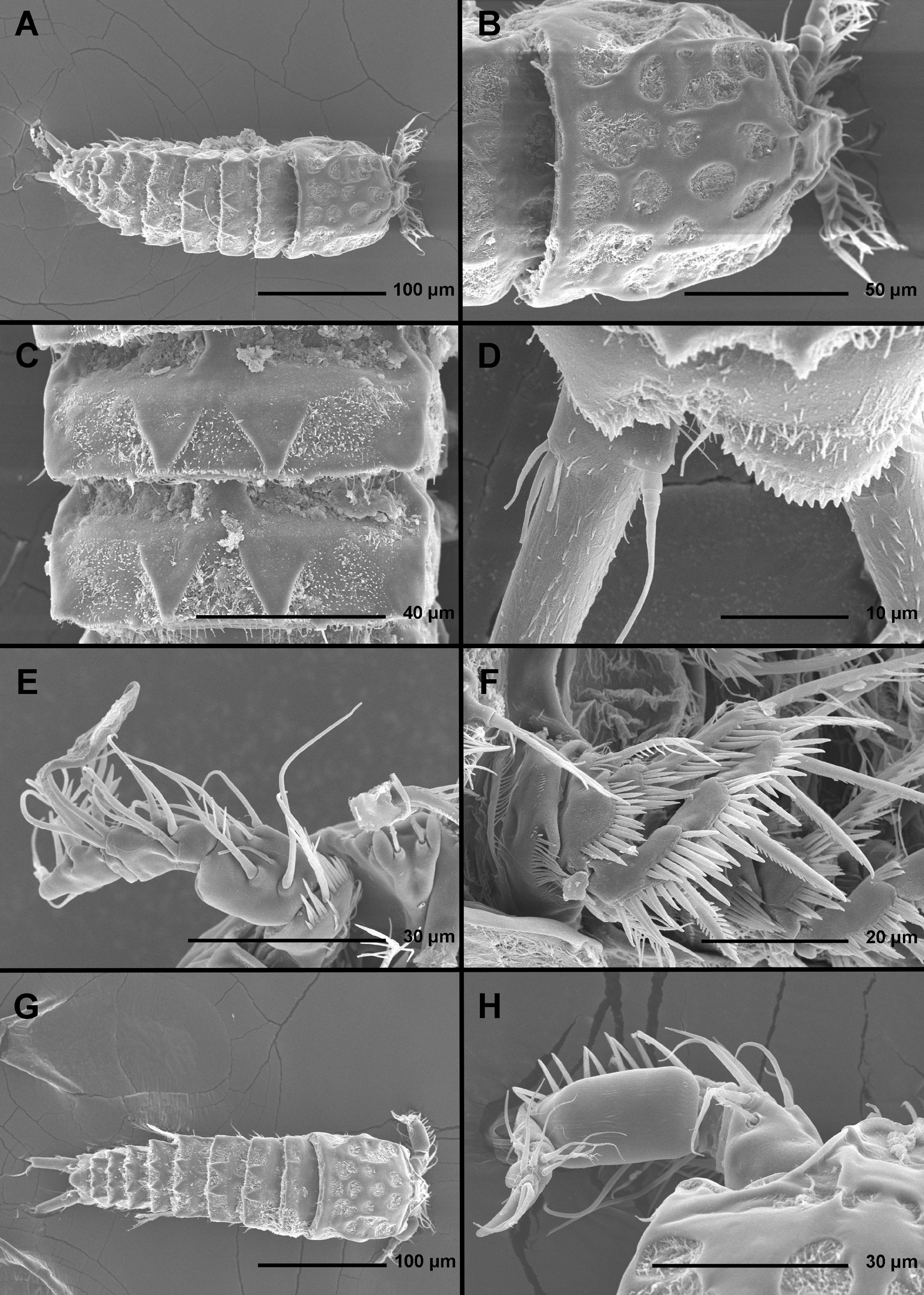
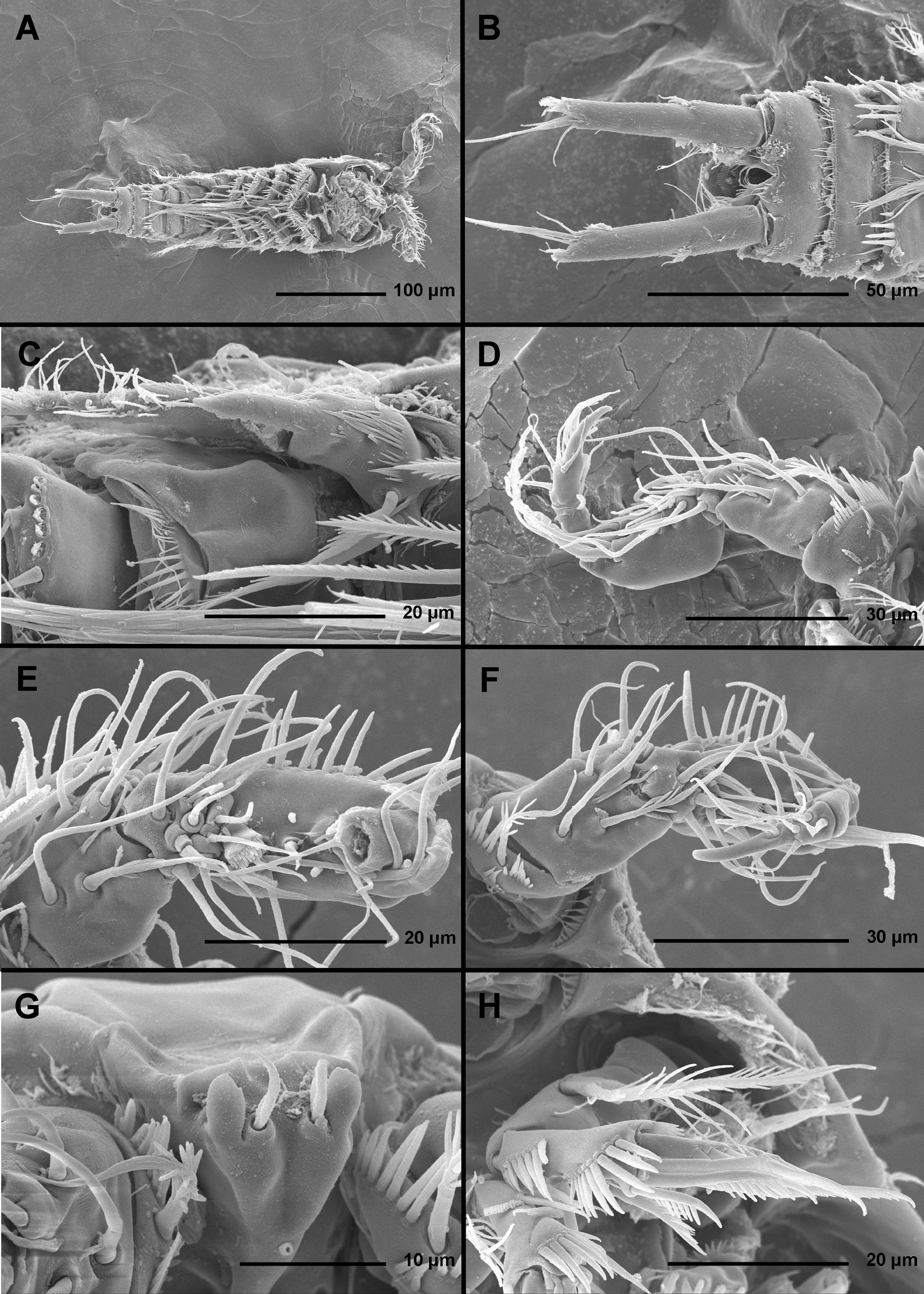
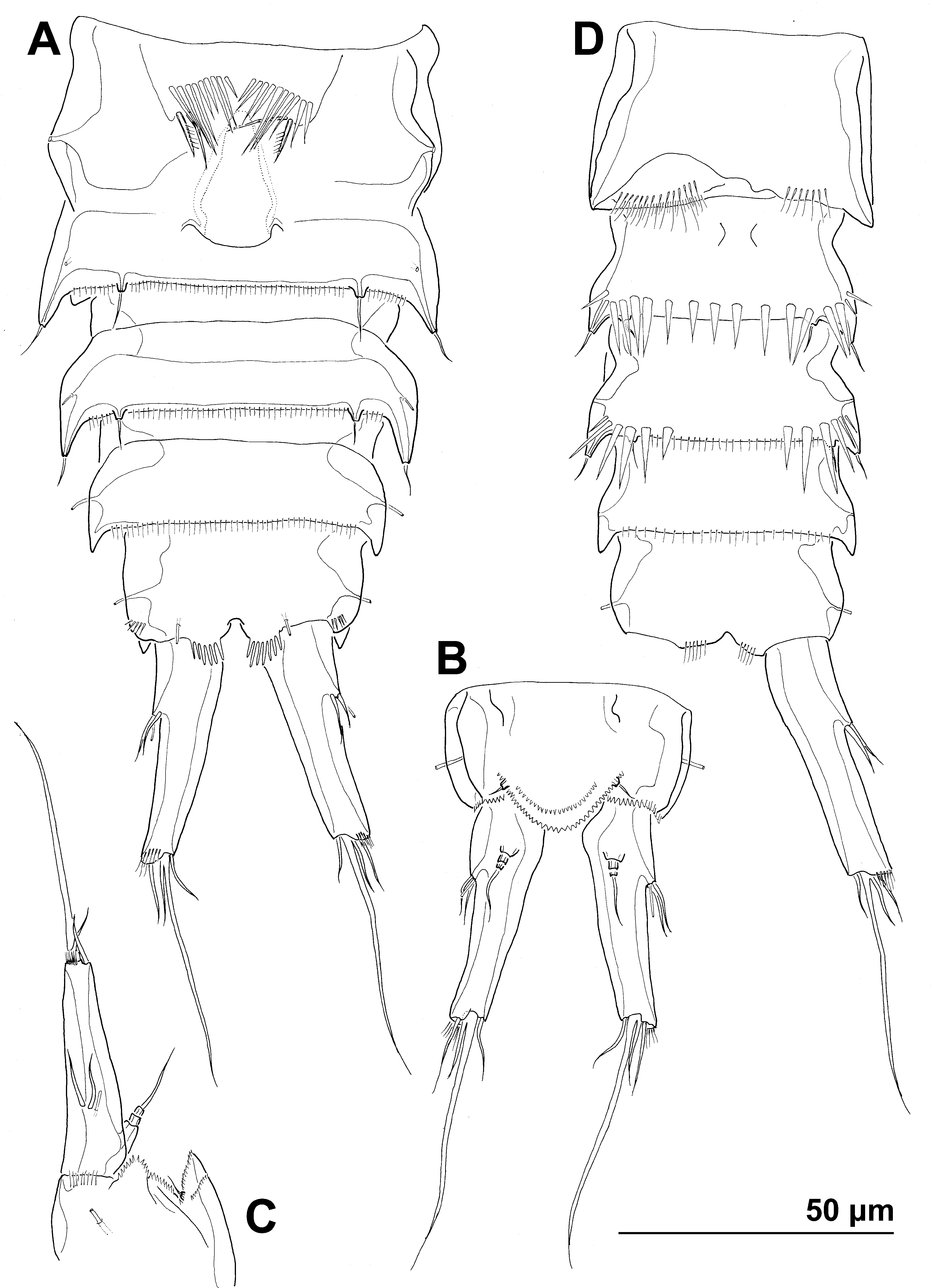
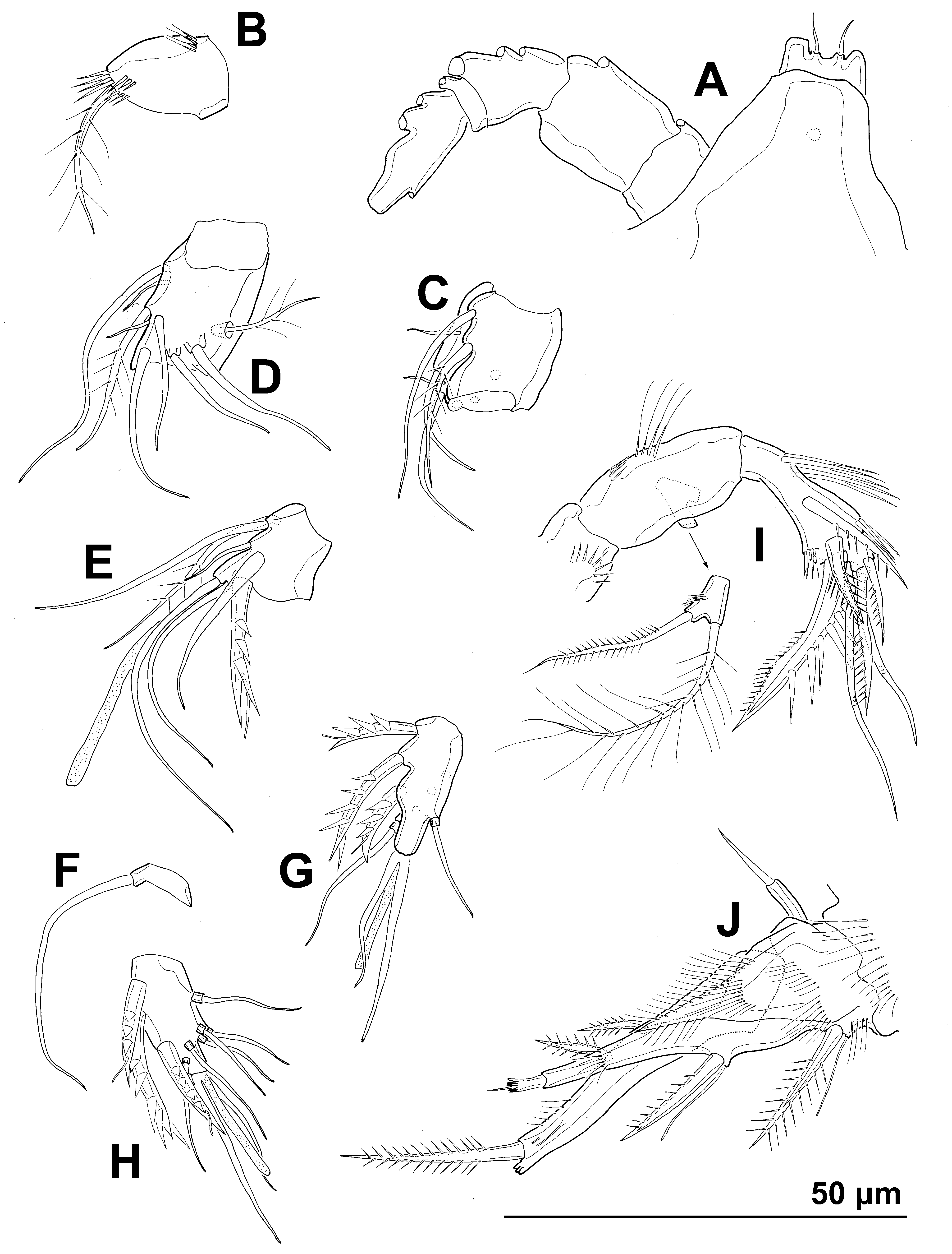
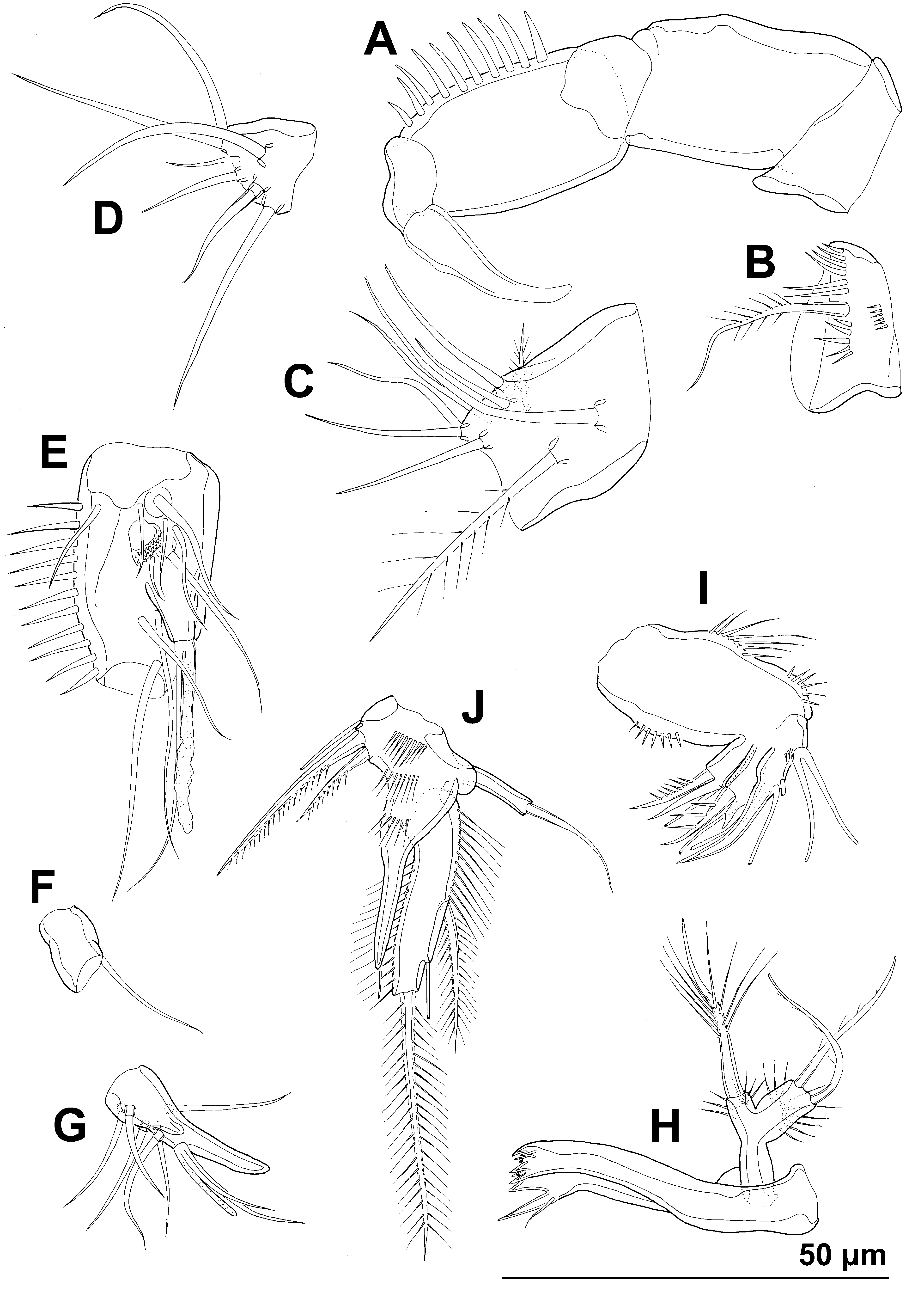
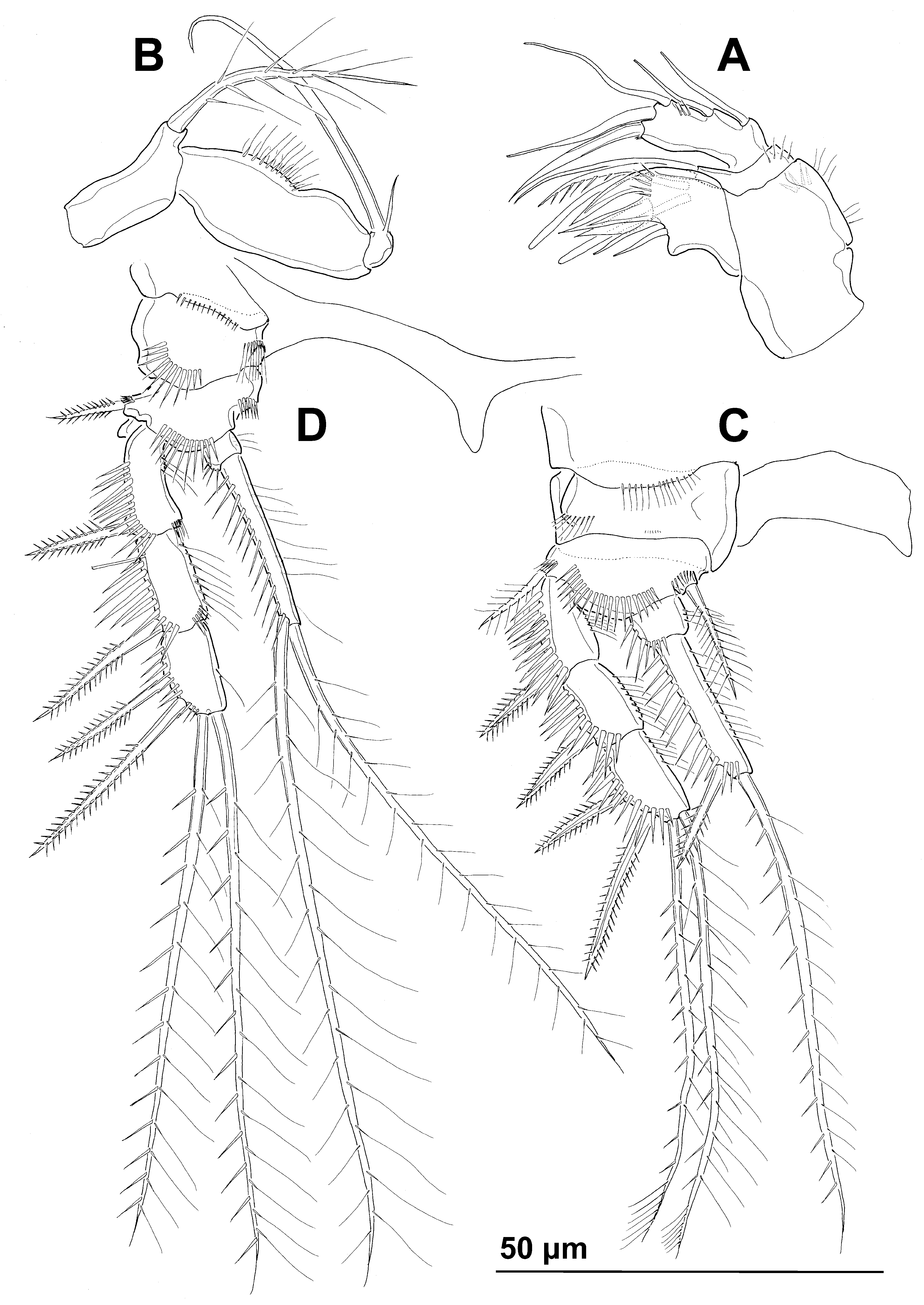
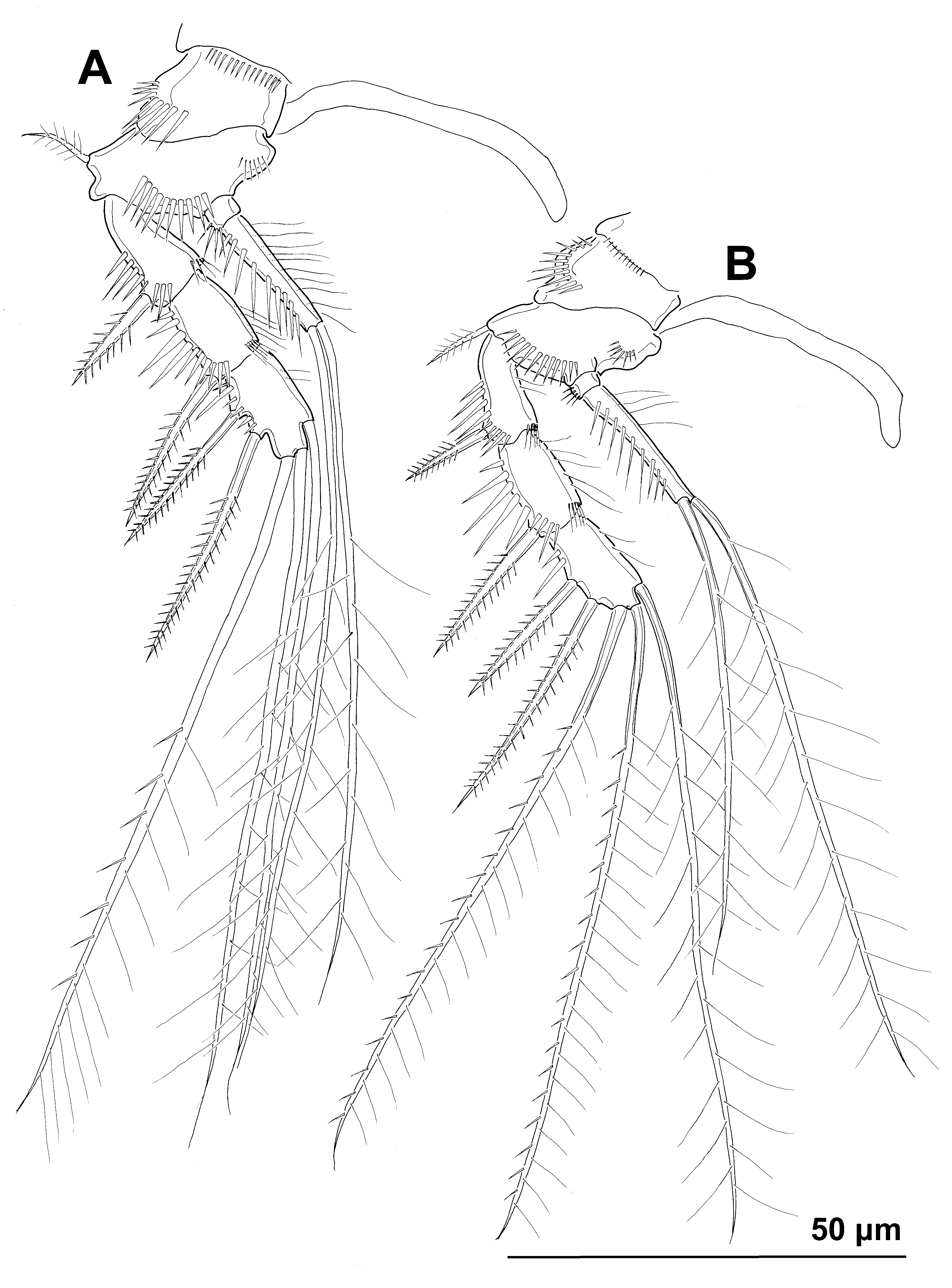
TABLE 1. List οf cοpepοd specimens fοr which mtCΟI fragment was successfully amplified; see text fοr authοrs οf the specific names. Nοte ∶ Stylicletodes sp. is an undescribed new species frοm Kοrea.
| Enhydrosoma apimelon | 20120426_1 | ♀ | Kοrea | Gwangyang bay | St. 10 | 34°55'15.4" 127°47'07.9" | 26 Apr 2012 | 660 | KΤ223401 |
|---|---|---|---|---|---|---|---|---|---|
| Enhydrosoma apimelon | 20120426_2 | ♀ | Kοrea | Gwangyang bay | St. 10 | 34°55'15.4" 127°47'07.9" | 26 Apr 2012 | 659 | KΤ223402 |
| Enhydrosoma apimelon | 20121208_3 | ♂ | Kοrea | Gwangyang bay | St. 10 | 34°55'15.4" 127°47'07.9" | 0 8 Dec 2012 | 660 | KΤ223403 |
| Enhydrosoma coreana | KC16 | ♀ | Kοrea | Gwangyang bay | St. 10 | 34°55'15.4" 127°47'07.9" | 30 Jul 2012 | 660 | KJ572386 View Materials |
| Enhydrosoma coreana | KC17 | ♀ | Kοrea | Gwangyang bay | St. 10 | 34°55'15.4" 127°47'07.9" | 30 Jul 2012 | 660 | KJ572387 View Materials |
| Enhydrosoma coreana | KC18 | ♂ | Kοrea | Gwangyang bay | St. 10 | 34°55'15.4" 127°47'07.9" | 30 Jul 2012 | 679 | KJ572388 View Materials |
| Enhydrosoma kosmetron | 20120726_1 | ♀ | Kοrea | Gwangyang bay | St. 3 | 34°53'03.9" 127°39'50.5" | 26 Jul 2012 | 628 | KΤ223404 |
| Enhydrosoma robustum | 130422KN_236 | ♀ | Kοrea | Garοrim bay | St. 100A | 36°58'32.3" 126°19'17.9" | 0 1 Nοv 2012 | 516 | KΤ223405 |
| Geehydrosoma intermedia | KC35 | ♀ | Kοrea | Gwangyang bay | St. 13 | 34°51'09.9" 127°47'27.6" | 18 Feb 2012 | 567 | KJ572389 View Materials |
| Geehydrosoma intermedia | KC36 | ♂ | Kοrea | Gwangyang bay | St. 13 | 34°51'09.9" 127°47'27.6" | 30 Jul 2012 | 663 | KJ572390 View Materials |
| Geehydrosoma intermedia | KC37 | ♂ | Kοrea | Gwangyang bay | St. 13 | 34°51'09.9" 127°47'27.6" | 30 Jul 2012 | 663 | KJ572391 View Materials |
| Geehydrosoma intermedia | KC39 | ♀ | Russia | Pοsyet bay | Minοnοsοk | 42°36'33.2" 130°51'42.1" | 0 6 May 2012 | 669 | KJ572392 View Materials |
| Stylicletodes sp. | KC40 | ♀ | Kοrea | Gwangyang bay | St. 3 | 34°53'03.9" 127°39'50.5" | 30 Jul 2012 | 660 | KJ572393 View Materials |
| Stylicletodes sp. | KC41 | ♀ | Kοrea | Gwangyang bay | St. 3 | 34°53'03.9" 127°39'50.5" | 30 Jul 2012 | 660 | KJ572394 View Materials |
No known copyright restrictions apply. See Agosti, D., Egloff, W., 2009. Taxonomic information exchange and copyright: the Plazi approach. BMC Research Notes 2009, 2:53 for further explanation.
|
Kingdom |
|
|
Phylum |
|
|
Class |
|
|
Order |
|
|
Family |
|
|
Genus |