
Rumarcanella vorstmani (Toriumi, 1952)
|
publication ID |
https://doi.org/ 10.5281/zenodo.276554 |
|
DOI |
https://doi.org/10.5281/zenodo.5697364 |
|
persistent identifier |
https://treatment.plazi.org/id/03CE87FE-FF90-FFE4-3FDA-F892FD10F0F1 |
|
treatment provided by |
Plazi |
|
scientific name |
Rumarcanella vorstmani (Toriumi, 1952) |
| status |
|
Rumarcanella vorstmani (Toriumi, 1952) View in CoL
( Fig. 8 View FIGURE 8 )
Plumatella vorstmani Toriumi, 1952a: 268 View in CoL , figs 1–19; Mukai 1984: 51, fig. 2; 1999: 56, figs 3B, 4C, 6D; Wood et al. 2006: 18, figs 28–29, 39.
Plumatella javanica: Vorstman 1928a: 6 View in CoL , fig. 4, pl. 2(6–7); 1928b: 163.
Hyalinella vorstmani: Lacourt 1968: 86 , pl. 12(g, i); Wiebach 1973: 546.
Material examined. Floatoblasts from Fukuji Dam, village of Higashi; several mature colonies with floatoblasts and sessoblasts, Kanna Dam, village of Ginoza.
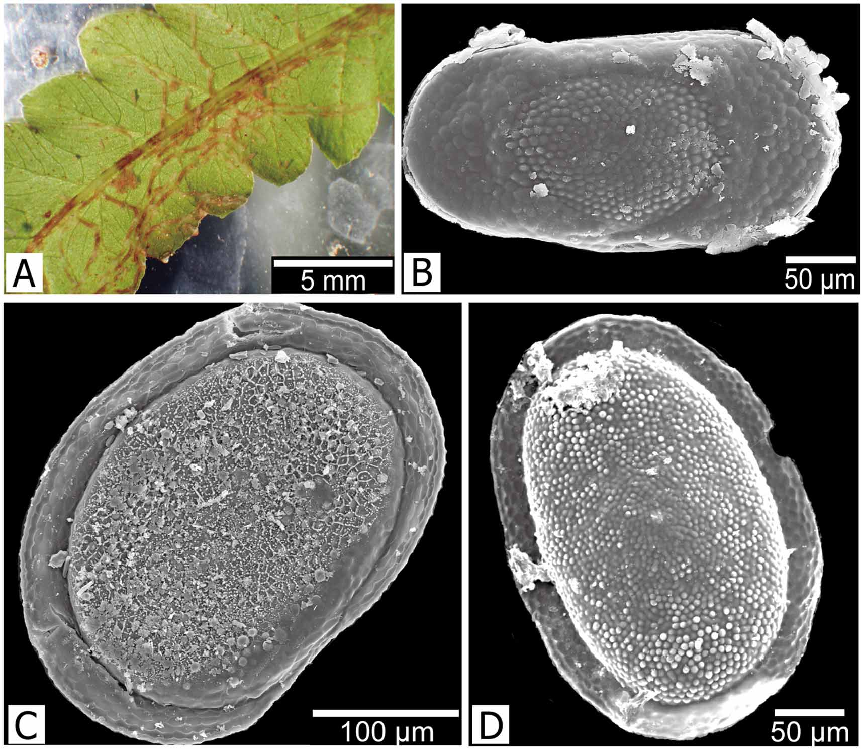
Description. Colony with narrow branches, transparent, almost entirely recumbent ( Fig. 8 View FIGURE 8 A). Tentacle number 20–27 ( Wood et al. 2006). Floatoblast ( Fig. 8 View FIGURE 8 B) oblong-elliptical, small, 295–322 (310±12) μm long by 155–182 (168±14) μm wide (n=4), with length/width ratio of about 1.8; symmetrical in lateral view; both ventral and dorsal fenestrae subcircular, both with tubercles bearing single hypertubercles. Sessoblast ( Figs 8 View FIGURE 8 C, D) small, 288–358 (324±28) μm long by 215–248 (229±14) μm wide (n=5); fenestra with tubercles, annulus with weak reticulation.
Distribution. Rumarcanella vorstmani has been reported from mainland Asia ( Bushnell 1973; Wood et al. 2006), India ( Lacourt 1968), Java and Indonesia ( Vorstman 1928a, b), and from Okinawa to Miyagi Prefecture in Japan ( Toriumi 1952a; Mukai 1984).
Remarks. Rumarcanella vorstmani was originally described as Plumatella javanica , but Toriumi (1952a) recognized the former as a distinct species on the basis of tentacle number, floatoblast size, and the surface microsculpture of the sessoblast. The transparent, weakly chitinized colony wall of R. vorstmani has made generic placement difficult. Lacourt (1968) included this species in Hyalinella owing to the soft, transparent ectocyst, but because the species produces sessoblasts, Wiebach (1973) transferred it to Plumatella . In a similar case, R. minuta had long been reported to produce only floatoblasts and was placed in Hyalinella . Toriumi (1972) reported the occurrence of sessoblasts, and Wood et al. (2006) transferred the species to Plumatella .
Toriumi reported tubercles on the sessoblast of R. vorstmani . When Wood & Wood (2000) reexamined specimens that Mukai (1984) had identified as P. vorstmani , they found the surface of the capsule to be reticulate rather than tuberculate. However, Wood et al. (2006) reported R. vorstmani sessoblasts with a tuberculate fenestra from many sites in Thailand. In our collection, the reticulation ( Fig. 8 View FIGURE 8 C) on the sessoblast fenestra is easily lost from the surface, and the strong tubercles underneath become apparent ( Fig. 8 View FIGURE 8 D). Thus, the reticulation may be associated with the membrane that covers the sessoblast surface.
No known copyright restrictions apply. See Agosti, D., Egloff, W., 2009. Taxonomic information exchange and copyright: the Plazi approach. BMC Research Notes 2009, 2:53 for further explanation.
|
Kingdom |
|
|
Phylum |
|
|
Class |
|
|
Order |
|
|
Family |
|
|
Genus |
Rumarcanella vorstmani (Toriumi, 1952)
| Hirose, Masato & Mawatari, Shunsuke F. 2011 |
Hyalinella vorstmani:
| Wiebach 1973: 546 |
| Lacourt 1968: 86 |
Plumatella vorstmani
| Wood 2006: 18 |
| Mukai 1984: 51 |
| Toriumi 1952: 268 |
Plumatella javanica:
| Vorstman 1928: 6 |