
Gibboletia tergata ( Mills, 1940 ) Espinasa & Smith, 2023
|
publication ID |
https://doi.org/ 10.11646/zootaxa.5228.3.6 |
|
publication LSID |
lsid:zoobank.org:pub:0EA12807-8881-4886-A775-E2722F22292F |
|
DOI |
https://doi.org/10.5281/zenodo.7544185 |
|
persistent identifier |
https://treatment.plazi.org/id/03CF3248-FFA1-3175-3EF9-F9C1FEE8878D |
|
treatment provided by |
Plazi |
|
scientific name |
Gibboletia tergata ( Mills, 1940 ) |
| status |
comb. nov. |
Gibboletia tergata ( Mills, 1940) comb. nov.
Figs. 1–41 View FIGURE 1 View FIGURE 2 View FIGURE 3 View FIGURES 4–14 View FIGURES 15–21 View FIGURES 22–30 View FIGURES 31–41
Nicoletia tergata Mills, 1940: 271
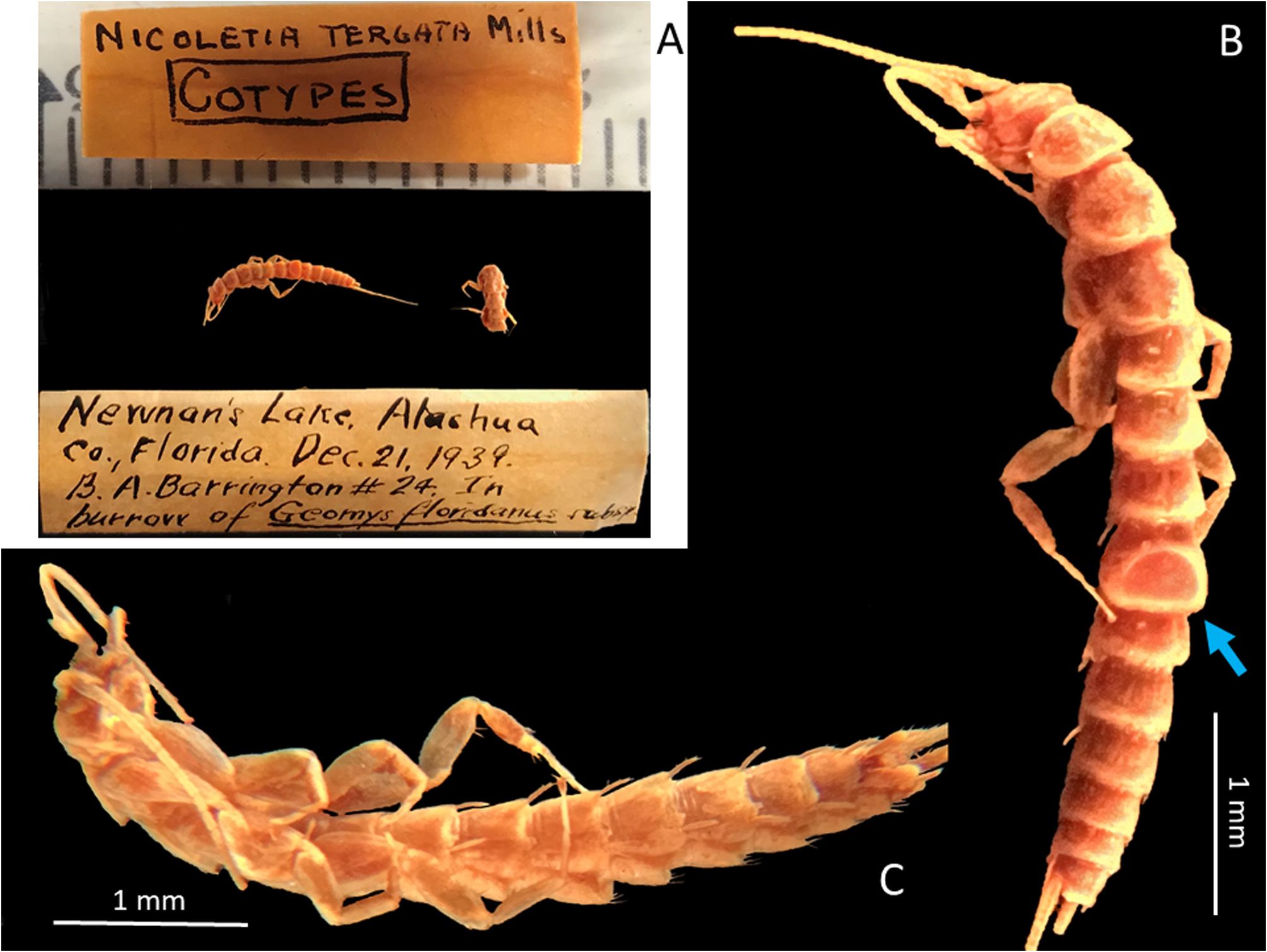
Type material. Cotypes, USA: Florida, Newman’s Lake, Alashua County, 21 December 1939, B.A. Barrington, in burrow of Geomys pinetus floridanus [ Geomyidae ] one complete Ô and a thorax ( INHS Insect Collection 1500901 in alcohol).
New material examined. USA: FLORIDA: Levy County, 3 miles SW Archer (29.5103°N, 82.6602°W), 6 July 2019 – 22 March 2020, Kyle E. Schnepp, underground column trap, 1Ô ( HW 0.93) in alcohol ( AMS K.377944); 1♀ ( HW 1.05 ) GoogleMaps in alcohol ( AMS K.377945); 1♀ ( HW 1.05 ) on two slides ( AMS K.541616); 1Ô ( HW1.10 ) on two slides ( AMS K.541615) on two slides, 1♀ ( HW 1.05 ) in alcohol ( AMS K.377946); 1Ô ( HW 0.83) in alcohol ( AMS K.377947); 1 subadult ♀ ( HW 0.83) in alcohol ( AMS K.377948); 1 juvenile ( HW 0.64), head, thorax and abdominal segments I– V, in alcohol ( AMS K.377949); pieces of appendages from all specimens from this collection event, loose in tube, one slide ( AMS K.541619) . USA: FLORIDA: Columbia County. 6 miles NW High Springs (29.8674°N, 82.6664°W), 25 September 2020 – 16 October 2020, Kyle E. Schnepp, underground column trap, 1Ô ( HW 1.18 ) GoogleMaps on two slides ( AMS K.541617); 1Ô ( HW 1.13 ) in alcohol ( AMS K.377950; 1Ô ( HW 1.13 ) in alcohol ( AMS K.377951); 1♀ ( HW 1.10 ) in alcohol ( AMS K.377952); 1Ô ( HW 1.05 ) in alcohol ( AMS K.377953); 1Ô ( HW 1.13 ) in alcohol ( AMS K.377954); 1Ô ( HW 1.15 ) in alcohol ( AMS K.377955); 1 juvenile ♀ ( HW 0.68) in alcohol ( AMS K.377956); pieces of appendages from all specimens from this collection event, loose in tube, on one slide ( AMS K.541618) .
Redescription. Medium sized, parallel-sided silverfish with elongate antennae and terminal filaments, shape typical for the subfamily. Alcohol-preserved specimens off-white. Pigment lacking. Head and body length in preserved specimens examined, most specimens in alcohol unnaturally distended and elongated, but one more usual looking specimen (K.541618) from the Levy County population with an H+B length of 6.7 mm (HW 1.10); specimens from the Columbia population generally larger with HW up to 1.18 and body lengths also about 7 mm long. Thorax length about one third H+B and width only slightly wider than HW; antennae incomplete in all specimens but at least half length of H+B; caudal filaments incomplete in all specimens, at least half as long as H+B.
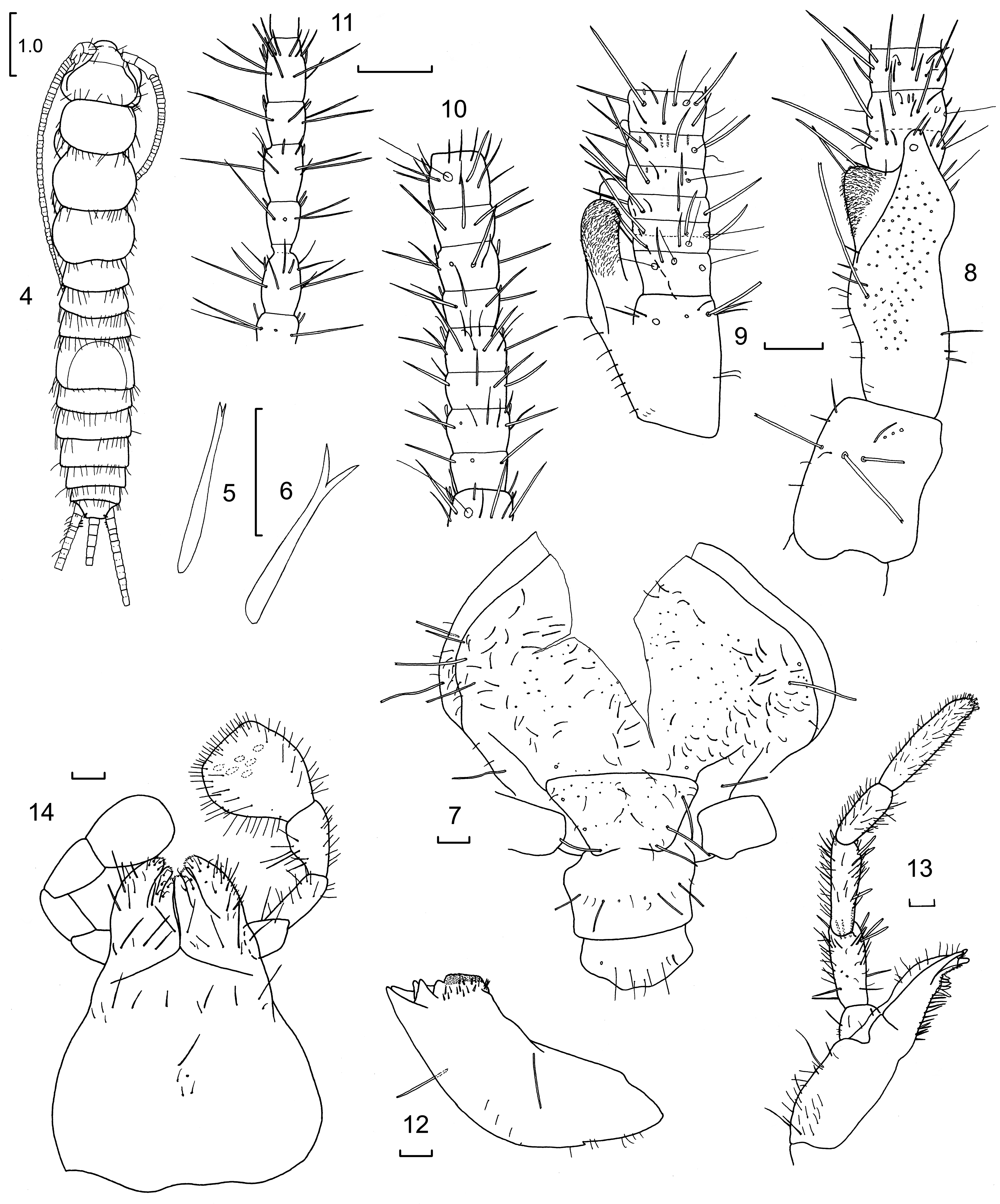
Macrochaetae. Mostly simple, parallel-sided or tapered with small apical bifurcations but on appendages some macrochaetae stout and carrot-shaped, sometimes with a small apical bifurcation ( Figs. 5, 6 View FIGURES 4–14 ). Some of the longer parallel-sided macrochaetae, when slide-mounted, with an artifactual distortion caused by the mounting medium (see Smith et al. (2012) and Molero-Baltanás et al. (2013)).
Head. Length and width about equal, not covered by prothorax at hind margin ( Fig. 7 View FIGURES 4–14 ), prognathous, vertex with distinct transverse post-antennal suture, postero-lateral corners of vertex with 2+2 dorsal and 2+2 infralateral macrochaetae; five larger macrochaetae in antero-lateral corners adjacent to the antennal bases, two of which are posterior to the transverse post antennal suture; disc with numerous scattered small fine setae. Clypeus with transverse row of six setae and with one or two small fine setae between them. Labrum with similar transverse row of setae, the most laterad being shorter and much more robust.
Mandibles ( Fig. 12 View FIGURES 4–14 ) strong with well-developed molar and incisor regions and one macrochaeta on the external face as well as a few longer and shorter simple setae; a group of three stout setae on the margin adjacent to the molar area and about 14 small setae on the inner face below the molar region. Maxillae ( Fig. 13 View FIGURES 4–14 ) elongate, galea only surpassing length of lacinia by the two distinct apical papillae, lacinia well sclerotized with a single strong apical tooth; pectinate prostheca a little shorter than lacinia with five or six lamellate processes and about 11 setae along margin, the more basal setae in groups of two; maxillary palp of moderate length, distal article with six branched papilla apically, other specialised sensilla not seen, second and third articles subapically with some stronger, carrotshaped, apically bifid setae; all articles with smaller setae. Labium ( Fig. 14 View FIGURES 4–14 ) much longer than wide, submentum with weak transverse row of thin setae, mentum also with weak row of long thin setae; glossae with some setae as well as small setulae apically, ultimate article of palp about 1.2–1.4 times longer than wide with six papillae of usual type; penultimate article with long setae on the inner face.
Antennae long but broken on all specimens. Scape of male slightly longer than wide, with several strong macrochaetae and some smaller setae ( Fig. 8 View FIGURES 4–14 ) both above and below. Pedicel of male with large, apically bifurcate apophysis on dorsal face ( Figs. 8, 9 View FIGURES 4–14 ), three times longer than wide, each apophysis reaching to about the third or fourth interval; the longest lobe armed subapically with a short, robust seta on the dorsal aspect, the shorter lobe densely covered in numerous setae, the dorsal face of the pedicel with scattered small setae often set on protuberances. Basal annulus of flagellum with four trichobothria, following five or six annuli each with two trichobothria; the seventh interval with two rings of setae with the trichobothria restricted to the distal ring; intervals further subdivided into four annuli by the eleventh interval, each interval with a ring of setae and two trichobothria in the most distal annulus, small basiconic sensilla visible subdistally from about the fifth interval, consisting of a mixture of typical “sausage-shaped” type C sensilla (see Adel, 1984) and much longer, finer setae with rounded tips; intervals around mid-antenna ( Fig. 10 View FIGURES 4–14 ) consisting of eight annuli with only a single trichobothrium in the most distal annulus; most annuli also with a ring of fine setae or basiconic sensilla subapically. The most distal surviving intervals (probably about ⅔ antennae length) also with similar pattern but the arrangement of fine setae and basiconic sensilla distad of the ring of setae ( Fig. 11 View FIGURES 4–14 ) less dense.
Thorax. Length about one third that of H+B and not wider than abdomen, pronotum significantly smaller than the meso and metanota ( Figs. 15–17 View FIGURES 15–21 ); pronotum rounded sub rectangular in shape with obvious collar of 8–10 longer macrochaetae and additional smaller setae, all nota with 2–5 strong submarginal macrochaetae along the lateral margins and some strong submarginal macrochaetae in the lateral parts of the posterior margin, all margins also with longer and shorter, simple tapered setae and setulae; disc of nota with many small but strong setae.
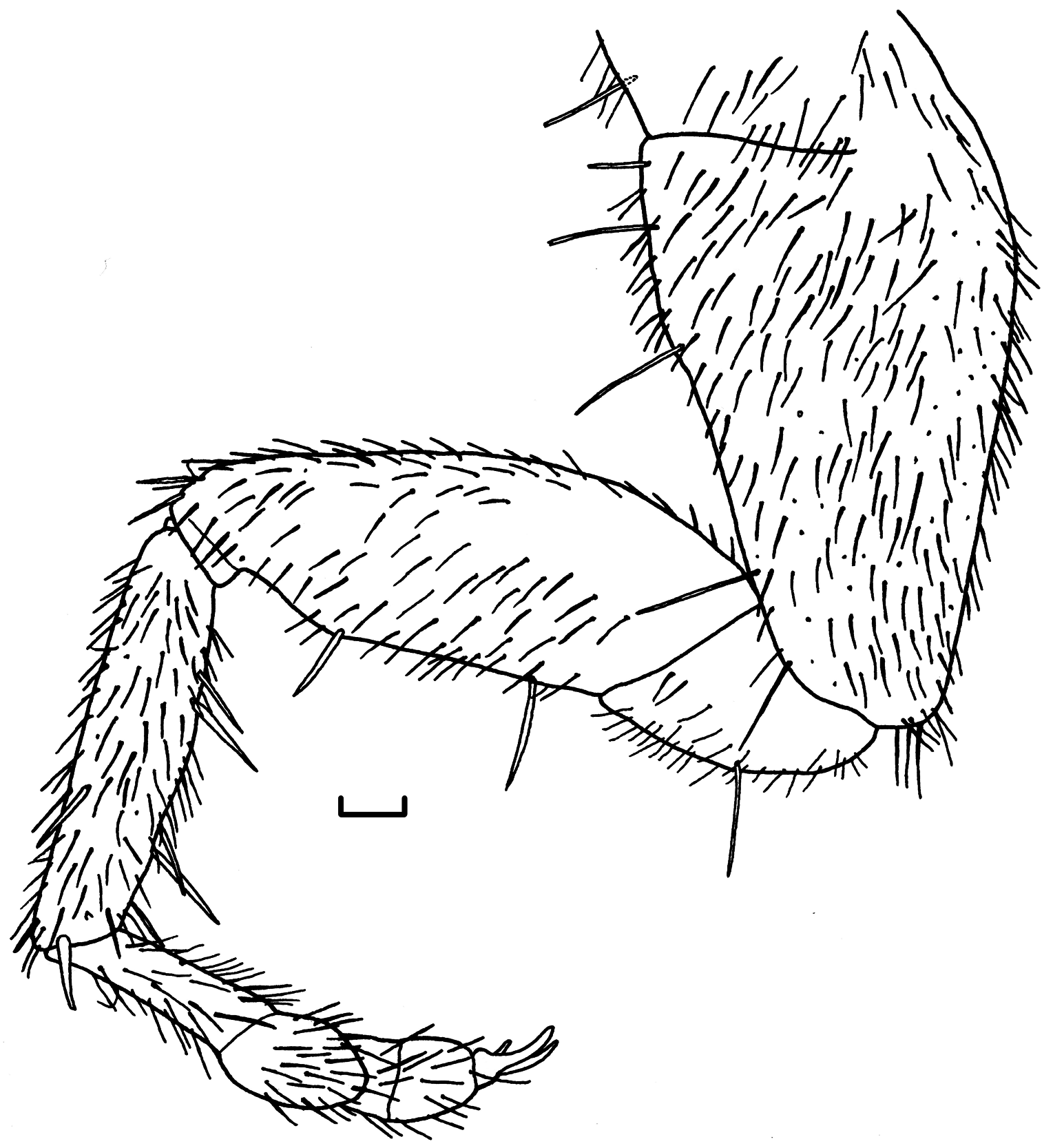
Legs typical for subfamily but quite elongate ( Figs. 18–20 View FIGURES 15–21 ). Tibial L/W ratio of foreleg 3.4–4.4, middle leg 3.6–5.2, hind leg 4.0–4.4. Tarsal L/W ratio of foreleg 7.7–8.7, middle leg 7.6–11.3, hind leg 11.9–13.8. On foreleg precoxae with line of setae, coxae with about five long macrochaetae spaced along the outer margin, with some stronger setae more distally along this margin and over the articulation with the trochanter, inner margin without macrochaetae, margins and face covered with scattered small setae; trochanter with one thin macrochaeta and several smaller setae; femur with a somewhat larger carrot-shaped macrochaeta about ¾ along the leading margin and another one or two macrochaetae over the articulation with the tibia, posterior margin with a proximal macrochaeta and two tapered macrochaetae on the distal bulge, all margins also with setae and setae scattered over the ventral face with the exception of the proximal anterior quarter; foreleg tibia without thicker macrochaetae near the dorsal margin but with one distally on the face about ¾ along the tibial length; ventral margin with two tapered macrochaetae about ¼ along the margin and two longer macrochaetae at about ⅔ along the margin, setae along distal end not noticeably stronger than other setae on face of tibia; distal spur long, slender and smooth, without small subapical teeth. Tarsus with four articles, the basal tarsomere about 40% of the total length of the foreleg, with stronger setae distally on basal tarsomere. Pretarsus with two strong curved outer claws and small medial empodium. Middle and hind legs similar except only two macrochaetae along the outer margin of the coxae and the tibia with two carrot-shaped macrochaetae in the more distal position on the posterior margin; basal article of the tarsi with a strong spine distally on the dorsal face.
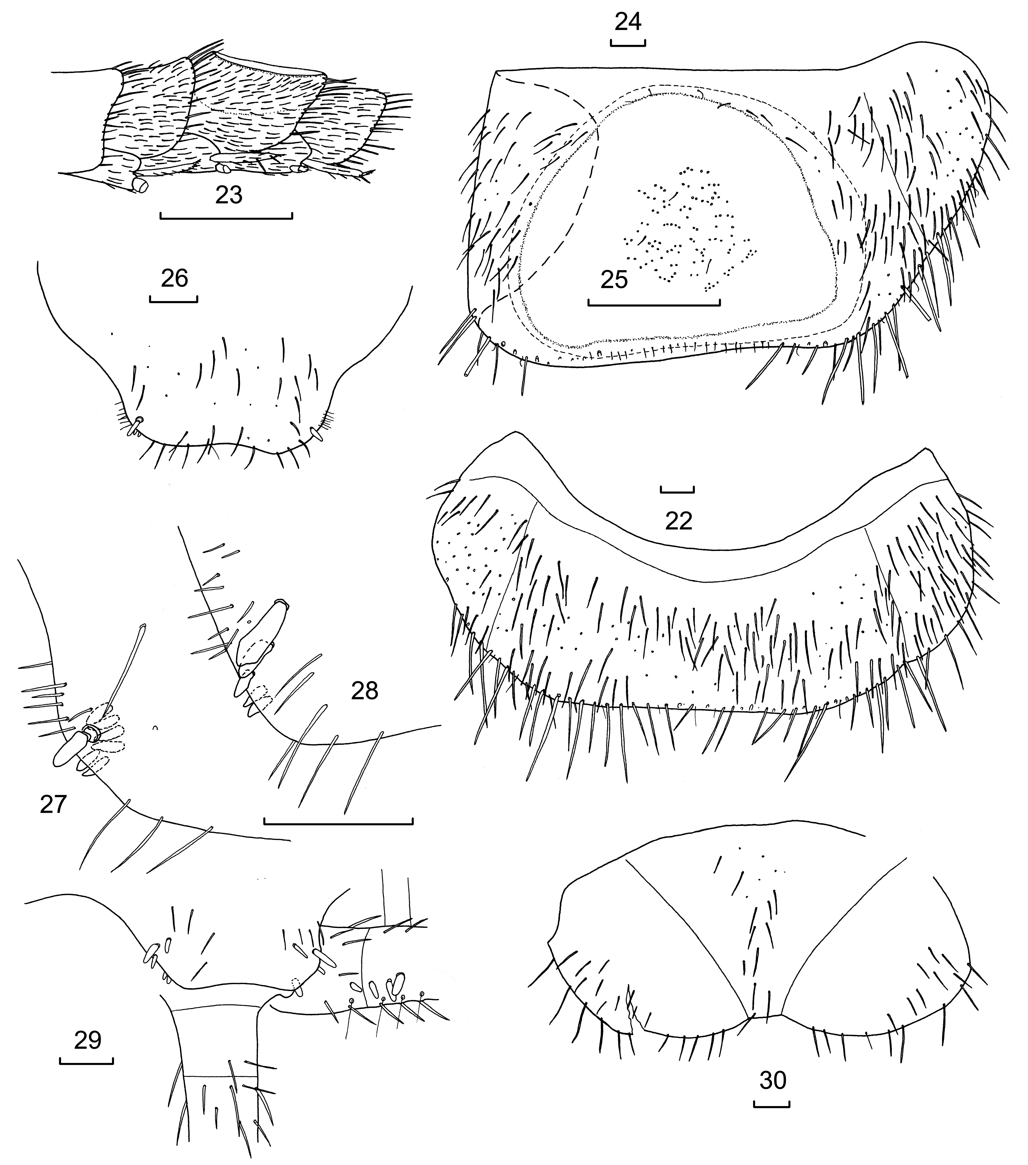
Abdomen. Slightly more slender than the thorax at its base. All urotergites wrapping around the body without a sharp lateral fold. Suture between the tergite and the paratergites visible on I–IX. Abdominal tergites I–III and V–IX ( Fig. 22 View FIGURES 22–30 ) with long, simple, marginal macrochaetae rather evenly spaced along the lateral and posterior margins as well as a few smaller setae and some setulae, the disc covered with numerous long, scattered setae. Abdominal tergite IV with semicircular raised region across most of the dorsum ( Figs. 23, 24 View FIGURES 22–30 ); outer rim raised, otherwise shallowly concave, surface granulated with a few minute, scattered setulae ( Fig. 25 View FIGURES 22–30 ); beneath the surface tissue darker than the surrounding tissue and adhering quite strongly to the cuticle so that it could not be separated from the cuticle during dissection without damaging the cuticle; posterior margin of the rim demarcated by small setae, longer macrochaetae and setae absent.
Urotergite X ( Figs. 26–28 View FIGURES 22–30 ) small, trapezoidal with concave posterior emargination, the rounded posterior corners dorsally with prominent, erect, 1+1 larger submarginal pegs and usually additional 1+1 smaller conules more postero-laterally close to margin; on one specimen (e.g. K.377947) additional 1+1 smaller pegs on the dorsal face of urotergite X medio-anteriorly of the larger ones, and groups of about 10 small setae on the margins just anterior to these pegs, the dorsal face with some small scattered setae ( Fig. 29 View FIGURES 22–30 ); ventral face with groups of 2–6 pegs below the rounded posterior corners, numbers variable between specimens and even from side to side.
Urosternite I divided into a sternum and two lateral coxites ( Fig. 30 View FIGURES 22–30 ), the sternum with several small setae along the median section of the disc, posterior margin without notable setae; posterior margin of the lateral coxites each with several small setae along the margins and on the more lateral parts of the discs. Urosternites II–VII entire ( Fig. 31 View FIGURES 31–41 ), not divided into separate coxites and sternum, each bearing 1+1 styli, the disc covered with many scattered setae; posterior margin without significant setae, the margins lateral of the styli with several stronger setae. Eversible vesicles on urosternites II–VI, each with about four or five setae on the vesicle; VII with pseudovesicles each bearing one or two setae ( Fig. 32 View FIGURES 31–41 ). Urosternite VIII entire ( Fig. 33 View FIGURES 31–41 ), without vesicles, without macrochaetae, face with scattered setae, posterior margin distinctly protruding, margins laterad of the styli with a few small setae. Apical spine of stylus with three barbs ( Fig. 34 View FIGURES 31–41 ).
Urosternite IX ( Fig. 35 View FIGURES 31–41 ) divided into separate coxites bearing styli and long, wide parameres (L/W 3.3–3.4). Parameres with many setae along their inner margin and apices with several hairs on tubercles with glands visible below the cuticle. External process of coxites with one small seta and a setula, internal process with two setae near the inner margin. Penis very long, extending beyond the apices of the inner processes of coxites IX, with longitudinal opening surrounded by numerous short hairs.
Appendix dorsalis of male ( Fig. 36 View FIGURES 31–41 ) without pegs, basal division with a few small setae, subsequent division indistinctly divided into five annuli, each with a ring of setae and trichobothria, the distal-most ring with a strong macrochaeta medially as well as a strong seta. Cerci with basal division bearing a ring of small setae and trichobothria; second division indistinctly divided into four annuli, each bearing strong setae and trichobothria and inner margin bearing 2–5 pegs ( Fig. 36 View FIGURES 31–41 ); one peg sometimes occurring on the more proximal division; following division with two rings of setae and trichobothria, fourth division with three annuli, the most distal bearing one wide macrochaeta. The number of pegs somewhat variable from one side to the other on the same individual.
Female. As for male except pedicel without apophyses ( Fig. 37 View FIGURES 31–41 ); urotergite X trapezoidal with 1+1 macrochaetae in the postero-lateral corners ( Fig. 38 View FIGURES 31–41 ), without pegs on ventral surface, dorsal surface with scattered setae only; cerci without pegs. Coxites on sternites VIII and IX separate, subgenital plate large, parabolic with setae scattered over the surface ( Figs. 39, 40 View FIGURES 31–41 ). Ovipositor ( Figs. 40, 41 View FIGURES 31–41 ) simple, elongate (up to 2.7 times HW) with about 16 divisions, surpassing the apices of styli IX by a little more than twice the length of the styli. The apex of the anterior valves with a typical acute triangular projection, that of the posterior valves rounded with the typical region of hooked processes on the penultimate division; both with simple fine setae only.
Remarks. Although the molecular data suggests at least two sister species, we have not found convincing, stable morphological characters (other than the swelling of the tarsi) that convinces us that these may be distinct species. Furthermore, we are not sure which represents the original Nicoletia tergata of Mills. We have selected the population from Levy Country, which lies slightly closer to the N. tergata type locality (about 35 km) for the illustrations and description, but also compared it to the material from Columbia County (50 km from the N. tergata type locality). The secondary sexual characters of many Nicoletiidae are usually considered as diagnostic for each species; however, we have not been able to separate these two populations based on the secondary sexual characters. With more specimens that allow a better understanding of variability, it might eventually be possible to separate these populations.


| INHS |
Illinois Natural History Survey |
| V |
Royal British Columbia Museum - Herbarium |
No known copyright restrictions apply. See Agosti, D., Egloff, W., 2009. Taxonomic information exchange and copyright: the Plazi approach. BMC Research Notes 2009, 2:53 for further explanation.
|
Kingdom |
|
|
Phylum |
|
|
Class |
|
|
Order |
|
|
Family |
|
|
Genus |
Gibboletia tergata ( Mills, 1940 )
| Espinasa, Luis & Smith, Graeme B. 2023 |
Nicoletia tergata
| Mills, H. B. 1940: 271 |