
Anthrenus latefasciatus Reitter, 1892
|
publication ID |
https://doi.org/ 10.5281/zenodo.280796 |
|
DOI |
https://doi.org/10.5281/zenodo.5697292 |
|
persistent identifier |
https://treatment.plazi.org/id/03CF87AA-FFE6-FFC2-FF64-4EADFD6EF9C8 |
|
treatment provided by |
Plazi |
|
scientific name |
Anthrenus latefasciatus Reitter, 1892 |
| status |
|
Anthrenus latefasciatus Reitter, 1892
Syn.: Anthrenus pimpinellae var. latefasciatus Reitter, 1892:13 Anthrenus pimpinellae ssp. latefasciatus: Mroczkowski, 1961:192 Anthrenus latefasciatus: Kalík & Ohbayashi, 1985:75
Material examined. 430 exuviae: KAZAKHSTAN W, Mangistauskaya Obl., fort Schewtschenko 30km NE, Karagan, 44º38’N / 50º35’E, 10-11VIII 0 7 near sea shore, leg R. Ruta. All material deposited in collection of Division of Invertebrate Biology, Evolution and Conservation, Department of Biology, Evolution and Ecology, University of Wrocław, ul. Przybyszewskiego 63/77, 51–148 Wrocław, Poland.
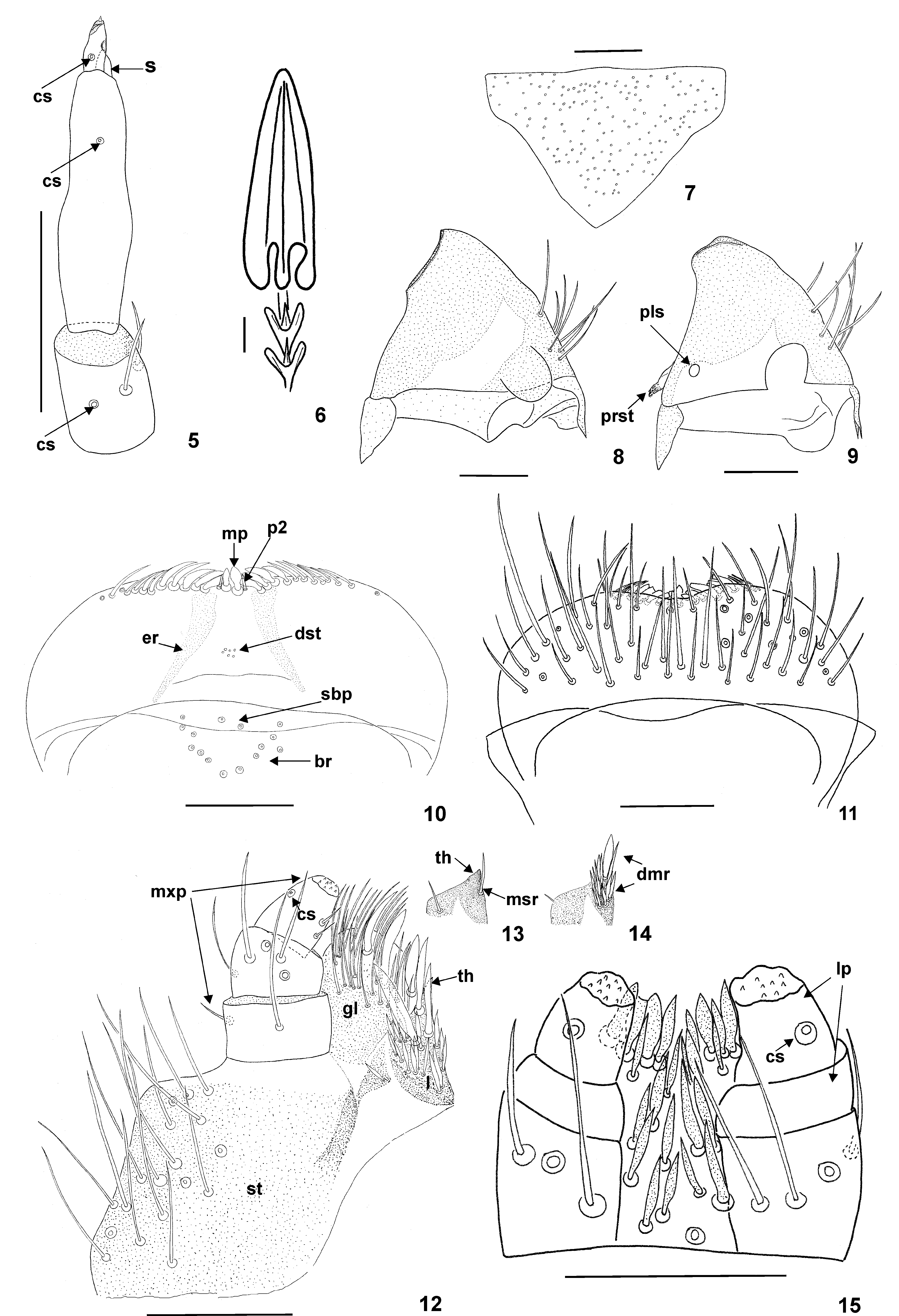
Description. Last larval instar (exuviae of final instar). Length 4.5–5.0 mm [along midline from head to last abdominal tergum]. Body fusiform, and relatively long [ratio of total length to width: 3:1], rather flat than concave. Integument of head dark brown; nota and terga light yellowish brown with dark brown or black pigmented areas (darkness varies; sometimes absent; Figs 1–2 View FIGURES 1 – 4 , 16, 18–20 View FIGURES 16 – 21 ); all tergal plates sclerotized ( Figs 1–2 View FIGURES 1 – 4 ), sternum I hyaline; central area of sterna II and III slightly sclerotized and heavily pigmented; abdominal sterna rather sclerotized and slightly pigmented ( Fig. 4 View FIGURES 1 – 4 ). Tufts of prominent dark brown spicisetae present between legs. Coxae, femora and tibiae light yellowish brown. Spicisetae and hastisetae on terga and sterna brown ( Figs 1–4 View FIGURES 1 – 4 ). Club–shaped, round–headed setae and urogomphi absent. Head protracted and hypognathous ( Figs 2–3 View FIGURES 1 – 4 ). Stemmata present. All stemmata arranged in two horizontal rows. Frons triangular, without frontal median tubercies; setal patterns as illustrated ( Fig. 7 View FIGURES 5 – 15 ). Antennae oriented anterolaterally and composed of 3 antennomeres ( Fig. 5 View FIGURES 5 – 15 ). Terminal antennomere 2 times as long as wide, with one small appendage on apex and one campaniform sensillum (cs) in proximal half of antennomere (at 1/3 of the length). Ratio of length of terminal antennomere to length of one and two antennomeres combined 0.1:1.0. Sensorium (s) arising from apex of antennomere 2, in apical position, and extending above apex. One campaniform sensillum (cs) at 2/3 total length of antennomere 2 under sensorium. Basal segment with 2 setae and one campaniform sensillum (cs) ( Fig. 5 View FIGURES 5 – 15 ). Gula separate from postmentum; epicranial stem present. Median endocarina absent. Labro-epipharyngeal margin with 9–14 setae in outer series. Mesal of labro–epipharyngeal setae (mp) spatulate while second pair (p2) slender. Ventral side of epipharynx ( Fig. 10 View FIGURES 5 – 15 ) with basal transverse row (br) of placoid sensillae. Epipharynx with 6–15 sensory cups in proximal transverse series (br), epipharyngeal rods (er) present and diverging proximally. Two sensory cups present in subproximal epipharyngeal sensillae (sbp). Distal epipharyngeal sensillae (dst) arranged in group of 6, but not enclosed/encircled by furrow. Lateral setae on epipharynx absent ( Fig. 10 View FIGURES 5 – 15 ). Dorsal surface of labro–epipharynx with numerous setae ( Fig. 11 View FIGURES 5 – 15 ). Mandibles brown with dark brown (almost black) apices; apical teeth and ventral accessory process absent. Apical half of mandible heavily sclerotized and sharply delineated from basal half ( Figs 8–9 View FIGURES 5 – 15 ). Mandibular mola and pseudomola absent. Hyaline lobe at ventral base of mandible absent. Prostheca (prst) slightly falciform with asperate surface structure ( Fig. 9 View FIGURES 5 – 15 ); mesal brush of setae near base of mandible absent. Placoid sensillum (pls) in basal 1/5 of structure. Maxillary palp (mxp) with 3 palpomeres; terminal palpomere longest. Ratio of length of terminal palpomere to the length of the two proceeding palpomeres combined 0.6:1.0. Basal palpomere with two setae, second palpomere with three to five setae and two campaniform sensillae; third palpomere with two short setae and one campaniform sensilla. Terminal palpomere with group of 8–11 small sensillae apically situated. Lacinia (l) with one (often unvisiable) slightly sclerotized small lacinial tooth (th) at apex ( Fig. 12 View FIGURES 5 – 15 ). Sclerotization of lacinia and stipes (st) as illustrated on Fig. 12 View FIGURES 5 – 15 . Mesal row of setae on lacinia (msr) composed of one thick basal seta ( Fig. 13 View FIGURES 5 – 15 ). Straight, thick to slender setae in dorsomesal row of setae (dmr) present ( Fig. 14 View FIGURES 5 – 15 ). Galea (gl) arising from stipes (st), extending above apex of lacinia and terminating near apex of terminal palpomere. Apical area of galea densely covered with setae ( Fig. 12 View FIGURES 5 – 15 ). Stipes with 13–20 long setae near anterio–lateral margin ( Fig. 12 View FIGURES 5 – 15 ). Hypopharynx hyaline. Bridge sclerite (i.e. central part of distal element of hypopharyngeal sclerome) connected medially. Anterior arms of bridge sclerite and distal lateral sclerites of hypopharynx absent. Anterior branch of suspensoria elongated. Ligula with ~24 spurlike setae; ~10 on apex, and ~12 on middle section between labial palpi ( Fig. 15 View FIGURES 5 – 15 ). Labial palp (lp) 2–segmented. Basal segment wider than terminal segment; 2.0 times as wide as long; lacking setae. Terminal segment 1.1 times as long as wide, with group of 8–11 small sensillae in apical area and one campaniform sensillum (cs) ( Fig. 15 View FIGURES 5 – 15 ) at half total length of segment.
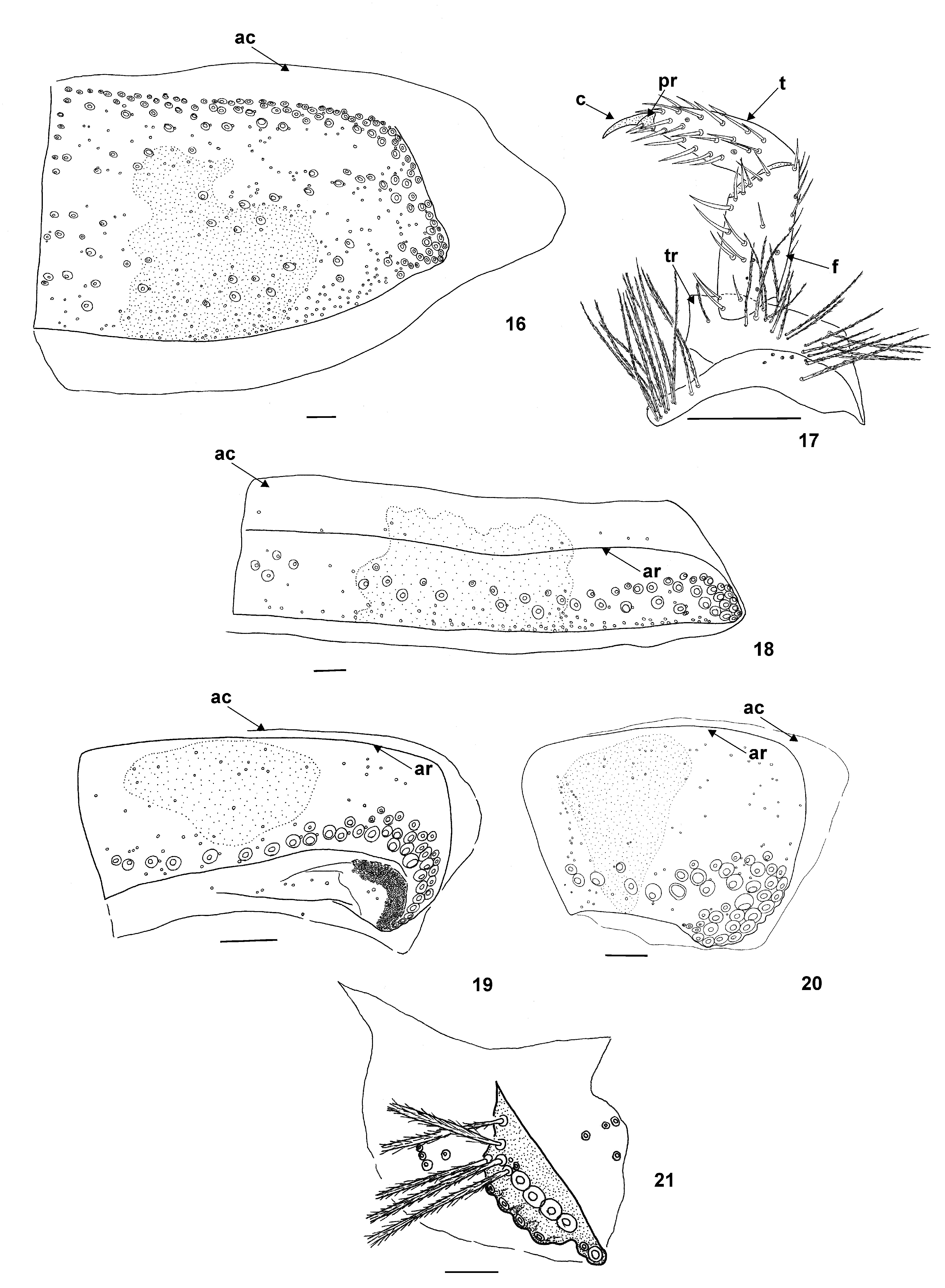
Antecostal suture (ar) on notum I absent, but distinct on nota II and III as well as abdominal terga I–IV; terga V–VIII without antecostal suture. Acrotergites (ac) of notum I without setae; acrotergites of other thoracic and abdominal segments with a few fragile and small spicisetae. Nota I with long, stout, large spicisetae along anterior (directed anteriorly under head), lateral and posterior margin (directed latero–posteriorly and vertically). Some spicisetae present on central area of disc of notum I; numerous hastisetae arranged among spicisetae on almost entire area ( Fig. 16 View FIGURES 16 – 21 ). Nota II and III and abdominal terga I–IV with median row of large spicisetae, mainly directed latero–posteriorly and vertically; hastisetae less numerous than spicisetae, on posterior half of terga, mainly along posterior margin ( Fig. 18 View FIGURES 16 – 21 ). Abdominal terga with V–VIII large spicisetae located along posterior margin and densely arranged on latero–posterior angle; hastisetae much less numerous than spicisetae, the former mainly located on anterior half of terga ( Figs 19–20 View FIGURES 16 – 21 ). Hastisetae of abdominal segments forming dense lateral brushes, particulary on posterolateral region of abdominal terga V, VI, VII, inserted on each side of membrane behind tergum ( Fig. 19 View FIGURES 16 – 21 ). Setal patterns of abdominal tergum I as illustrated ( Fig. 18 View FIGURES 16 – 21 ); abdominal tergum VII as illustrated ( Fig. 19 View FIGURES 16 – 21 ). Abdominal tergum VIII without pair of abdominal pits (oval apertures); setal patterns as illustrated ( Fig. 20 View FIGURES 16 – 21 ). Abdominal tergum IX reduced in size with numerous long spicisetae creating caudal brush and arranged as illustrated ( Fig. 21 View FIGURES 16 – 21 ). Legs covered with many dark brown setae as illustrated on Fig. 17 View FIGURES 16 – 21 . Tarsal claws (c) dark brown. Ratio of length of tibia (t) to length of femur (f) 1:1. Pretarsus (pr) with two narrow lanceolate setae inserted at base. Anterior pretarsal seta the same length as pretarsus. Length of posterior pretarsal seta the same length as anterior pretarsal seta ( Fig. 17 View FIGURES 16 – 21 ).
Pupa unavailable, but probably develops within last larval exuvium which is split lengthwise from head to abdominal nota I (often to abdominal nota VIII) ( Figs 1–2 View FIGURES 1 – 4 ).
Distribution. Afghanistan; Caucasus; China; Iran; Kazakhstan; North Korea; Kyrgyzstan; Mongolia; Syria; Tadzhikistan; Turkmenistan; Uzbekistan ( Háva 2011).

No known copyright restrictions apply. See Agosti, D., Egloff, W., 2009. Taxonomic information exchange and copyright: the Plazi approach. BMC Research Notes 2009, 2:53 for further explanation.