
Tafana Simon, 1903
|
publication ID |
https://doi.org/ 10.5852/ejt.2021.742.1291 |
|
publication LSID |
lsid:zoobank.org:pub:23424EC3-8440-4C72-AD9E-C1EC3087F7CA |
|
DOI |
https://doi.org/10.5281/zenodo.4658596 |
|
persistent identifier |
https://treatment.plazi.org/id/03CF87AD-FFA1-6622-FDEE-9014FA8CFE7F |
|
treatment provided by |
Plazi |
|
scientific name |
Tafana Simon, 1903 |
| status |
|
Genus Tafana Simon, 1903 View in CoL
Tafana Simon, 1903:124 View in CoL
(type species by original designation: T. riveti Simon, 1903: 124 View in CoL ).
Tafana View in CoL – Petrunkevitch 1911: 514; 1928: 174. — Roewer 1954: 546. — Bonnet 1959: 4232. — Brescovit 1997: 80, 90, figs 225–238.
Diagnosis
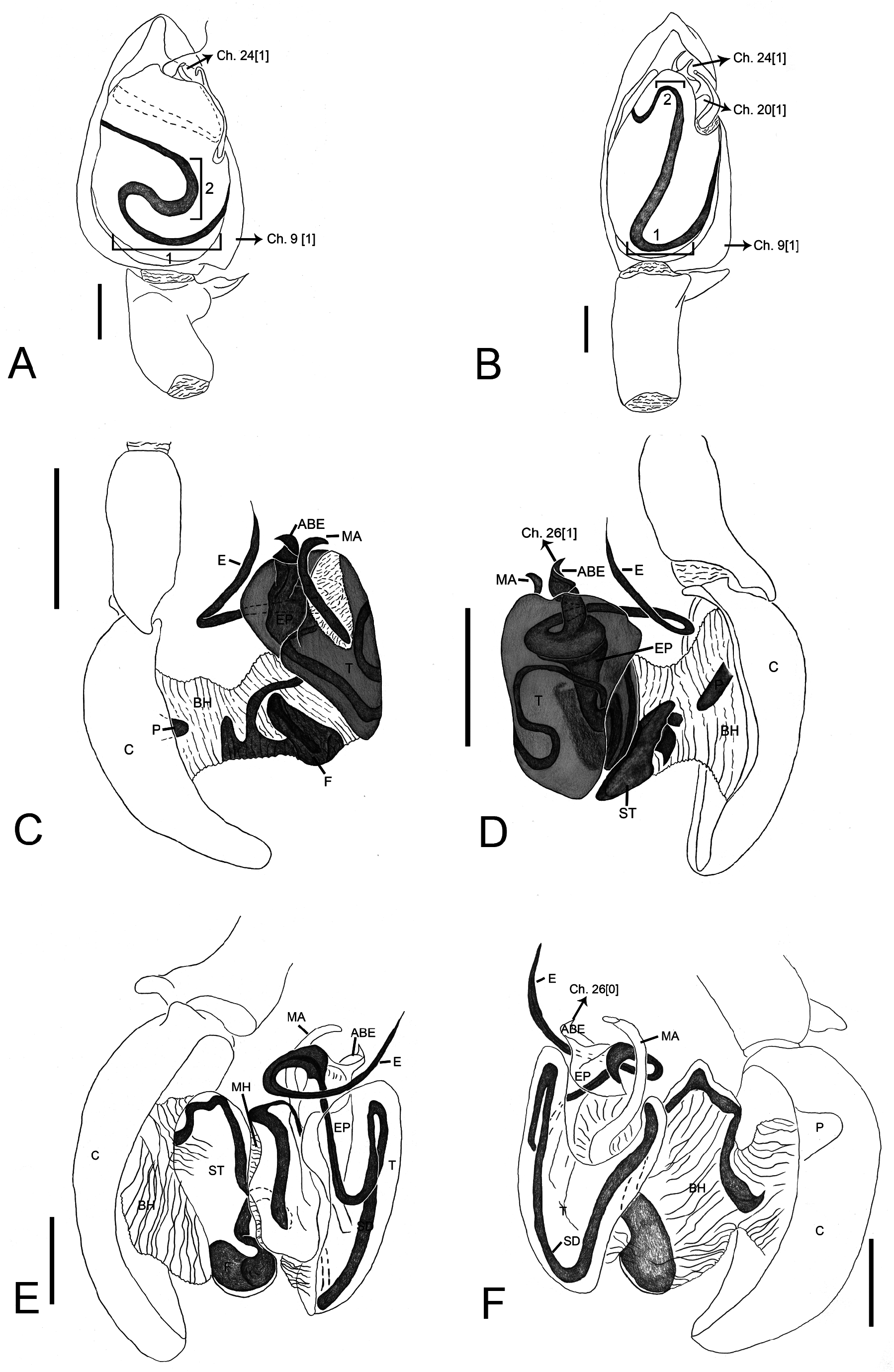
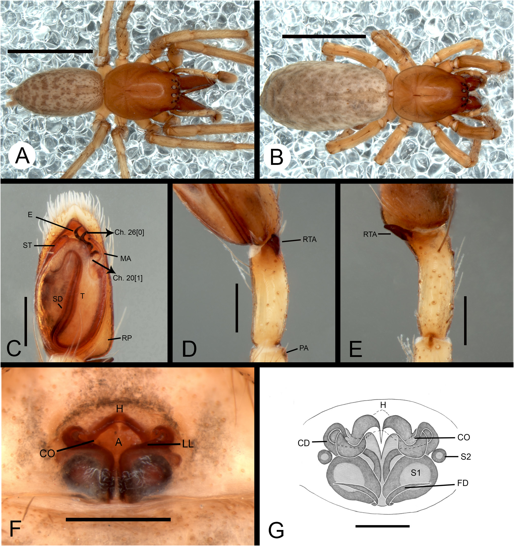
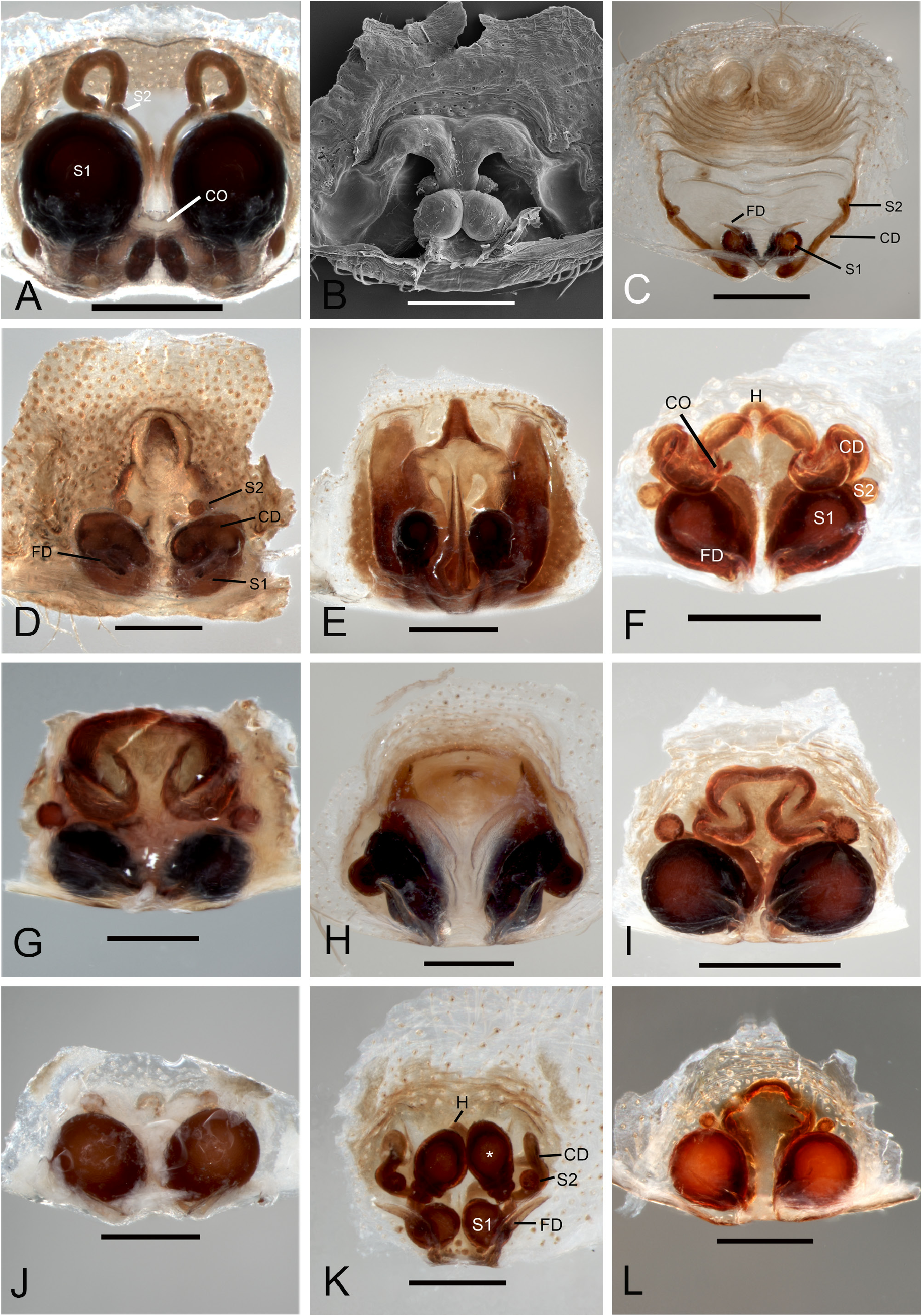
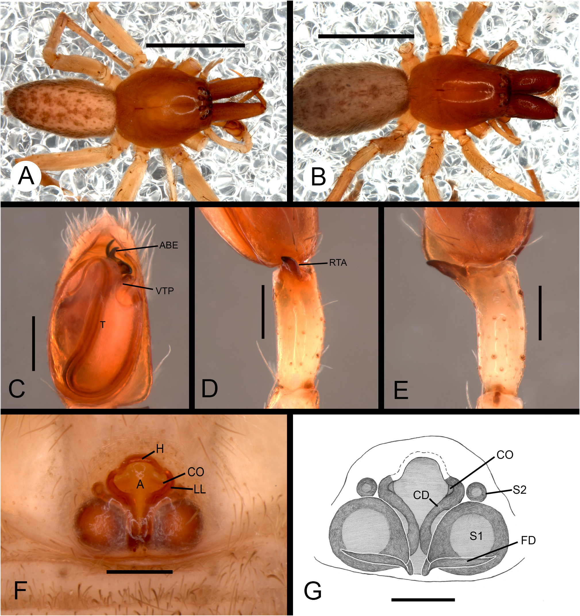
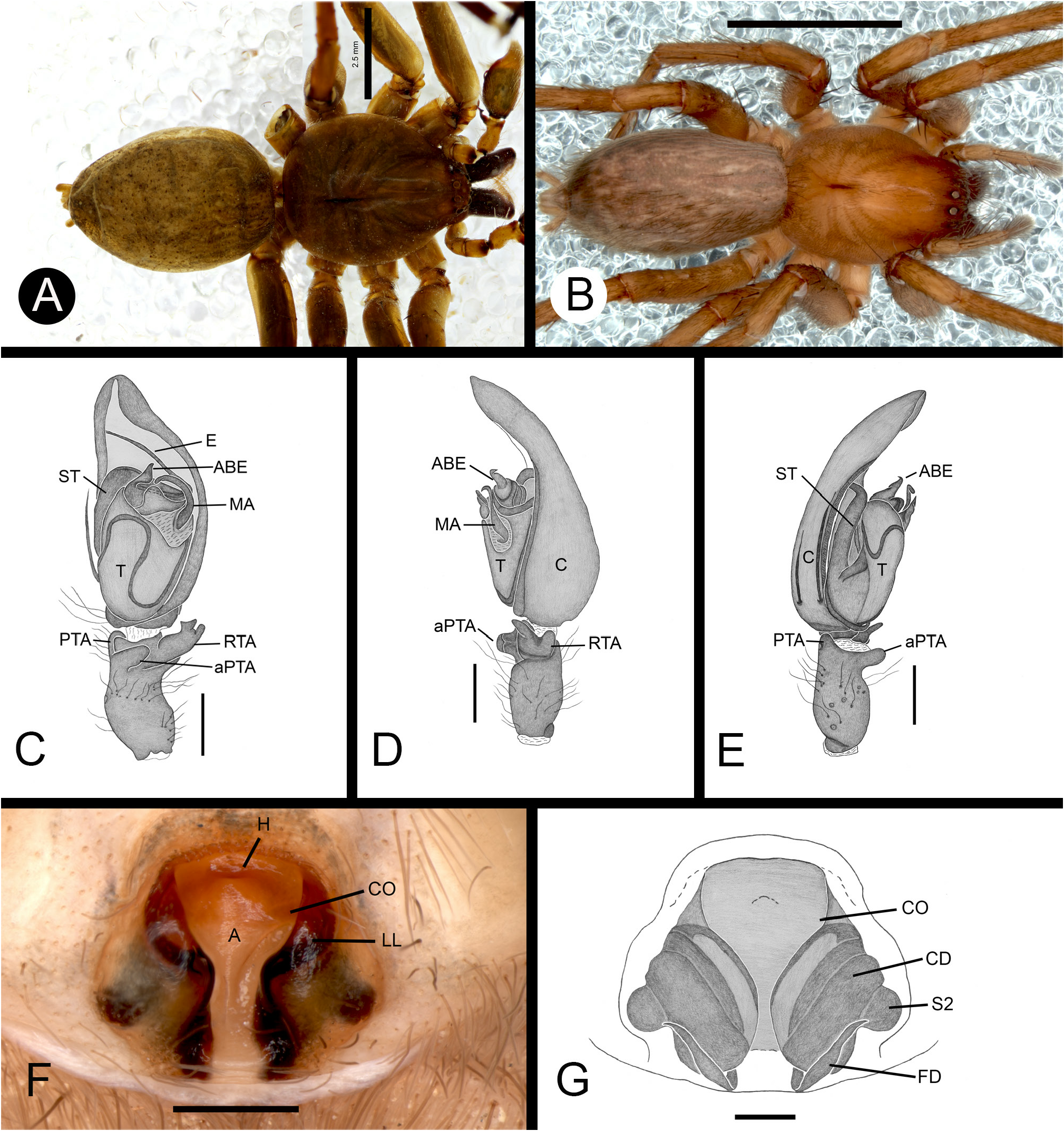
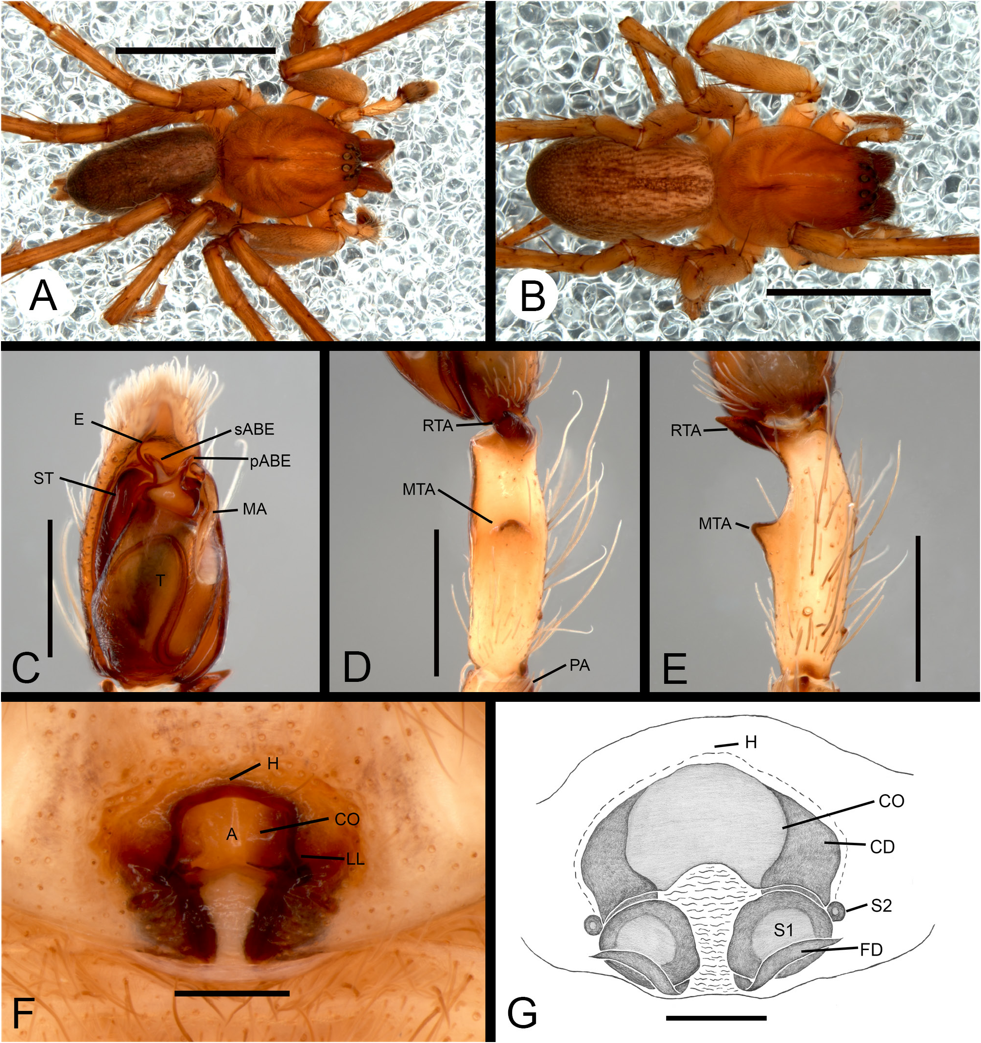
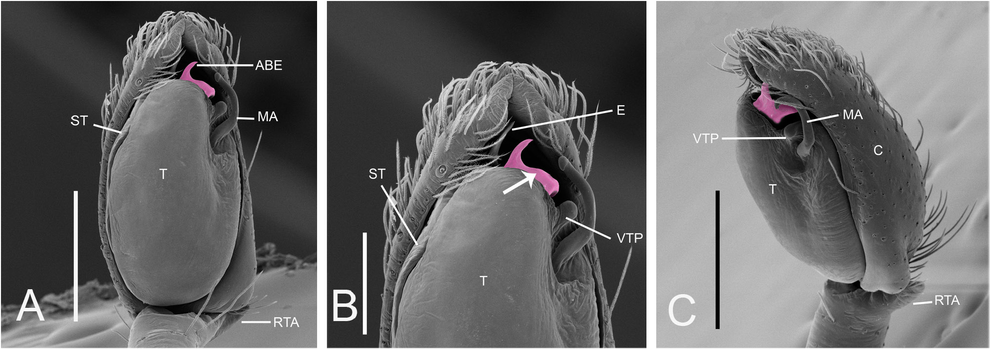
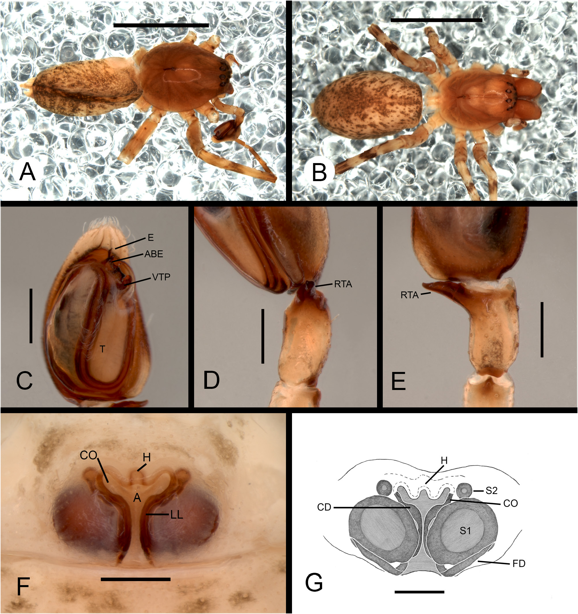
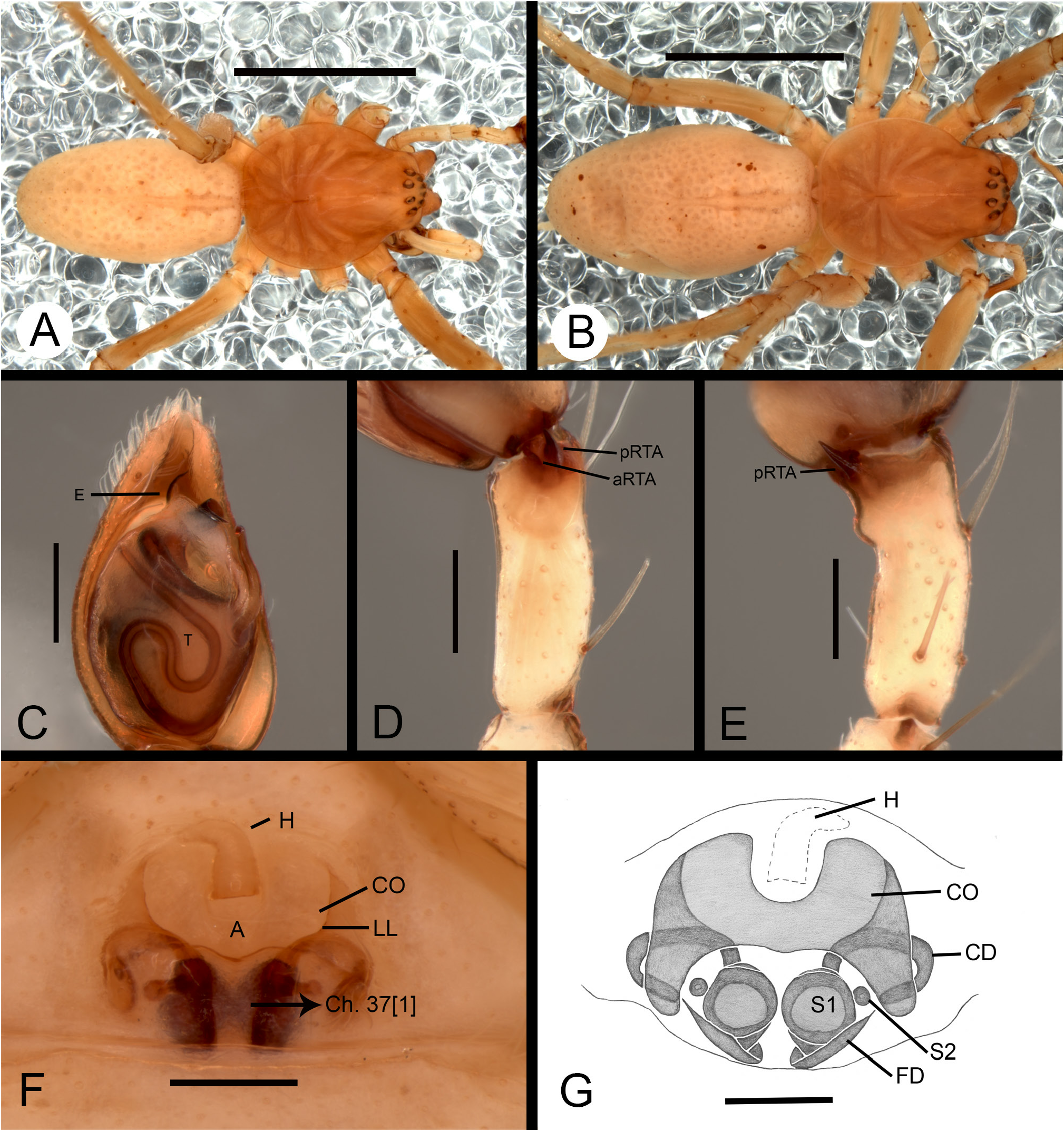
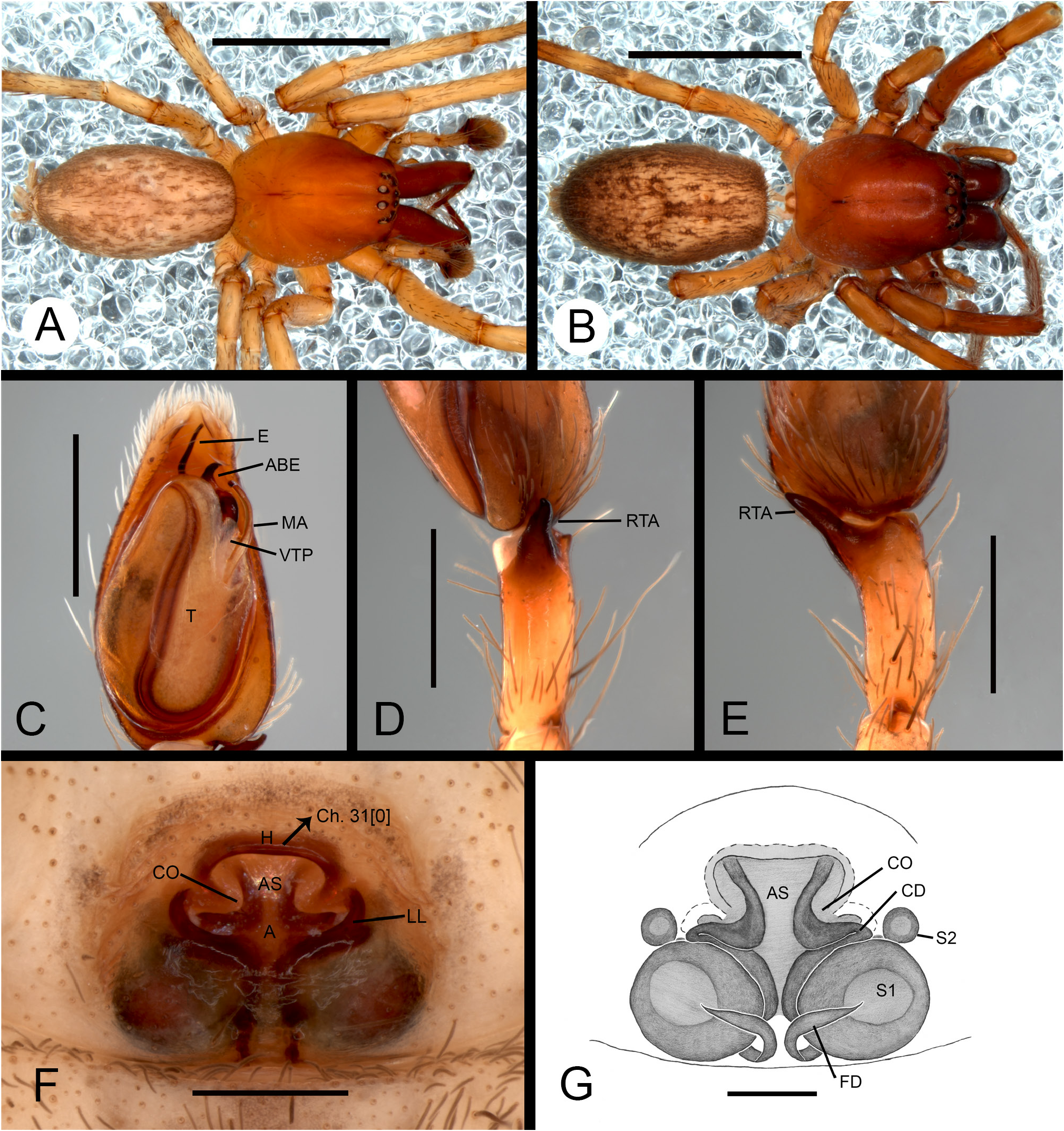
Males of species of the genus Tafana resemble those of the genera Aljassa Brescovit, 1997 , Aysha Keyserling, 1891 , Hibana Brescovit, 1991 , Hatitia Brescovit, 1997 , Osoriella Mello-Leitão, 1922 , Pippuhana Brescovit, 1997 , Temnida Simon, 1896 , Umuara Brescovit, 1997 and Xiruana Brescovit, 1997 by having an embolic process in the internal male palp (see Brescovit 1997: 176–187, figs 221, 247, 265, 272, 280), an apophysis at the base of the embolus (see Oliveira & Brescovit 2015a: 209, fig. 5a–b) and further resemble Umuara by having a ventral tegular process near the median apophysis in the male palp (see Oliveira & Brescovit 2015b: 438, figs 2a–b), but differ by the presence of a conical or coiled laminar apophysis at the base of the embolus ( Figs 9C–F View Fig , 12B, C View Fig , 23B View Fig , 26A–B View Fig ). The females resemble those of Anyphaena , Patrera Simon, 1903 , Iguarima Brescovit, 1997 , Umuara and Pippuhana (see Brescovit 1997: 139–187), by having a hood on the anterior fold of the epigynum, but differ by having the posterior extension forming the lateral lobes ( Figs 7D View Fig , 10F View Fig , 22F View Fig , 32E–F View Fig , 33D–E View Fig ), lateral lobes sinuous ( Figs 17F View Fig , 19F View Fig ) and posterior region of the epigynum covered by cuticle ( Figs 10F View Fig , 17F View Fig , 21F View Fig ).
Description
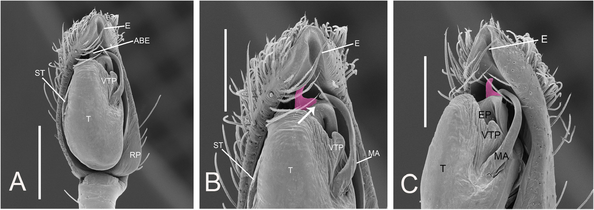
Body from pale white to orange and reddish brown ( Figs 10A View Fig , 11A View Fig , 22A View Fig ). Legs orange or pale and darker at tarsi ( Figs 22B View Fig , 25A View Fig ). Carapace sub-rectangular, narrow in anterior region, and enlarged near coxae I–III, cephalic region high, highest behind the posterior eye row ( Figs 8A View Fig , 10A View Fig ). Eyes, in dorsal view, anterior row slightly recurved and posterior row procurved ( Figs 8A View Fig , 10B View Fig ). Chelicerae robust and projected, with vertical length approximately half the length of the carapace, basal condyle conspicuous ( Fig. 11A–B View Fig ), with 3–5 promarginal teeth and 4–7 retromarginal ( Fig. 8B View Fig ). Two rows of trichobothria with striated base in dorsal view on the metatarsus and tarsus I–IV ( Fig. 8J View Fig ). Tarsal organ with teardrop shape on the distal region of tarsus and male palp ( Figs 8I View Fig , 20D View Fig ), slit sensilla elongated on the tarsus ( Fig. 8K View Fig ). Paired tarsal claws with 8–14 teeth ( Fig. 8G–H View Fig ). Male palp: patellar apophysis short and rounded ( Figs 10D View Fig , 16D–E View Fig , 24D View Fig ); distal prolateral tibial apophysis short ( Fig. 13C, E View Fig ); retrolateral tibial apophysis with large base and conical apex ( Fig. 22D–E View Fig ), usually exhibiting anterior branches and posterior branches ( Fig. 13C–D View Fig ) or only one branch ( Figs 10D View Fig , 11D View Fig , 24D–E View Fig ); median tibial apophysis branched ( Fig. 29D–E View Fig ) or unique ( Fig. 30D–E View Fig ); cymbium oval with retrolateral projection ( Figs 9A–B View Fig , 10C View Fig , 12A View Fig , 22C View Fig , 26A, C View Fig ), with small furrow under this projection, to accommodate the retrolateral tibial apophysis, or without ( Figs 29C View Fig , 30C View Fig ); chemosensory setae on the cymbium ( Fig. 20D View Fig ); subtriangular and ypsiloid petiole ( Fig. 9C–D, F View Fig ); subtegulum with 5–6 semicircular rings, short and exposed distal projection on the prolateral side on the palp when not expanded ( Fig. 9D–E View Fig ); tegulum with ventral tegular process narrow near the median apophysis ( Figs 9B View Fig , 10C View Fig , 12B–C View Fig , 18B View Fig ) and bordering the apex of the tegulum ( Figs 9A View Fig , 22C View Fig , 26A–B View Fig , 28C View Fig ); sclerotized median apophysis long, sharp and with a curved apex ( Figs 18B View Fig , 20B View Fig , 23C View Fig ); embolic process conical, sclerotized, covered by the tegulum, inserted at the base of the embolus ( Fig. 9C–F View Fig ); sperm duct presenting four or five loops in palp expanded ( Fig. 9C–F View Fig ) and in palp not expanded three loops in ventral view ( Fig. 9A–B View Fig ), internally traversing the embolic process reaching the embolus ( Fig. 9E View Fig ); embolus long ( Figs 9D View Fig , 22C View Fig , 24C View Fig ) or short ( Figs 10C View Fig , 15C View Fig , 17C View Fig ), filiform helical and curved retro-apically, connected to the embolic process ( Figs 9D–E View Fig , 15C View Fig , 22C View Fig ); apophysis at the base of the embolus, conical ( Figs 9B, E View Fig , 10C View Fig , 12C View Fig , 18A–B View Fig ) or laminar ( Figs 23B View Fig , 24C View Fig , 26A–B View Fig ). Oval abdomen, tracheal spiracle curved in the middle of the abdomen in ventral view ( Figs 7B View Fig , 8L View Fig ); anterior spinnerets bisegmented, with basal article large and distal article circular, with several spigots of piriform gland and one spigot of the ampullate gland ( Fig. 8C–D View Fig ); median spinnerets unsegmented, with several spigots of the aciniform gland and not presenting spigots of the cylindrical gland ( Fig. 8C, E View Fig ); posterior spinnerets bisegmented with basal and distal article cylindrical, one third longer than the others, with several spigots of the aciniform gland and one spigot of the ampullate gland without spigots of the cylindrical gland ( Fig. 8C, F View Fig ); colulus substituted by 8–10 setae ( Fig. 8C View Fig ). Epigynum with sinuous lateral lobes, lobe parallel in the posterior region ( Figs 10F View Fig , 22F View Fig ); hood approximately triangular ( Figs 10F View Fig , 16F View Fig , 25F View Fig , 27F View Fig ), semicircular ( Figs 11F View Fig , 14F View Fig , 32D View Fig ) or circular ( Figs 15F View Fig , 17F View Fig , 30F View Fig , 32J View Fig , 33G– H View Fig ) in the anterior region; posterior region with cuticle covered the lateral lobes ( Figs10F View Fig , 11F View Fig , 17F View Fig , 21F View Fig , 27F View Fig ) or without ( Figs 13F View Fig , 15F View Fig , 16F View Fig , 28F View Fig ); atrium with septum in the anterior region ( Figs 11F View Fig , 14F View Fig , 21F View Fig ) or without ( Figs 13F View Fig , 15F View Fig , 22F View Fig , 25F View Fig ); copulatory ducts sinuous ( Figs 10G View Fig , 11G View Fig , 19G View Fig , 32F–I View Fig ) or coiled ( Figs 13G View Fig , 16G View Fig , 32H, K View Fig ), running along margin of the lateral lobes, reaching the spermathecae, visible through transparency; secondary spermathecae rounded ( Figs 10G View Fig , 11G View Fig , 14G View Fig , 32F–G, I View Fig , 33B–C View Fig ) but may be irregular or inconspicuous ( Figs 13G View Fig , 28G View Fig , 29G View Fig , 32H View Fig , 33F–G View Fig ); secondary spermathecae short, near the primary spermathecae ( Figs 15G View Fig , 17G View Fig , 22G View Fig , 26D View Fig , 32F, J, L View Fig ), wide ( Figs 11G View Fig , 13G View Fig , 14G View Fig , 32G, I View Fig ) or in the middle of the copulatory ducts ( Figs 16G View Fig , 21G View Fig , 29G View Fig , 32K View Fig , 33G View Fig ); primary spermathecae oval, positioned together ( Figs 10G View Fig , 15G View Fig , 20E View Fig , 22G View Fig , 26D View Fig , 32F–G View Fig ) or separated from each other by almost a third of their diameter ( Figs 13G View Fig , 28G View Fig , 29G View Fig , 33F–H View Fig ); fertilization ducts near the epigastric furrow, originating from base of spermathecae ( Figs 10G View Fig , 11G View Fig , 20E View Fig , 22G View Fig , 26D View Fig , 29G View Fig , 32F–L View Fig , 33A–H View Fig ).


No known copyright restrictions apply. See Agosti, D., Egloff, W., 2009. Taxonomic information exchange and copyright: the Plazi approach. BMC Research Notes 2009, 2:53 for further explanation.
|
Kingdom |
|
|
Phylum |
|
|
Class |
|
|
Order |
|
|
Family |
Tafana Simon, 1903
| De Oliveira, Luiz Fernando M. & Brescovit, Antonio Domingos 2021 |
Tafana
| Brescovit A. D. 1997: 80 |
| Bonnet P. 1959: 4232 |
| Roewer C. F. 1954: 546 |
| Petrunkevitch A. 1928: 174 |
| Petrunkevitch A. 1911: 514 |
Tafana
| Simon E. 1903: 124 |
| Simon E. 1903: 124 |