
Entomophthalmus rufiolus (LeConte, 1866)
|
publication ID |
https://doi.org/ 10.1649/0010-065x-68.2.331 |
|
persistent identifier |
https://treatment.plazi.org/id/03CFD643-FFDE-5F6A-AD93-F338FC03FACF |
|
treatment provided by |
Valdenar |
|
scientific name |
Entomophthalmus rufiolus (LeConte, 1866) |
| status |
|
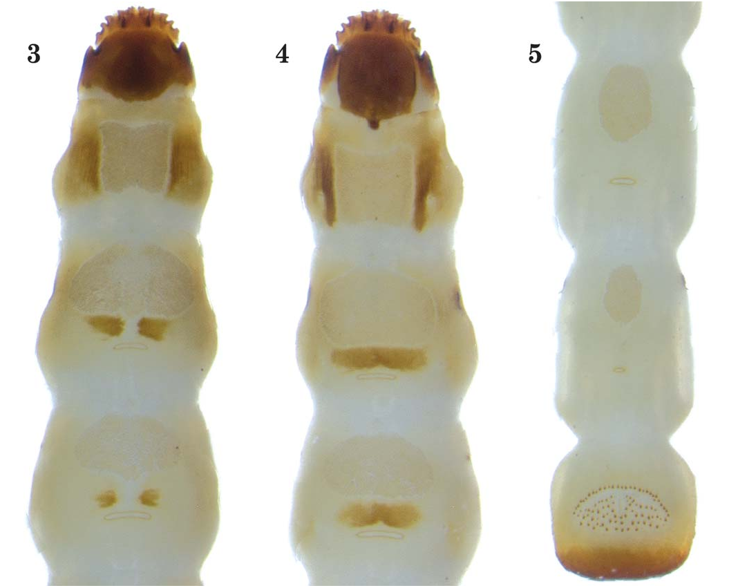
Entomophthalmus rufiolus (LeConte, 1866) fifth instar ( Figs. 1–5 View Fig View Fig View Figs )
Diagnosis. The dorsal and ventral thoracic scleromes, along with the shape of the areoles, as well as the abdominal microtrichial patches should distinguish this species from Rhagomicrus bonvouloiri (Horn, 1886) .
Specimens Examined. Twelve larvae collected at USA: WISCONSIN: Dane Co., Black Hawk Ridge , 2 mi. S. Sauk City, 22 April 1995, Robert L. Otto, collected under bark of fermenting oak log (1 larva) ; Turville Point Preserve , N43°02.897′, W-89°22.243′, 7 October 2012, Robert L. Otto, in rotten Quercus limb (4 larvae) GoogleMaps , N43°03.197′, W-89°22.887′, 11 October 2012, Robert L. Otto, in rotten Basswood limb (6 larvae); Oconto Co., Rueckert’ s property, T29 N R17 E sec. 16, 21 April 2007, Robert L. Otto, in white rotten maple limb (1 larva) .
Description. Length 10.0–13.0 mm, width 1.00– 1.25 mm. Orthosomatic. Body ( Fig. 1 View Fig ): Subcylindrical, parallel-sided, cream-yellow with head, prothoracic sclerome patches, and caudal end of abdominal segment IX dark brown. Setae reduced or absent. Pair of small legs reduced to very short setae near posterolateral areas of each thoracic segment. Dorsal and ventral microtrichial patches slightly darker in color than their surrounding areas. Head ( Fig. 2 View Fig ): Strongly flattened, prognathus, and inserted into prothorax. Dorsal cephalic disc subcircular. Slightly elevated median carina present on dorsal cephalic disc. Ventral cephalic disc similar to dorsal side, unmodified. Anterior portion of head capsule heavily sclerotized. Lateral sides of head capsule unsclerotized. Each lateral side of head capsule consists of five projections. Basal lateral projection enlarged. Second through 5th lateral projections directed anterolaterally. Antennae minute, arising between 4th and 5th lateral projections. Scape not visible. Pedicel elongate. Sensorum and flagellum subequal in length. Sensory papillae indistinct. Mandibles minute, resting in mesal acumination of head capsule between 5th lateral projections. Each mandible heavily sclerotized, distinct, oval, longer than wide, bifid. Labial and maxillary palpi extremely small, near indistinct. Ligula, mala, lacinia, and galea absent. Hypostomal rods also absent. Prothorax ( Figs. 3, 4 View Figs ): Length subequal to following 2 thoracic segments. Tergum with pair of triangular scleromes with undefined craniad and lateral sides. Trapezoidal microtrichial patch between the scleromes. Small, oval areole present near posterior end, below the microtrichial patch. Ventroanterior aspects consisting of 2 sclerotized bars extending down length of segment, converging toward posterior end. Sternal micrortrichial patch and areole similar to those on tergum. Mesothorax: Tergum with elliptical microtrichial patch; 2 short, diverging, sclerotized lines present beneath the patch. Sternum with similar microtrichial patch; horizontal, sclerotized line present beneath patch. Both surfaces with caudally bent, oval areole near base. Metathorax: Tergum with elliptical microtrichial patch near anterior end; pair of small, subcircular scleromes present below the patch. Sternum very similar to tergum, except horizontal sclerotized line much shorter. Caudally bent, oval areole present near base of segment. Abdomen: Segments I-IX subequal in length and width. Terga I-VIII with large, elliptical microtrichial patch near anterior end and small, oval areole at caudal end of segment. Microtrichial patch and areole on sterna I-VIII similar to those on terga. Tergum IX without patches, sparsely punctate near caudal end. Sternum IX ( Fig. 5 View Figs ) heavily sclerotized at caudal half with prominent, semicircular, circumanal asperities. Urogomphi absent on segment IX. Spiracles annular-biforous.
Distribution. Entomophthalmus rufiolus is known from Canada: Ontario, Québec, New Brunswick; and USA: Alabama, Arkansas, Florida, Georgia, Indiana, Illinois, Kentucky, Louisiana, Maine, Maryland, Massachusetts, Minnesota, Missouri, New Hampshire, New Jersey, New York, North Carolina, Ohio, Pennsylvania, Texas, Virginia, and Wisconsin ( Muona 2000; Webster et al. 2012). All specimens used in this study came from Wisconsin.
Biology. Entomophthalmus rufiolus is an uncommon and widespread species, found largely in eastern North America. In Wisconsin, E. rufiolus was found in a variety of forest systems. I collected adults and larvae in northern dry-mesic forest, northern hardwood swamp, northern mesic forest, northern wet-mesic forest, floodplain forest, oak barrens, oak opening, oak woodland, southern dry-mesic forest, southern hardwood swamp, and southern mesic forest. Other collectors in Wisconsin have taken specimens by Malaise traps, as well as purple prism traps (Synergy Semiochemicals, British Columbia) deployed throughout Wisconsin. Muona (2000) reported E. rufiolus was taken only on hickory ( Carya sp. ; Juglandaceae ), at blacklight traps, at window trap, and at a Coleman ® lantern light trap.
Webster et al. (2012) obtained four adults in various forest systems in New Brunswick during July and August. All specimens were collected from Lindgren funnel traps. The beetles were taken in an old silver maple ( Acer saccharinum L.; Aceraceae ) swamp, an old red oak ( Quercus rubra L.; Fagaceae ) forest with scattered white pine ( Pinus strobus L.; Pinaceae ), and a red spruce ( Picea rubens Sargent ; Pinaceae ) forest with red maple ( Acer rubrum L.) and balsam fir ( Abies balsamea (L.) Miller; Pinaceae ).
During my collections, all E. rufiolus larvae were extracted exclusively from maples, oaks, or basswoods in Wisconsin during 1995, 2007, and 2012. All extracted larvae were found in drier, firm, hard sections of white rotten tree limbs with little moisture, except on one occasion when a single specimen was collected in a moist, white rotten oak log. Larvae were positioned parallel with the grain of the sapwood and burrowing between layers of wood fibers, leaving no galleries behind them. Their unique head capsule is useful as a wedge, allowing the larva to move through the sapwood. Mature larvae were observed constructing pupal chambers approximately 2.5 cm beneath the bark layer. Larvae assumed a U-shaped position as they molted to the pupa in the chamber. Fourteen adults emerged during 2013 from wood pieces collected at a wooded preserve in Madison the previous fall. The forest was a southern mesic forest dominated with basswood along with oaks, maples, and walnut ( Juglans nigra L.; Juglandaceae ) as associates. Behaviors were not recorded nor were attempts to breed specimens to obtain further information on the species’ life cycle.


No known copyright restrictions apply. See Agosti, D., Egloff, W., 2009. Taxonomic information exchange and copyright: the Plazi approach. BMC Research Notes 2009, 2:53 for further explanation.
|
Kingdom |
|
|
Phylum |
|
|
Class |
|
|
Order |
|
|
Family |
|
|
Genus |