
Pentasetacus plicatus, Chetverikov, Philipp E. & Petanović, Radmila U., 2016
|
publication ID |
https://doi.org/ 10.11646/zootaxa.4144.2.4 |
|
publication LSID |
lsid:zoobank.org:pub:C560E771-F103-4A22-948A-D78EFEBD9D0A |
|
DOI |
https://doi.org/10.5281/zenodo.5616018 |
|
persistent identifier |
https://treatment.plazi.org/id/03D05F7E-FFDC-FFE7-FF0D-8832FAFAFB53 |
|
treatment provided by |
Plazi |
|
scientific name |
Pentasetacus plicatus |
| status |
sp. nov. |
Pentasetacus plicatus n. sp. Chetverikov & Petanović
( Fig. 1 View FIGURE 1 )
Female holotype. Idiosoma vermiform, 264 long, 61 wide at the level of seta c2. Prodorsal shield subpentagonal 32 long x 49 wide, with short, subtriangular frontal lobe 3 long x 7 wide basally. Shield ornamentation weak and includes three short faint grooves (=median and two admedian lines) and two distinct ridges (=submedian I lines) in posterior half of shield. Indistinct medioposterior fovea (“median pit” sensu Keifer 1962, p. 16) near posterior prodorsal shield margin. Unpaired seta vi (10) inserted near base of frontal lobe, directed forward; ve 13, directed anterolaterad, tubercles 30 apart; sc 15, directed anterolaterad, tubercles 24 apart. Distance between tubercles vi–ve 15, vi–sc 18, ve–sc 12. Two indistinct eye-like structures (oval areas of thin cuticle) present anterolaterally to tubercules of sc. Gnathosoma elongate, directed forward and slightly ventrad, 29 long. Basal segment of palp 12 wide; palpcoxal apodeme 7 long; pedipalp coxal seta ep 5, pedipalp genual seta d 14, subapical pedipalp tarsal seta ν 1. Subcapitular (suboral) plate subtriangular, rounded anteriorly, 13 long, 20 wide, with two longitudinal lateral ridges. Leg I 27, tibia 5, l' 10, tibial solenidion φ 7; tarsus 6, u’ 4, ft’ 10, ft’’ 20, ω 8 without knob; empodium 4/4- rayed, 6 long, each ray of three basal pairs with two additional secondary branches, terminal paired rays without additional branching; l” 24; bv 7. Leg II 23, tibia 4, l' absent; tarsus 5, u’ 4, ft’ 9, ft’’ 20, ω 8 without knob; empodium 4/4-rayed, 5 long; l” 24; bv 5. Coxae ornamented with several short lines; coxal II apodemes distinct, extending up to the level of lateral angles of genital area. Setae 1b 15 long, 12 apart; 1a 39 long, 10 apart; 2a 38 long, 25 apart. Prosternal apodeme not discerned; 4 coxigenital annuli (2 posterior ones complete, 2 anterior incomplete, i.e. segregated from lateral annuli) before epigynium. Genital coverflap semi-circular, smooth, 14 long x 21 wide; setae 3a 10 long, 17 apart; pregenital plate absent; posterior genital cuticle framed by dense bandlike genital flange. Opisthosoma vermiform (nearly parallel-sided for most of its length), widest at the level of c2 tubercles; 71 dorsal and 70 ventral annuli bearing small, pointed microtubercles. Setal lengths: c1 10, c2 14, d 13, e 7, f 30, h1 17, h2 67; 13 annuli between rear prodorsal shield margin and c1 tubercles; 8 annuli from rear shield margin to c2; 13 annuli between c2–d; 17 annuli between d and e; 26 annuli between e and f; and 6 annuli between f and h1.
Male (n=3). Only three male specimens in good condition were available for measurement. In comparison to females, males are smaller; they have in average about 10 dorsal and 10 ventral annuli less than females, slightly shorter legs and opisthosomal setae ( Table 1 View TABLE 1 ) and similar ornamentation of prodorsal shield ( Fig. 2 View FIGURE 2 ). Genital area ellipsoid, 12–14 long, 16–18 wide, flanked anteriorly by ribbon-like genital coverflap and laterally by two arc-like genital folds forming together genital rim ( Fig. 3 View FIGURE 3 ). In one male genital coverflap was thrown back so that gonopore was available for observation ( Fig. 3 View FIGURE 3 B). Tubercles of 3a situated immediately behind posterior extremity of arc-like folds, 3a 14–15 long. Genital opening situated under rounded medial flap of genital coverflap and limited posteriorly by three cuticular folds forming genital cone ( Fig. 3 View FIGURE 3 A,B); 3‒5 paired rounded ridges situated behind genital cone between tubercules of 3a. CLSM indicated that setae eu were absent. Anterior genital apodeme is a thin narrow plate situated in frontal plane. Two posterior coxigenital annuli are thickened forming two pregenital ridges protruding inside from ventral cuticle ( Fig. 3 View FIGURE 3 C,D). Remnants of soft genital organs (ductus ejeculatorius, testis and paired vas deferens) similar to those recently described in Pentasetacus araucariae and Trisetacus sp. (Chetverikov et al. 2014, figs. 3, 12) and other eriophyoids (Chetverikov 2015, figs. 1,2) were observed under CLSM in one male ( Fig. 3 View FIGURE 3 E).
Immature instars ( Fig. 4 View FIGURE 4 , Table 1 View TABLE 1 ). Only three dorso-ventrally oriented and appropriately cleared nymphs and one larva were available for investigation. The larva has notably broader and fewer ventral annuli than nymphs. A progenital chamber or any other structure resembling it (e.g. a fovea) is absent in immatures. In comparison to almost equally dorso-ventrally annulated females and males, immatures have considerably fewer numbers of ventral annuli (49‒53 in nymphs and 33 in larva) than number of dorsal annuli (54‒65 in nymphs and 57 in larva); additionally the frontal lobe is absent in immatures. Eye-like structures are more distinct in immatures than in adults, but the medio-posterior fovea on the prodorsal shield is more clearly seen in adults than in immatures.
Host plant and relation to the host. Araucaria araucana (Molina) K.Koch , commonly known as “monkey puzzle tree”. Twigs of A. araucana were inspected under a stereomicroscope; neither eriophyoid mites nor distinct damages of leaves, buds or stem were found. However when the plant material was processed using extraction method numerous P. plicatus n. sp. mites were found, suggesting that these mites are probably needle vagrant.
Type locality. Chile, Valparaíso Region, Vina del Mar , City Park.
Type material. About 60 mites (females, males and immatures) in 50 slides collected in Chile, Vina del Mar, 28 March 2012, coll. Ž. Tomanović and V. Žikić. Type material is divided in two parts: half of the slides (including holotype) are kept at the Zoological Institute of RAS and the others are deposited in the collection of Prof. R. Petanović in Belgrade University.
Additional material. Five females of P. plicatus n. sp. in five slides collected from twigs of A. araucana collected Peru, Cusco Region, Calca Prov., Pisac , 25 March 2012, coll. Ž. Tomanović and V. Žikić.
Etymology. The specific epithet, plicatus , is an adjective, gender masculine, corresponding to the uncommon cuticular folds in male postgenital area in this species.
Differential diagnosis. P. plicatus n. sp. is the second described species of the genus Pentasetacus . It differs from the type species of the genus ( P. araucariae ) in ornamentation of prodorsal shield, topography of male genital area, presence/absence of progenital chamber in immatures and several morphometrics ( Table 2 View TABLE 2 ).
Characters Mite species
1. Indicated as “pregenital plate” in redescription of P. araucariae ( Fig. 7 View FIGURE 7 on page 133 in Chetverikov et al. 2014)

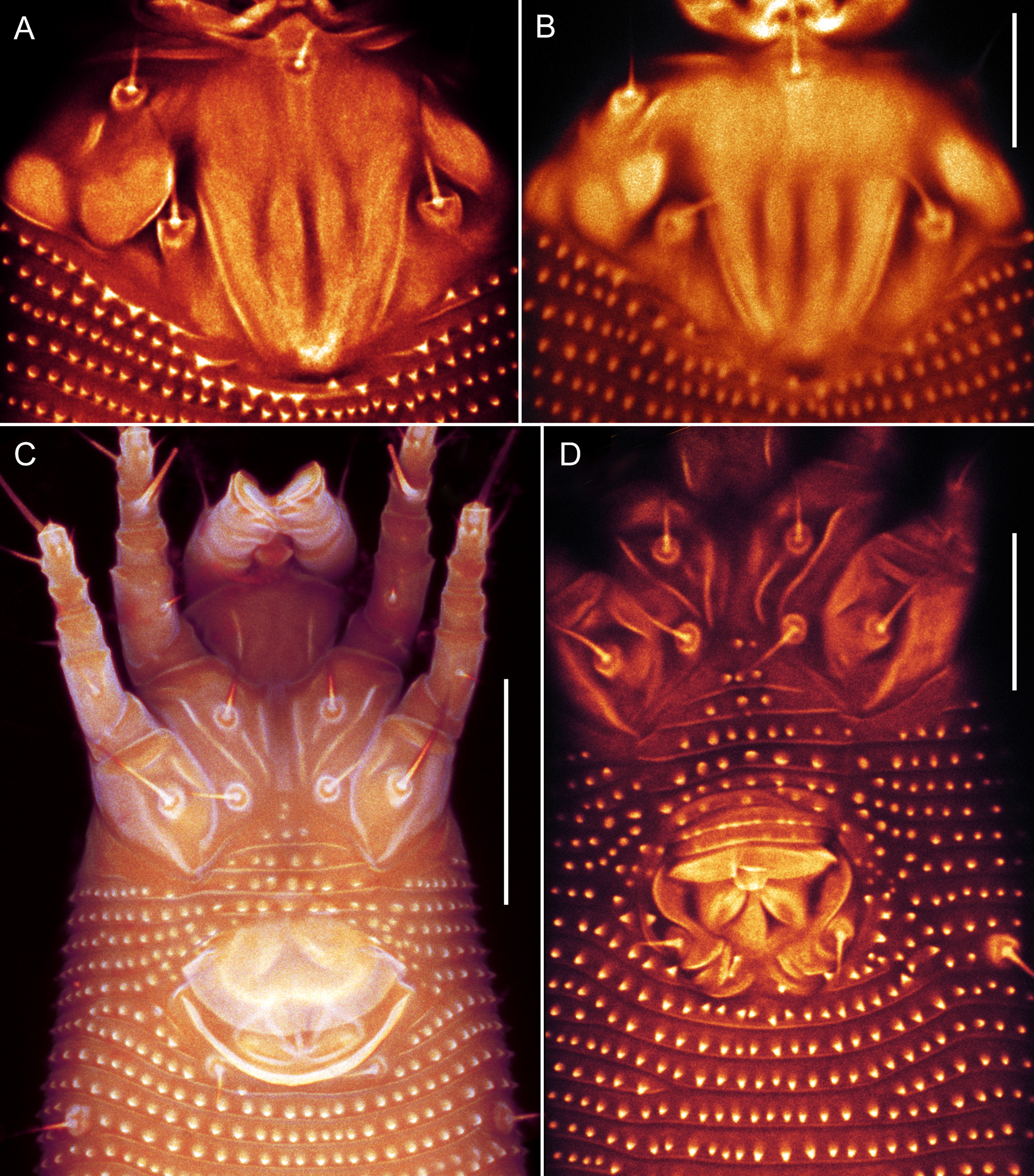
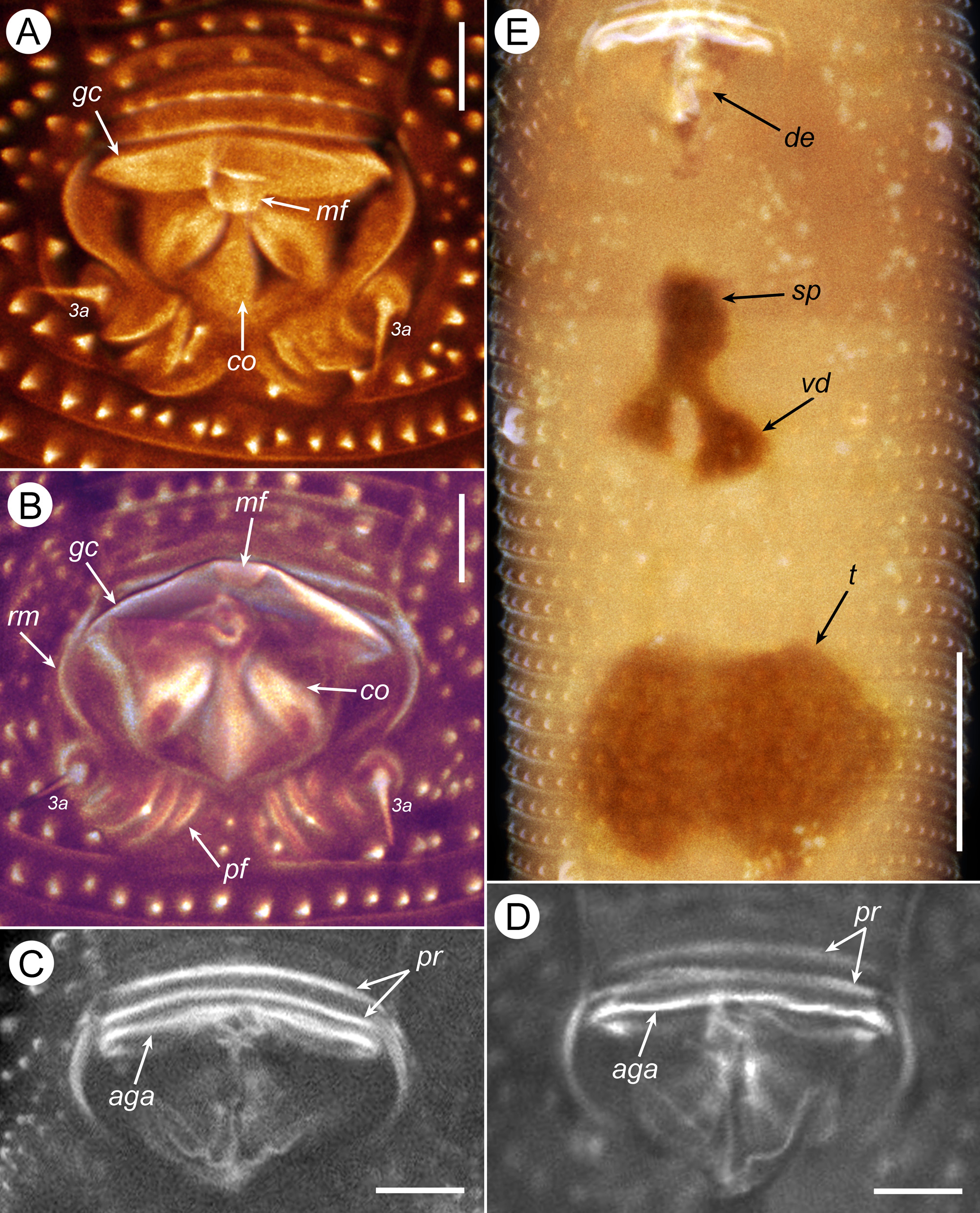
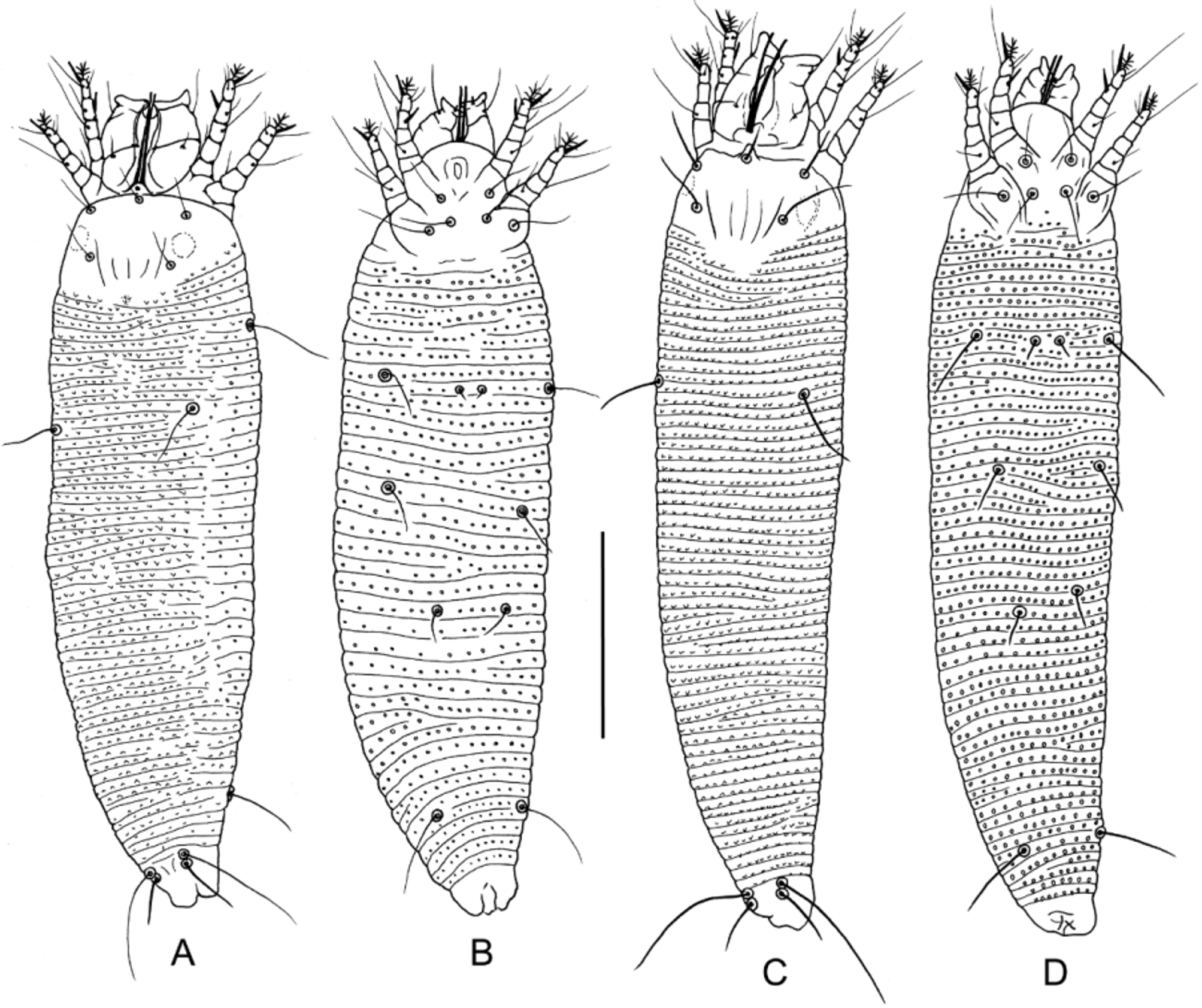
TABLE 1. Measurements of Pentasetacus plicatus n. sp. (L—length, W—width, N—number).
| Characters L of body | Females (n=7) Mean±sd Min─Max 255±17.1 233–287 | Males (n=3) Mean Min─Max 216 209–223 | Nymphs (n=3) Larva Mean Min─Max (n=1) 191 185–197 191 |
|---|---|---|---|
| W of body L of prodorsal shield W of prodorsal shield | 57±4.1 53–65 31±1.3 29–33 47±3.6 42–51 | 49 44–52 29 28–30 41 35–44 | 42 38–44 50 26 25–26 24 35 32–37 41 |
| L of frontal lobe W of frontal lobe L of gnathosoma | 3±0.4 3–4 8±1.0 6–9 27±1.6 24–29 | 3 3–3 7 7–7 25 24–27 | - - - - - - 24 22–26 24 |
| L of suboral plate W of suboral plate L of d (s. antapic.) | 12±0.7 11–13 19±1.5 18–22 13±1.4 12–16 | 11 11–12 19 17–20 14 13–14 | 11 10–12 10 17 15–20 17 6 6–6? |
| L of v (s. apic.) L of ep (s. basal.) L of vi (s.f.) | 1±0.0 1–1 5±0.0 5–5 10±1.1 8–12 | 1 1–1 4 4–4 9 7–12 | 1 1–1 1 4 4–4 1 7 6–8 6 |
| L of ve (s.d.1) L of sc (s.d.2) D between vi & ve | 13±1.7 10–16 16±1.3 15–19 14±1.0 12–15 | 9 9–10 15 13–19 14 13–14 | 9 8–9 8 13 12–14 11 11 8–14 12 |
| D between vi & sc D between ve D between sc | 17±1.0 15–18 27±1.8 25–30 22±1.7 18–24 | 17 16–19 27 27–28 22 21–24 | 19 16–22 15 24 23–26 25 20 20–21 18 |
| D between ve & sc L of leg I L of leg II | 11±1.4 8–12 26±0.6 25–27 24±0.6 23–25 | 11 11–12 24 23–24 23 22–24 | 12 12–13 12 18 16–19 16 18 17–18 15 |
| L of tarsus I L of tarsus II L of tibia I | 6±0.5 5–6 5±0.3 5–6 5±0.3 4–5 | 5 4–5 4 4–4 5 5–5 | 4 4–4 4 4 4–5 3 4 4–4 3 |
| L of tibia II L of ω I L of ω II | 4±0.5 4–5 8±0.5 7–9 8±0.7 7–9 | 5 4–5 8 7–8 8 7–9 | 3 2–3 2 6 6–6 6 7 7–7 6 |
| L of em I L of em II L of ft' I | 7±0.5 6–8 7±0.5 6–8 9±1.4 7–11 | 7 7–7 7 7–7 8 7–9 | 6 5–6 5 6 5–7 5 14 7–20 5 |
| L of ft' II L of ft'' I L of ft'' II | 10±1.0 9–11 20±1.3 19–23 20±1.0 18–21 | 7 6–8 20 19–20 17 14–20 | 16 15–17 4 9 6–11 8 9 6–14 8 |
| L of u' I L of u' II L of tibial solenidion φ I | 4±0.5 3–4 4±0.5 3–4 8±0.5 7–8 | 4 3–4 4 3–4 7 6–7 | 2 2–3 2 2 2–3 1 4 3–4 5 |
| L of l’ I (s.tib.I) L of l'' I (s.pat.) L of l'' II (s.pat.) | 8±1.1 7–10 23±1.4 21–25 23±1.3 22–25 | 6 4–7 21 18–23 21 18–23 | 6 4–7 2 16 16–16 8 19 15–23 12 |
| L of bv I (s.fem.) | 6±1.0 4–7 | 5 4–6 | 5 5–7 4 ......continued on the next page |
TABLE 2. Differences between Pentasetacus plicatus n. sp. and P. araucariae.
| Pentasetacus plicatus n. sp. | Pentasetacus araucariae | |
|---|---|---|
| Ornamentation of prodorsal shield in adults | Median and admedian lines present; medioposterior fovea present | Median and admedian lines absent; medioposterior fovea absent |
| length of vi morphometrics length of ve length of sc length of 3a Female length of c1 length of d length of e | 10 (8─12) 13 (10─16) 16 (15─19) 10 (8─11) 11 (10─12) 15 (13─16) 9 (7─10) | 16 (15─19) 17 (16─18) 31 (27─37) 17 (14─20) 19 (15─22) 22 (18─25) 22 (18─26) |
| number of dorsal annuli | 69 (66─73) | 90 (85─96) |
| number of ventral annuli | 70 (67─72) | 92 (85─100) |
| Topography of male genital coverflap1 and genital cone | Coverflap ribbon-like, with rounded medioposterior flap covering gonopore, median ridge on genital coverflap absent; genital cone is formed posteriorly by three cuticular folds | Coverflap ribbon-like, bearing a short median ridge, medioposterior flap absent; genital cone is formed posteriorly by entire bilobed plate (Chetverikov et al. 2014, fig. 7) |
| Topography of male postgenital region | 3‒5 paired rounded ridges behind genital cone between tubercules of 3a | Irregular microtubercles scattered between and around tubercles of 3a (Chetverikov et al. 2014, fig. 7) |
| Progenital chamber in immatures | Absent | Present (Chetverikov et al.2014, fig. 4) |
| Relation to host ( Araucaria araucana ) | Presumably needle vagrant causing no visible damages | Induces large blister-like galls on the upper and lower sides of leaves and clusters of bulge-like amorphous galls on the green parts of stems |
| RAS |
Union of Burma Applied Research Institute |
No known copyright restrictions apply. See Agosti, D., Egloff, W., 2009. Taxonomic information exchange and copyright: the Plazi approach. BMC Research Notes 2009, 2:53 for further explanation.