
Carolinensis sp.
|
publication ID |
https://doi.org/10.11646/zootaxa.5357.2.3 |
|
publication LSID |
lsid:zoobank.org:pub:A8932459-5A17-4812-8557-B9613DE69CEB |
|
DOI |
https://doi.org/10.5281/zenodo.10018365 |
|
persistent identifier |
https://treatment.plazi.org/id/03D07B6E-FFF2-0E66-E0CB-8DDAFBE8F878 |
|
treatment provided by |
Plazi (2023-10-18 12:08:10, last updated 2024-11-29 15:27:20) |
|
scientific name |
Carolinensis sp. |
| status |
|
Site of infection: Small intestine
Host species: Peromyscus yucatanicus
Localities: Dzununcan farm, Aak ecological park, Papam ranch, Kuncheil cattle ranch (Yucatan) and Jolie Jungle eco-hotel (Quintana Roo)
Specimens deposited: CNHE 12010‒12013
GenBank accession numbers: OR271666, OR271680
Comments: The morphological characters observed in the synlophe and the caudal bursa of the specimens agree with the description of the genus Carolinensis ( Durette-Desset 2009) . Our specimens had a synlophe with 16 ridges at midbody in both sexes ( Figure 4F View FIGURE 4 ); subsymmetrical bursa with rays 8 arising from base of dorsal ray; bifid extremities of dorsal ray; and spicules 210–285 long, characters shared with Carolinensis peromysci (Durette-Desset) . However, our specimens exhibit some differences, such as a pattern of type 1-4 t 1-3-1, the level of divergence of the dorsal ray (second half) and the spicule tips (curved). The latter two features are similar to those reported in Carolinensis dikmansi (Durette-Desset) but this species had a larger spicule (370) and a well-developed genital cone. Based on these differences, we cannot assign Carolinensis to the species level.
This finding expands the geographical distribution of Carolinensis to Quintana Roo.
Durette-Desset, M. - C. (2009) Strongylida. Trichostrongyloidea. In: Anderson, R. C., Chabaud, A. G. & Willmott, S. (Eds.), Keys to the nematodes parasites of vertebrates. CAB International, Wallingford, pp. 110 - 177. https: // doi. org / 10.1079 / 9781845935726.0110
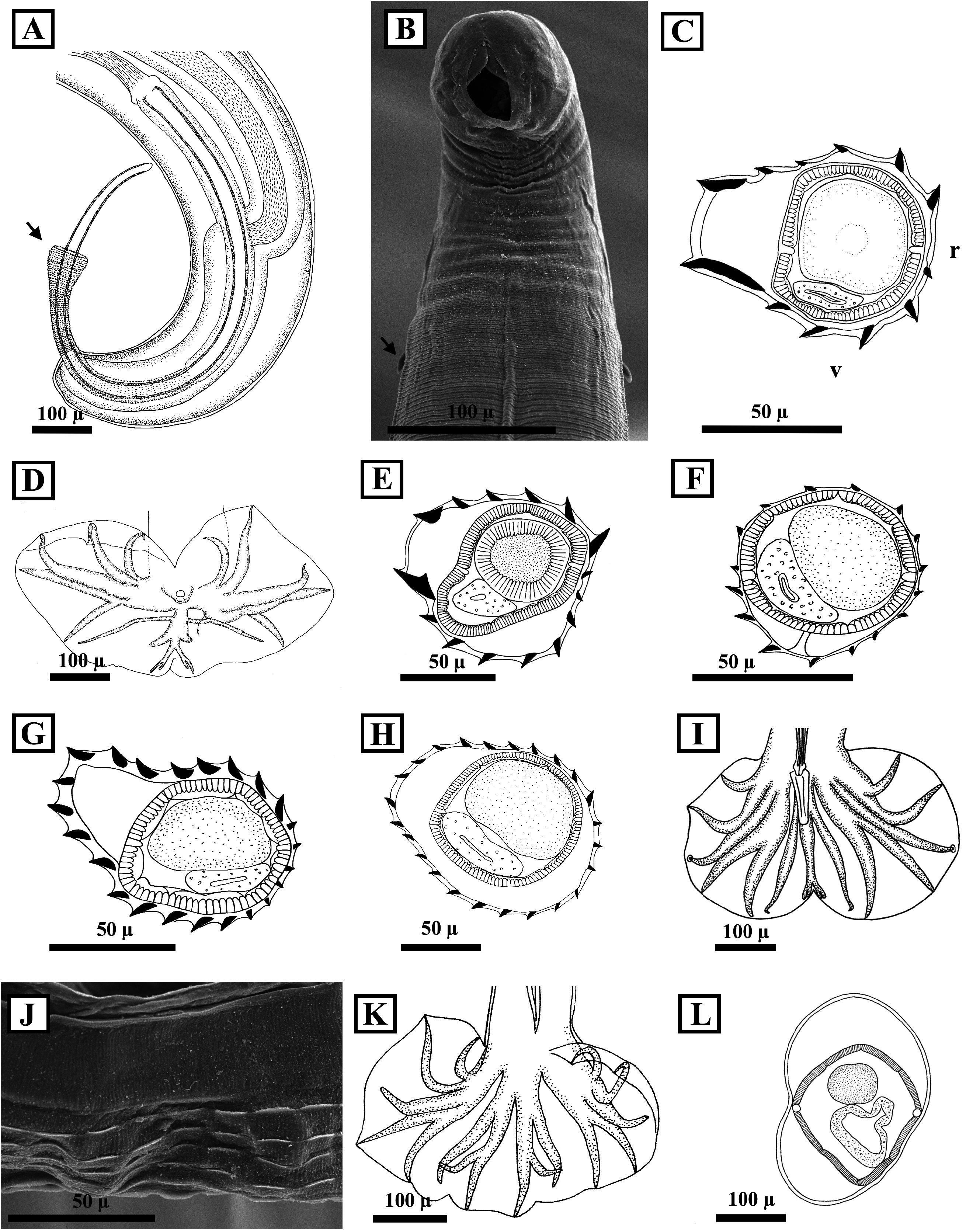
FIGURE 4. A. Posterior end of male specimen of Trichuris silviae from Heteromys gaumeri showing the slightly campanulate spicular sheath (arrow), lateral view. B. SEM micrograph of the anterior extremity of Monodontus sp. from Sigmodon toltecus showing the mouth opening, paired cutting plates and cervical papillae (arrow), ventral view. C. Cross section at midbody showing features of the synlophe of female Vexillata vexillata from Heteromys gaumeri. D. Male caudal bursa of Vexillata vexillata from Heteromys gaumeri, ventral view. E. Cross section at midbody showing features of the synlophe of male Heligmosomoidea gen. sp. cf. Vexillata from Ototylomys phyllotis. F. Cross section at midbody showing features of the synlophe of female Carolinensis sp. from Peromyscus yucatanicus. G. Cross section at midbody showing features of the synlophe of male Hassalstrongylus aduncus from Sigmodon toltecus. H. Cross section at midbody showing features of the synlophe of female Hassalstrongylus musculi from Oligoryzomys fulvescens. I. Male caudal bursa of Hassalstrongylus musculi from Ototylomys phyllotis, dorsal view. J. SEM micrograph of female Heligmostrongylus sp. from Peromyscus yucatanicus showing the discontinuous ridges, lateral view. K. Male caudal bursa of Heligmostrongylus sp. from Peromyscus yucatanicus, ventral view. L. Cross section at midbody of female Heligmonellidae gen. sp. from Sigmodon toltecus showing the absence of synlophe. Abbreviations: r, right side; v, ventral side. All cross sections of the synlophe orientated as in figure C.
No known copyright restrictions apply. See Agosti, D., Egloff, W., 2009. Taxonomic information exchange and copyright: the Plazi approach. BMC Research Notes 2009, 2:53 for further explanation.
|
Kingdom |
|
|
Phylum |
|
|
Class |
|
|
Order |
|
|
SubOrder |
Trichostrongylina |
|
SuperFamily |
Heligmosomoidea |
|
Family |
|
|
SubFamily |
Nippostrongylinae |
|
Genus |