
Aeshna persephone Donnelly
|
publication ID |
https://doi.org/ 10.5281/zenodo.195502 |
|
DOI |
https://doi.org/10.5281/zenodo.5695689 |
|
persistent identifier |
https://treatment.plazi.org/id/03D08789-FFEE-FFE3-7090-F907FC7CFD5A |
|
treatment provided by |
Plazi |
|
scientific name |
Aeshna persephone Donnelly |
| status |
|
Aeshna persephone Donnelly View in CoL
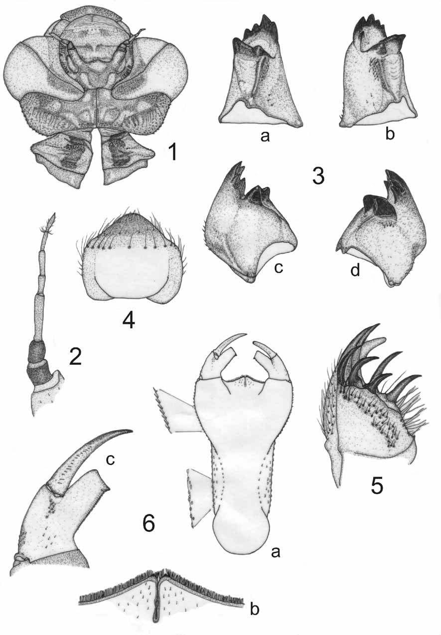
Figs. 1–10 View FIGURES 1 – 6 View FIGURES 7 – 11
Material. —Six exuviae (2 ɗ + 2 Ψ reared), nine larvae. MEXICO, Estado de México; Municipality of Tejupilco, road 134 Temascaltepec-Tejupilco, Río Chilero, N18° 58' 58"; W100° 03' 54", elevation 1643 m, pine-oak forest, 19–IX–2009, 1 Ψ (emerged 28–IX–2009), R. Novelo leg; deposited at Colección Entomológica del Instituto de Ecología, A.C. ( IEXA), Xalapa; USA, ARIZONA, Cochise County, Southwest Research Station, 5 mi. W of Portal, 1 larva, VII–1956, 1 ɗ (“emerged at Gainesville”, no date), VII–1958, M. J. Westfall, Jr. leg.; Cochise County, Scotia Canyon, Huachuca Mountains, 18–IX–2002, 1 Ψ exuvia, S.
Upson; Cochise County, Ft. Huachuca, stream, upper Garden Canyon, 2–VIII–2007, 1 Ψ exuvia, R. A. Behrstock; Cochise County, stream in Scotia Canyon, Coronado National Forest Road 4759, N31 26' 56" W110 24' 3", elevation 1810 m, 26–VII–2007, 4 larvae, K. J. Tennessen; ARIZONA, Coconino County, Oak Creek, Hwy. 89A, Pine Flat Campground, 25–VI–2003, 1 ɗ (emerged 15–VII–2003), 1 Ψ (emerged 18–VII– 2003), 4 larvae, K. J. Tennessen; all U.S. specimens deposited in FSCA.
DESCRIPTION: Larva dark brown when alive; exuviae light brown, granulated, and hairless. Femora and tibiae ringed with pale and dark bands. Abdomen lanceolate with a complex color pattern ( Fig. 8 View FIGURES 7 – 11 ).
Head. — Wider than long ( Fig. 1 View FIGURES 1 – 6 ), with moderately abundant, minute, spiniform setae except on anteclypeus, which is notoriously striated; lateral margins behind compound eyes straight and convergent; occipital margin slightly concave, cephalic lobes rounded with longitudinal rows of spiniform setae. Compound eyes large. Antennae 7–segmented ( Fig. 2 View FIGURES 1 – 6 ), the third segment the longest, relative length of antennomeres: 0.40, 0.40, 1.0, 0.45, 0.55, 0.50, 0.50, scape and pedicel dark brown with minute spiniform setae mainly on pedicel, flagellomeres yellowish brown and bare. Labrum granulose with a bare, central, oval area ( Fig. 1 View FIGURES 1 – 6 ), inferior margin setose, maximum distal width about 3 mm. Mandibles ( Fig. 3 View FIGURES 1 – 6 ) with formula L 1 2 3 4 0 a (m) b ( Figs. 3 View FIGURES 1 – 6 a, c) / R 1 2 3 4 y a (m) b ( Figs. 3 View FIGURES 1 – 6 b, d) ( Watson 1956); both mandibles with a dense brush of short, stout, stiff setae along the ventrointernal border ( Figs. 3 View FIGURES 1 – 6 a, b). Ventral pad of hypopharynx subpentangonal ( Fig. 4 View FIGURES 1 – 6 ), densely setose on anterior and lateral margins. Maxilla: galeolacinia with seven long, robust, incurved teeth ( Fig. 5 View FIGURES 1 – 6 ), and a row of long, stiff setae; a short, basal, blunt spine at inner margin; palp incurved, ending in a robust spine, with numerous stiff long setae on its external surface. Labium: prementumpostmentum articulation reaching the anterior margin of metacoxae. Prementum ( Fig. 6 View FIGURES 1 – 6 a) 1.4 – 1.6 times as long as its widest part, lateral margins more or less straight along basal 0.60 and appearing granular, apical 0.40 convex and serrulate. Ligula moderately prominent ( Fig. 6 View FIGURES 1 – 6 a), as long as 0.25 of its width; its distal margin ( Fig. 6 View FIGURES 1 – 6 b) covered with abundant piliform setae and two small blunt spines, one on each side of median cleft (best seen in ventral view); medial cleft deep, closed. Apical lobe of palp ( Fig. 6 View FIGURES 1 – 6 c) squarely truncate, rounded on external angle, with a stout, blunt end-spine at the internal angle; apical margin bare, internal margin serrate; a group of 14–16 setulae extending medially from near base of movable hook to near articulation of palp. Movable hook long, as long as palp; sharply pointed, with a row of 15–17 small, inwardly directed, stiff setae on basal 0.60 of its dorsal surface ( Fig. 6 View FIGURES 1 – 6 c).
Thorax. — Pronotum ( Figs. 1 View FIGURES 1 – 6 , 7 View FIGURES 7 – 11 a) granulose with a transverse, elongated bare area to each side of midline; pale, with two dark wide bands, one to each side of middle; lateral margins produced in a blunt, conical protuberance with minute spiniform setae, posterior margin sinuate. Proepisterna and proepimera projected laterally forming V-shaped, bluntly tipped apophyses, or “prothoracic apophyses” ( Fig. 7 View FIGURES 7 – 11 b); proepimeral apophysis the largest; most of proepisterna yellowish brown, most of proepimera dark brown ( Fig. 7 View FIGURES 7 – 11 a). Synthorax with a complex color pattern ( Fig. 7 View FIGURES 7 – 11 a). Anterior and posterior wing sheaths parallel (when alive), mostly yellowish brown with a longitudinal, dark brown band along costal margin, the anterior ones scarcely surpassing the posterior margin of abdominal segment 3, the posterior ones reaching basal 0.70 of segment 4. Legs yellowish brown, with abundant spiniform setae. Femora and tibiae with alternating dark and pale rings, well defined when alive but hardly visible in exuviae; apexes of all tibiae and ventral surfaces of all tarsi with tridentate, scale-like setae; tarsal claws simple with a pulvilliform empodium.
Abdomen. — Lanceolate ( Fig. 8 View FIGURES 7 – 11 ), finely granulose on dorsal and ventral surfaces, reaching its maximum width on abdominal segment 6; lateral margins of 2–9 with small spiniform setae, lateral margins of 6–9 ending in a robust spine, largest on 8; posterior margins of tergites 1–10 and postero-lateral margins of sternites 3–10 with a row of obtuse dark serrations that increase in size gradually from front to rear. Color pattern complex ( Fig. 8 View FIGURES 7 – 11 ). Female gonapophyses well developed ( Fig. 9 View FIGURES 7 – 11 ), reaching basal 0.3 to 0.5 of sternite 10, median valvae slightly longer than lateral valvae, lateral valvae obtusely pointed and slightly convergent; median valvae parallel, sharply pointed. Caudal appendages ( Figs. 8–10 View FIGURES 7 – 11 ): cerci long ( Figs. 8, 10 View FIGURES 7 – 11 ), tapering caudad, sharply pointed, internal margins, in dorsal view ( Figs. 10 View FIGURES 7 – 11 a–b), sinuate, tips slightly convergent; yellowish brown on basal 0.80, apical 0.20 reddish; surface covered with minute, scattered spiniform setae. Epiproct dark brown on basal 0.80, apical 0.20 yellowish brown, parallel-sided, ending in two sharp, reddish points widely separated by a median U-shaped cleft ( Figs. 8, 10 View FIGURES 7 – 11 a); dorsal margin carinate on apical 0.50, covered with small, robust setae. Paraprocts pyramidal ( Figs. 9, 10 View FIGURES 7 – 11 a), dorsal and internal margins serrate, serrations low and truncate; tips red, sharply pointed, very slightly convergent; external and ventral surfaces mostly yellowish brown, internal surface mostly dark brown, all surfaces covered with short, stiff setae, more abundant on internal surface. Average size proportions (lengths measured dorsally), with paraproct adjusted to a value of 1.0: epiproct 0.82: cerci 0.69: paraprocts 1.0.
Measurements (in mm): (exuviae N = 6, larvae N = 9): Total length (including appendages), exuviae 42.0–46.5, larvae 37.5–41.0; abdomen length (ventral), exuviae 29– 32.5, larvae 25.0–28.0; maximum width of head 8.7–9.4; hind femur (dorsal) 6.8–7.5. Lateral spines on abdominal segments 6–9 (S6–S9; measured in ventral view as shown in Fig. 11 View FIGURES 7 – 11 ): on S6, 0.63–0.75; on S7, 0.98–1.22; on S8, 1.09–1.42; on S9, 0.86–1.09. Caudal appendages (dorsal): epiproct 3.57–4.00; cerci 2.86–3.41; paraprocts 4.2–4.9.
Remarks. — Larvae of Aeshna persephone were found in open rocky streams running through altered pine-oak forest, in reaches 5–10 m wide, with abundant boulders and riffles. The F-0 larvae were at stream’s edge among roots of riparian herbaceous vegetation near large boulders; water flow was very slow. Several specimens had broken structures, such as one of the antennae, movable hook of palp, S9 posterolateral spine, and tip of paraproct, indicative of a physically challenging environment.

| FSCA |
Florida State Collection of Arthropods, The Museum of Entomology |
No known copyright restrictions apply. See Agosti, D., Egloff, W., 2009. Taxonomic information exchange and copyright: the Plazi approach. BMC Research Notes 2009, 2:53 for further explanation.