
Icerya viraktamathi Joshi, 2021
|
publication ID |
https://doi.org/ 10.11646/zootaxa.4920.2.2 |
|
publication LSID |
lsid:zoobank.org:pub:093C6F08-5F63-41F1-9169-0D73098009F7 |
|
DOI |
https://doi.org/10.5281/zenodo.4519301 |
|
persistent identifier |
https://treatment.plazi.org/id/C158091B-3598-4A8C-839C-0735EE455A79 |
|
taxon LSID |
lsid:zoobank.org:act:C158091B-3598-4A8C-839C-0735EE455A79 |
|
treatment provided by |
Plazi |
|
scientific name |
Icerya viraktamathi Joshi |
| status |
sp. nov. |
Icerya viraktamathi Joshi sp. n.
urn:lsid:zoobank.org:act:C158091B-3598-4A8C-839C-0735EE455A79
Type material.
Holotype adult ♀: INDIA, Meghalaya / Bhoirymbong, MDR 30 / Magnolia champaca , 09.XII.2019 / Omprakash Navik leg. / [ICAR/ NBAIR /MONO/ Icerya /091219–01].
Paratypes: 6 ♀♀, same data as holotype; all mounted singly on individual slides [ICAR/NBAIR/MONO/ Icerya /091219–02 to 06].
Description of the adult female.
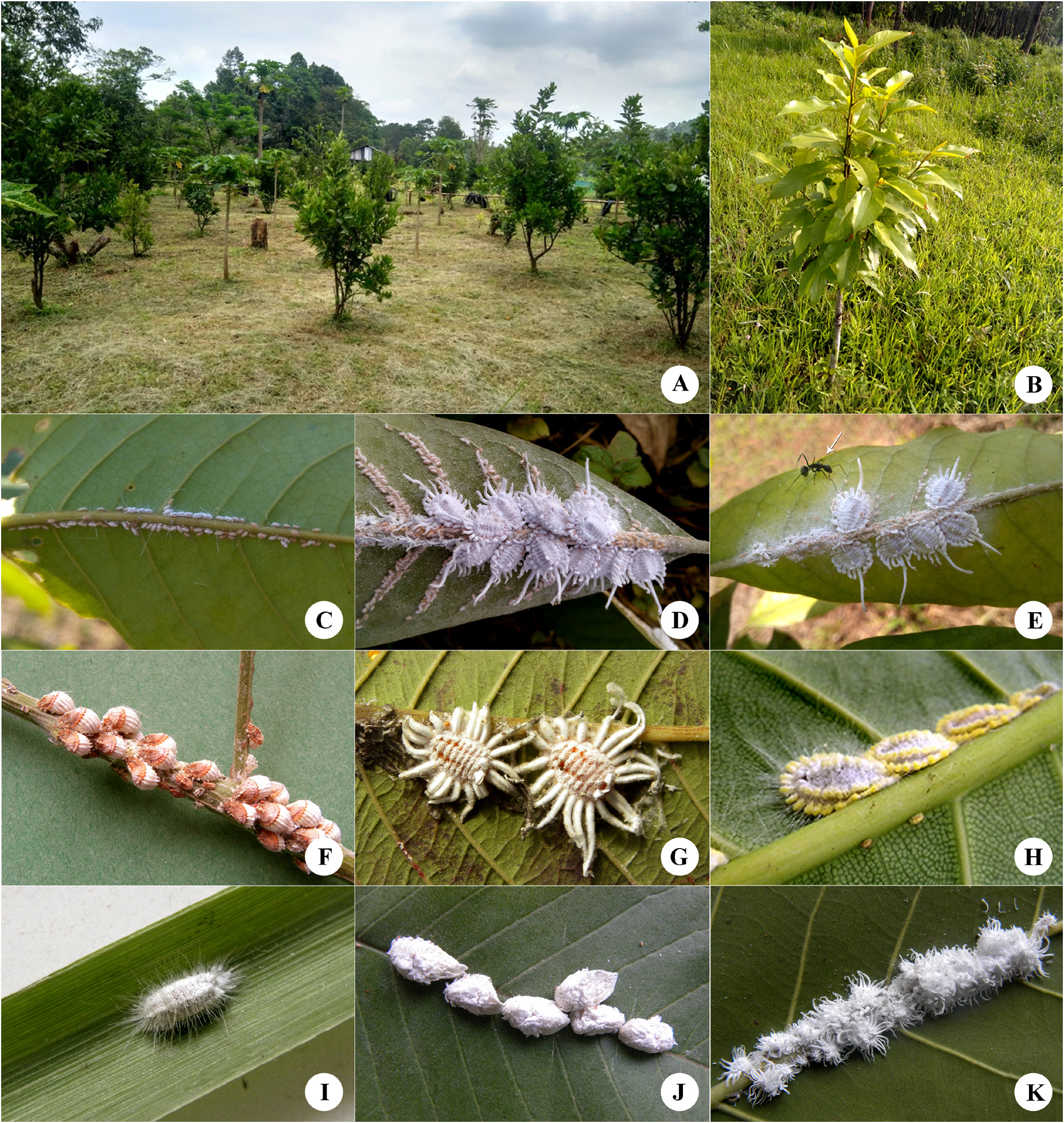
Live appearance. Crawler ( Fig. 1 C View FIGURE 1 ) body orange, coated with white wax, with a long glassy anal rod. Body of adult female elliptical, broadest at anterior of abdomen, narrowing towards head ( Fig. 1 D View FIGURE 1 ); in ethanol, body colour bright orange with legs and antennae dark brown; wax around ovisac band remains intact in ethanol, even after several months of preservation. Mid-dorsal and marginal area elevated, forming two submedian longitudinal furrows beside mid-dorsal ridge. Body covered dorsally by white wax, with one long caudal filament as long as body, curving upwards, and each side with a marginal row of nine wax filaments, all wax filaments curving upwards, anterior filaments shorter than posterior; mealy wax abundant around dorsal submargin, forming a thick, elevated submarginal ridge on each side. Last segment of abdomen with two short wax filaments on mid dorsal ridge; also one short stub-like filament on preceding abdominal segment, and one on head. Ovisac absent (the firstinstar nymphs were observed emerging from and settling around the female). Found on lower leaf surface, around veins. Ants ( Fig. 1 E View FIGURE 1 ) were found attending the scale insects.
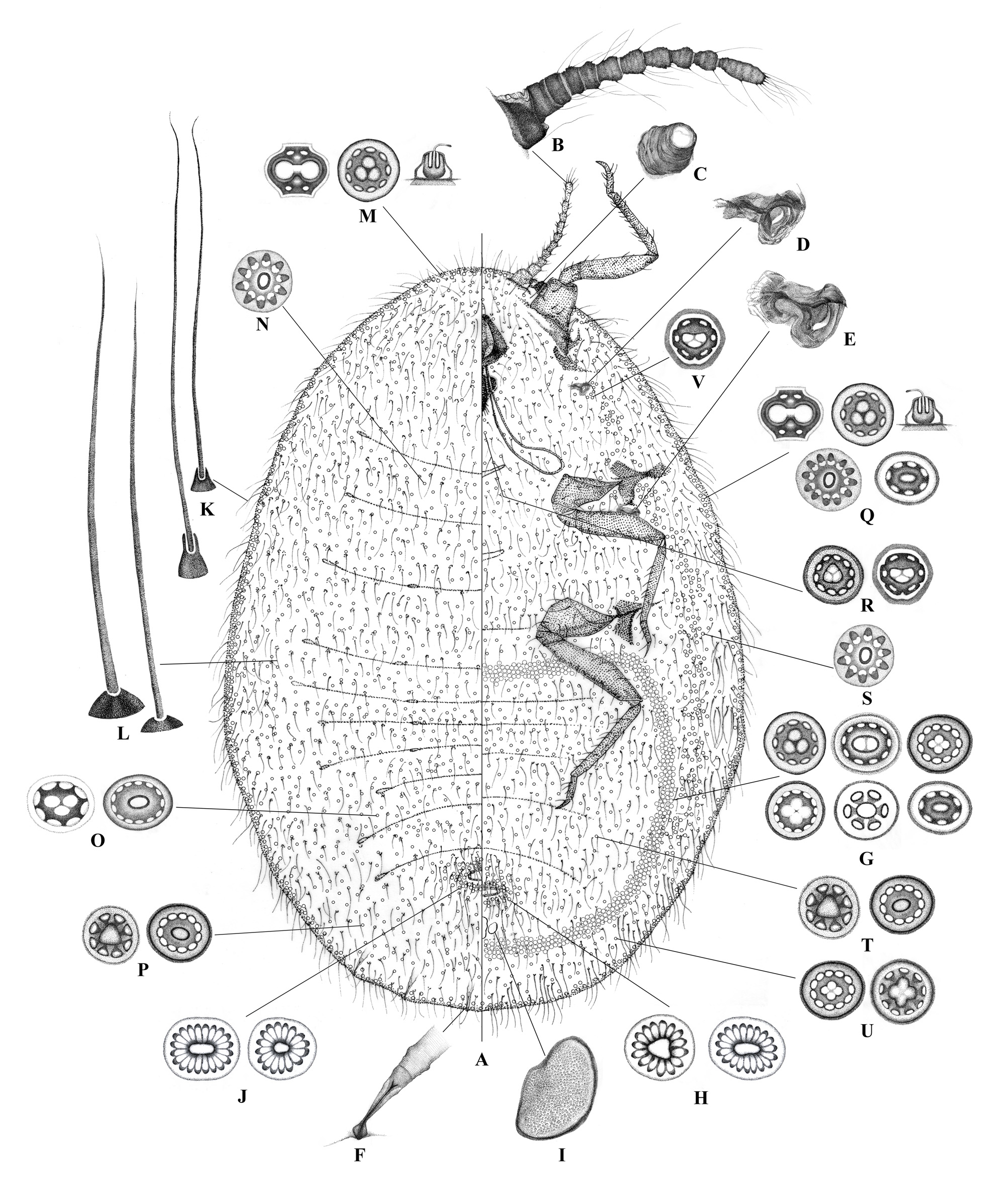
Slide-mounted adult female ( Fig. 2 View FIGURE 2 ; types of pores summarized in Table 1 View TABLE 1 ). Body elongate-oval to oval ( Fig. 2 A View FIGURE 2 ), 6.7–8.9 mm long, 4.8–6.3 mm wide. Antennae each 11 segmented ( Fig. 2 B View FIGURE 2 ), 1110–1170 μm long. Antennal segment lengths in μm: segment I 190–200 (the widest segment); segments III and IV subequal and shortest, each 65–70; segments II and V subequal in length, each 75–80; similarly, segments VI–X equal in length, each 95–110; apical segment longest, 210–220 long, 60–70 wide. Antennal setae all hair-like: segments I and II each with about 8 setae; segments III–V each with 6 or 7 setae; segments VI–X each with 5 setae; and segment XI with 13–14 setae. Eyes situated beside antennal bases, each dark, circular ( Fig. 2 C View FIGURE 2 ), 75–90 μm in diameter. Labium three segmented, 290–375 μm long, 250–350 μm wide; apex with 2 pairs of lateral hair-like setae, 1 pair of spatulate setae in the middle and 7 pairs of truncate setae between the hair-like and spatulate setae. Clypeolabral shield 400–500 μm long, 300–400 μm wide. Legs well developed; forelegs 2.61–2.83 mm long, shorter than middle legs (2.87–3.06 mm) and hind legs (3.20–3.45 mm long); tibia with robust setae towards apex; tarsus with an inner longitudinal band of robust setae, these increasing in length towards apex; claw with one pair of digitules, acute and shorter than claw. Leg measurements in μm: foreleg: coxa 375–390 long, 350–375 wide; trochanter 300–310 long, 200– 235 wide; femur 770–875 long, 200–245 wide; tibia 800–860 long, 87–100 wide; tarsus 280–300 long, 65–75 wide; claw 85–90 long, 25–30 basal width. Middle leg: coxa 370–395 long, 500–525 wide; trochanter 400–435 long, 300–385 wide; femur 850–900 long, 275–300 wide; tibia 875–900 long, 100–125 wide; tarsus 300–350 long, 70–80 wide; claw 75–85 long, 35–45 basal width. Hind leg: coxa 600–675 long, 500–550 wide; trochanter 200–260 long, 190–250 wide; femur 950–1000 long, 300–315 wide; tibia 970–1000 long, 100–125 wide; tarsus 400–425 long, 80–85 wide; claw 80–90 long, 40–50 basal width. Trochanters each with 4 campaniform sensilla on each surface; all trochanters each with a distal trochanteral seta 280–300 μm long. Mesothoracic spiracle ( Fig. 2 D View FIGURE 2 ) 1160–2000 μm long, atrial opening 1550–1700 μm wide. Metathoracic spiracle ( Fig. 2 E View FIGURE 2 ) 2200–2800 μm long, atrial opening 2180–2500 μm wide. Abdominal spiracles ( Fig. 2 F View FIGURE 2 ) numbering three pairs; atrium of each abdominal spiracle 15–18 μm wide. Ovisac band well developed, 6–8 pores wide at anterior edge, narrowing to 5 or 6 pores wide at lateral edge and again widening to 6–8 pores at posterior edge, formed by multilocular pores of five types ( Fig. 2 G View FIGURE 2 ): (i) large pores each with a stalked profile, 10–12 μm in diameter, with a trilocular centre and 9–10 outer loculi; (ii) large pores, each 10–11 μm in diameter, with a bilocular centre and 10 outer loculi; (iii) large pores, each 12–13 μm in diameter, with a quadrilocular centre and 11–13 outer loculi, which are rare, but were found on all the specimens examined; (iv) small pores, each 4–5 μm in diameter with a unilocular centre and 6 outer loculi; and (v) small pores, each 5–6 μm in diameter with a unilocular centre and 7 outer loculi. Vulvar opening situated on ventromedial abdomen, surrounded by multilocular pores ( Fig. 2 H View FIGURE 2 ), each pore 10–12 μm in diameter, with a triangular or an irregularly oval centre without clear division, and 10–20 elongate outer loculi. Ventral cicatrices numbering 3, arranged in an arc across midline posterior to vulva; largest, central cicatrix 2800–3000 μm long and 1200–1400 μm wide, lateral smaller cicatrices each 1500–1600 μm long and 1000–1100 μm wide. All cicatrices with central surface rough with a nodular texture ( Fig. 2 I View FIGURE 2 ). Dorsum with anal opening situated approximately at same level as vulva, surrounded by vulvar multilocular pores each 10–12 μm in diameter, each with a triangular or irregularly oval centre without clear locule formation and 10–20 elongate outer loculi; anal area surrounded by thick and stiff setae 200–250 μm; internal anal ring simple, sclerotized, 1500–1700 μm wide and 900–1000 μm long.
...Continued on the next page ...Continued on the next page
Dorsum. Hair-like setae ( Fig. 2 K View FIGURE 2 ) scattered over entire surface, densest on head and thorax and longest on abdominal margins. Flagellate setae ( Fig. 2 L View FIGURE 2 ) less numerous, scattered, longest on abdomen. On abdominal margins, hair-like setae each 300–400 μm long, and flagellate setae each 180–200 μm long; on submedial areas of abdomen, hair-like setae each 210–230 μm long, and flagellate setae each 100–150 μm long; and on thorax and head, hairlike setae each 280–300 μm long and flagellate setae each 90–100 μm long. Spiniform setae absent. Distribution and types of multilocular pores as follows. Marginal area ( Fig. 2 M View FIGURE 2 ) with (i) oblong simple multilocular pores each about 10 μm long and 12 μm wide, with a dumbbell-shaped bilocular centre and 4 outer loculi, and (ii) simple multilocular pores each with a stalked profile, 10–12 μm in diameter, with a trilocular centre and 9–10 outer loculi. Thoracic region ( Fig. 2 N View FIGURE 2 ) with simple multilocular pores each 12–13 μm in diameter with a unilocular centre and 8 or 9 outer loculi. Medial region ( Fig. 2 O View FIGURE 2 ) with (i) simple multilocular pores, each 8–9 μm in diameter with a trilocular centre and 8 outer loculi, (ii) oblong multilocular pores, each about 9 μm wide and 11 μm long with a unilocular centre and 13 outer loculi. Submedial region of abdomen ( Fig. 2 P View FIGURE 2 ) with (i) simple multilocular pores, each 8–9 μm in diameter with a triangular unilocular centre and 8 outer loculi, and (ii) simple oblong multilocular pores, each about 6 μm wide and 8 μm long with a unilocular centre and 11 outer loculi. Compound multilocular pores and open-centre pores absent. No dorso-medial cluster of multilocular pores was found.
Venter. Hair-like setae, each 600–700 μm long, present between antennae and in marginal clusters on all body segments; also, hair-like setae, each 300–400 μm long, on ventromedial abdomen, longest in vulvar area. Flagellate setae, each 50–200 μm long, scattered on head, thorax and ventromedial abdomen, forming irregular transverse rows. Spiniform setae absent. Distribution and types of multilocular pores as follows. Marginal area ( Fig. 2 Q View FIGURE 2 ) with (i) oblong simple multilocular pores each about 10 μm wide and 12 μm long, with a dumbbell- shaped bilocular centre and 4 outer loculi; (ii) simple multilocular pores, each 10–12 μm in diameter, with a stalked profile, a trilocular centre and 9–10 outer loculi; (iii) simple multilocular pores, each 14–15 μm in diameter, with an unilocular centre and 12 outer loculi; and (iv) simple small multilocular pores, each about 5 μm in diameter, with an unilocular centre and 7 outer loculi. Area posterior to mouthparts ( Fig. 2 R View FIGURE 2 ) with (i) simple multilocular pores, each about 12 μm in diameter with a trilocular centre and 11 outer loculi; and (ii) simple multilocular pores, each 6–7 μm in diameter with a reniform central loculus weakly sectioned into 3 poorly formed loculi, and 6 outer loculi. Submarginal region ( Fig. 2 S View FIGURE 2 ) forming a band around all the coxae, with simple multilocular pores, each about 13 μm in diameter with an unilocular centre and 10 outer loculi. Medial region ( Fig. 2 T View FIGURE 2 ) with (i) simple multilocular pores, each 8–9 μm in diameter with a triangular unilocular centre and 8 outer loculi; and (ii) simple oblong multilocular pores, each about 6 μm wide and 8 μm long with an unilocular centre and 11 outer loculi. Submedial region of abdomen ( Fig. 2 U View FIGURE 2 ) with (i) simple multilocular pores, each about 9 μm in diameter with a trilocular centre and 11 outer loculi, and (ii) simple multilocular pores, each about 13 μm in diameter with a quadrilocular centre and 9 outer loculi. Spiracular atrial opening ( Fig. 2 V View FIGURE 2 ) surrounded sparsely with simple multilocular pores, each 6–7 μm in diameter, with a reniform central loculus weakly sectioned into 3 loculi, and 6 outer loculi. Compound multilocular pores and open-centre pores absent. No ventromedial cluster of multilocular pores was found.
Taxonomic comments. Icerya viraktamathi belongs to the I. jacobsoni group. At present, the group consists of four species: I. assamensis , I jacobsoni , I jaihind and I. zimmermani . These species all have multilocular pores with a stalked profile. The adult female of I. viraktamathi is similar to that of I. zimmermanni Green in having simple multilocular pores with a stalked profile as well as simple multilocular pores each with a dumbbell-shaped bilocular centre and 4 outer loculi in a highly sclerotized rim on the margin, but the new species can be distinguished by possessing (characters of I. zimmermanni given in parenthesis): an ovisac band 6–8 pores wide (only 3–5 pores wide); an ovisac band formed of five types of simple multilocilar pores (four types); simple multilocular pores with quadrilocular centres present in the ovisac band and submedial region (absent); vulvar and anal pores restricted only to genital and anal areas (also present on venter of thoracic segments, forming a submarginal band); body contents orange in life (brown); margin of the body with shorter anterior and longer posterior wax filaments (wax filaments same size all round margin); caudal filament as long as body (caudal filament absent). The adult female of Icerya viraktamathi is also similar to that of I. siamensis in possessing simple multilocular pores with trilocular centres that appear stalked in profile, and simple multilocular pores each with a dumbbell-shaped bilocular centre and 4 outer loculi in a highly sclerotized rim on margin. However, I. viraktamathi can be differentiated from I. siamensis in possessing (characters of I. siamensis given in parenthesis): a complete ovisac band (incomplete); ovisac band 6–8 pores wide (only 4 or 5 pores wide); and by lacking crown-shaped pores on ventral margin (present).
Etymology. The species is named after Prof. C.A. Viraktamath (Gandhi Krishi Vigyan Kendra, University of Agricultural Sciences, Bengaluru, Karnataka, India) in recognition of his contributions to insect taxonomy in India; the name is in the masculine genitive.
Biological notes. The surveyed farm had more than 100 trees of Magnolia champaca ( Fig.1A View FIGURE 1 ) grown from seedlings obtained from the Government Nursery, Shillong; the trees are grown mainly for timber. The scale infestation was found on three trees, one growing near and two on the farm boundary. Invariably, the infestation was restricted to veins on the lower leaf surfaces and the plant did not exhibit any symptoms of damage ( Fig. 1B View FIGURE 1 ). No parasitoids or predators of the scale insect were obtained from the collections made during the present study. In the field, the ant Camponotus compressus (Fabricius) ( Hymenoptera : Formicidae ) was found attending the scale insects ( Fig. 1E View FIGURE 1 ). Heavy honeydew deposits and sooty mould development were not observed, possibly because the infestation was at its initial stage.
The record of this new species as a pest of a species of Magnolia is very important, as four Magnolia species viz., M. campbellii Hook. f. & Thomson, M. champaca , M. punduana Hook. f. & Thomson and M. rabaniana (Hook. f. & Thomson) D.C.S. Raju & M.P. Nayar are considered to be the main tree species in East Himalayan wet temperate forests. Of these, M. punduana is endemic to Meghalaya and M. rabaniana , which is now considered to be an endangered tree species, is endemic to North-East India ( Mir et al. 2017). Further surveys in North-East India, to record the presence of I. viraktamathi on other species of Magnolia , are vital from an ecological viewpoint.
| NBAIR |
NBAIR |
No known copyright restrictions apply. See Agosti, D., Egloff, W., 2009. Taxonomic information exchange and copyright: the Plazi approach. BMC Research Notes 2009, 2:53 for further explanation.
|
Kingdom |
|
|
Phylum |
|
|
Class |
|
|
Order |
|
|
Family |
|
|
Genus |