
Antennapeachia jambio, Izumi, Fujita & Yanagi, 2017
|
publication ID |
https://doi.org/ 10.12782/sd.22_109 |
|
publication LSID |
lsid:zoobank.org:pub:DDD14619-4742-4EE9-9675-127654BE19F5 |
|
persistent identifier |
https://treatment.plazi.org/id/A68CA32E-4FF2-4161-82A1-8F4109BB6C47 |
|
taxon LSID |
lsid:zoobank.org:act:A68CA32E-4FF2-4161-82A1-8F4109BB6C47 |
|
treatment provided by |
Felipe |
|
scientific name |
Antennapeachia jambio |
| status |
sp. nov. |
Antennapeachia jambio View in CoL sp. nov.
[New Japanese name: Misaki-no-antena] ( Figs 2–4 View Fig View Fig View Fig , Table 1)
a biological dredge onboard the R/V Rinkai-Maru, during the twelfth Coastal Organism Joint Survey of JAMBIO. The specimen was processed by the completely same way as the holotype except the place where that was preserved; the all process was undergone in Misaki Marine Biological Station.
The holotype specimen was deposited at the CMNH ( CMNH-ZG 06546 ) . The paratype one was at the National Museum of Nature and Science, Tsukuba (NSMT-Co 1596) .
Preparation of histological sections. The holotype specimen (CMNH-ZG 06546) was processed to histological sections by standard methods ( Presnell and Schreibman 1997). The dissected specimen preserved in 5% formalin was dehydrated with ethanol, dissected some tissues by tweezers and scissors, dehydrated by xylene, embedded in paraffin, sliced into serial sections (8 µm thick) by using a microtome, mounted on glass slides, and stained with hematoxylin and eosin.
Observation of cnidae. Cnidae on the tentacles (both antenna tentacles and marginal tentacles), column, actinopharynx (above tissues were from both specimens), and filaments (only from CMNH-ZG 6456, because the NSMT- Co 1596 was too damaged in proximal end to take tissue of filament for analysis) were observed. Images of the cnidae were obtained by differential interference contrast microscopy ( Yanagi et al. 2015), and the length and width were measured using the software ImageJ v. 1.49 ( Rasband 1997 – 2012). Cnida nomenclature followed Mariscal (1974).
Material examined. Holotype: CMNH-ZG 06546 ; dissected specimen, embedded tissues in paraffin, histological sections (3 slides), prepared nematocysts (5 slides); February 19, 2014, off Jogashima , Kanagawa Prefecture, 35°06.082′N, 139°34.232′E, at 238–309 m in depth collected by Kensuke Yanagi. GoogleMaps Paratype: NSMT-Co 1596; whole specimen, somewhat damaged in proximal end; February 15, 2017, off Miura Peninsula , Kanagawa Prefecture, 35°08.0383′N, 139°33.731′E, at 95–97 m in depth collected by Takato Izumi GoogleMaps . GoogleMaps
Etymology. This species was named, because the type specimens were collected in the second and twelfth JAMBIO Coastal Organism Joint Survey. JAMBIO stands for “Japanese Association for Marine Biology.”
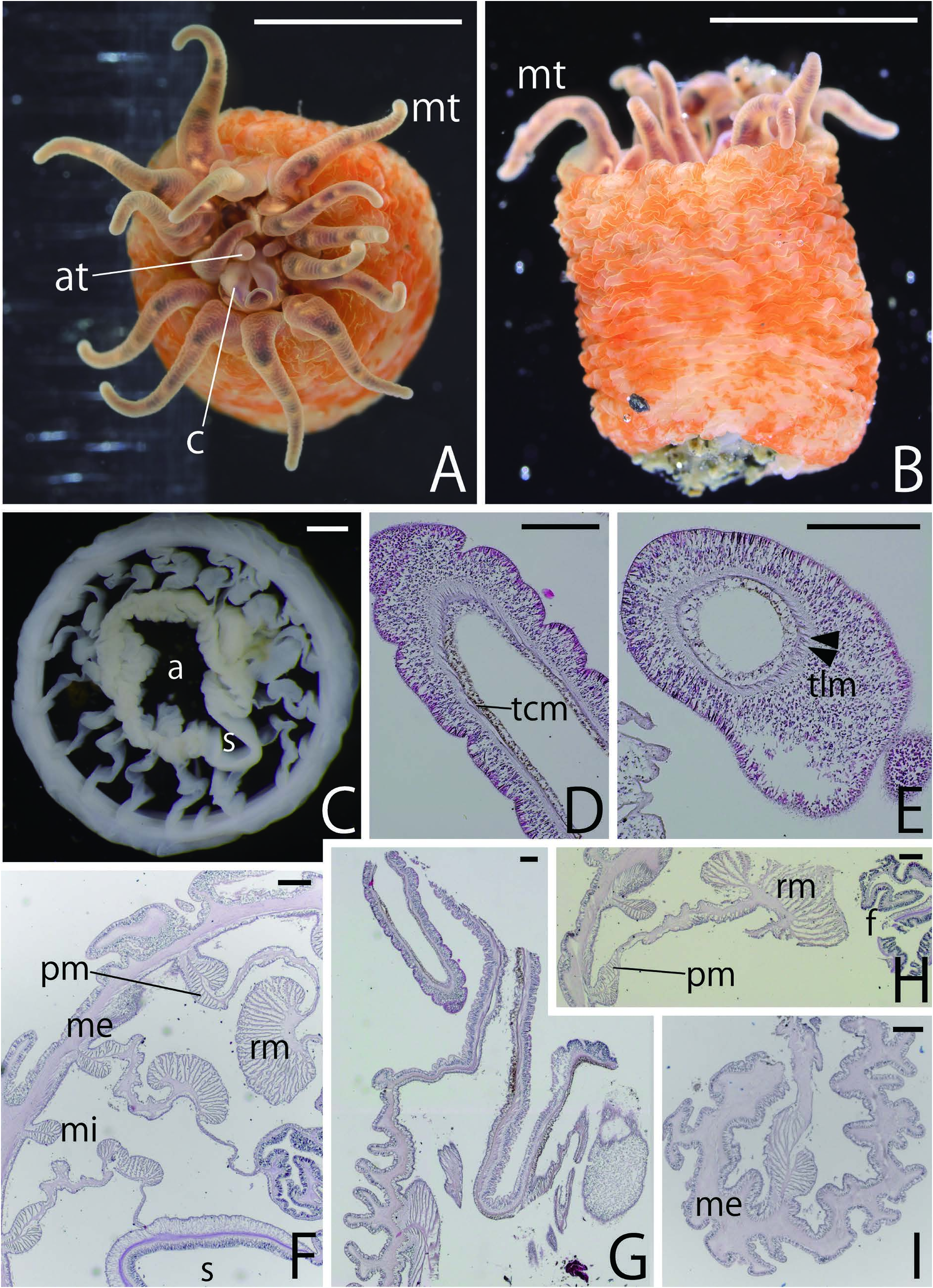
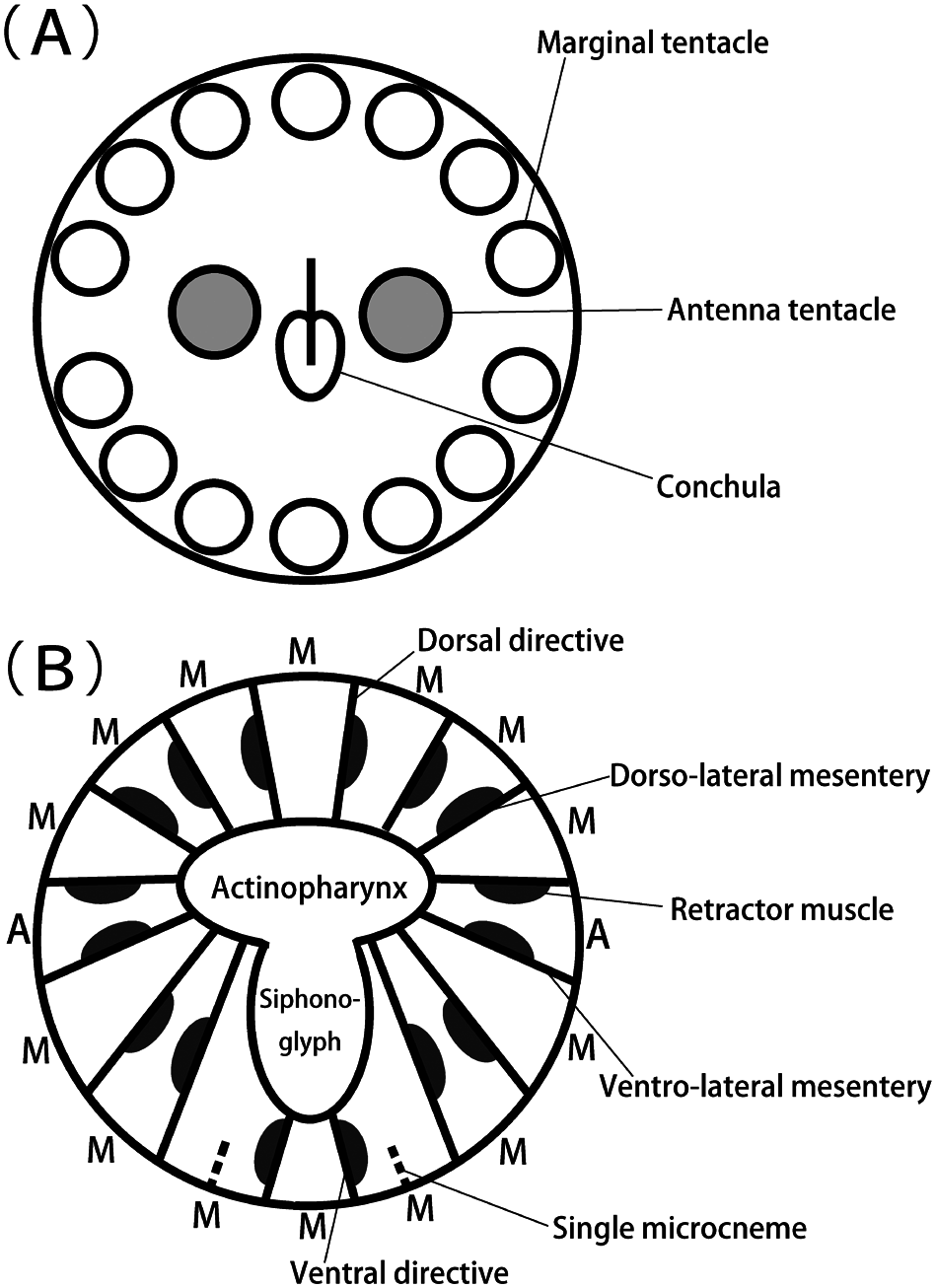
Description. External anatomy. Column smooth, cylindrical or barrel-like, rich in expansibility, length ca. 9 mm, diameter ca. 7 mm in alive, and length 8 mm diameter 6 mm in preserved specimens. Color of body orange ( Fig. 2A, B View Fig ). Column surface smooth, papillae absent, with numerous discontinuous wavy wrincles running direction of transversal and pale white patches ( Fig. 2B View Fig ). Aboral end flattened or slightly concaved, like a shape of donut, with tiny hole in center, semitransparent, so that line of mesenteries visible from outer side. Pedal disk absent, but somewhat sticky so that particles of mud adhered to column or aboral end ( Fig. 2B View Fig ). Two circles of tentacles on oral disc; 16 tentacles. Fourteen marginal tentacles in outer circle; two antenna tentacles in inner circle ( Fig. 3A View Fig ). Marginal tentacles protruding outside or hanging along column. Antenna tentacles rising straight upward. Marginal tentacles approximately 4–5 mm in length when fully expanded. Antenna tentacles 1–2 mm in length, far shorter than marginal tentacles. Both tentacles simple, without acrospheres. Coloration of marginal tentacles brownish, obscure cross-bands of yellow and brown; surface complicatedly wrinkled. Antenna tentacles without cross-bands; surface wrinkled ( Fig. 2A View Fig ). Oral disk diameter ca. 4 mm, same color as tentacles. Mouth at center of oral disk, a little swelled, with well-extended and robed conchula on ventral side ( Fig. 2A View Fig ).
Internal anatomy. Eight pairs of macrocnemes, six of first cycle and extra two pairs between ventro-lateral mesenteries and ventral directives; two independent microcnemes between ventro-lateral mesenteries and ventral directives ( Figs 2C View Fig , 3B View Fig ). All macrocnemes perfect and continuing along whole length of body. Microcnemes present from proximal end to middle part of body, but absent near oral end. Antenna tentacles between ventro-lateral endocoels ( Fig. 3B View Fig ). Seven marginal tentacles of outer cycle on dorsal half of body; one of them arised from dorsal endocoel; two (one on each side) from dorso-lateral exocoels; two from dorso-lateral endocoels; other two from lateral exocoels. Other seven on ventral half of body; one of them arised from ventral endocoel; two from endocoel between two extra pairs of macrocnemes; the other four in ventrolateral exocoel ( Figs 2A View Fig , 3B View Fig ). Tentacular circular muscle endodermal ( Fig. 2D View Fig ); tentacular longitudinal muscle ectodermal ( Fig. 2E View Fig ). Retractor muscles strongly circumscribed, limited at the center of each macrocneme ( Fig. 2C, F View Fig ), with approximately 15–30 a little branched muscular processes; parietal muscles of macrocnemes distinct, with around ten simple muscular processes ( Fig. 2F, H View Fig ). Mesoglea in body wall far thicker than ectoderm and endoderm, thinner in actinopharynx and siphonoglyph ( Fig. 2F View Fig ). Actinopharynx smooth, comparatively long in column, with huge siphonoglyph on ventral side, which always connected to it ( Fig. 2C View Fig ). Sphincter muscle absent ( Fig. 2G View Fig ). On aboral end, basilar muscle absent ( Fig. 2I View Fig ). Filaments at the tips of macrocnemes, small and limited near aboral end. Matured gametes not observed, so sexuality of specimens not clear.
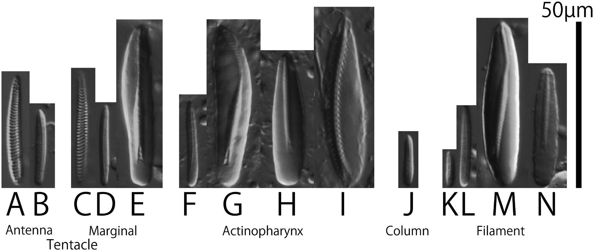
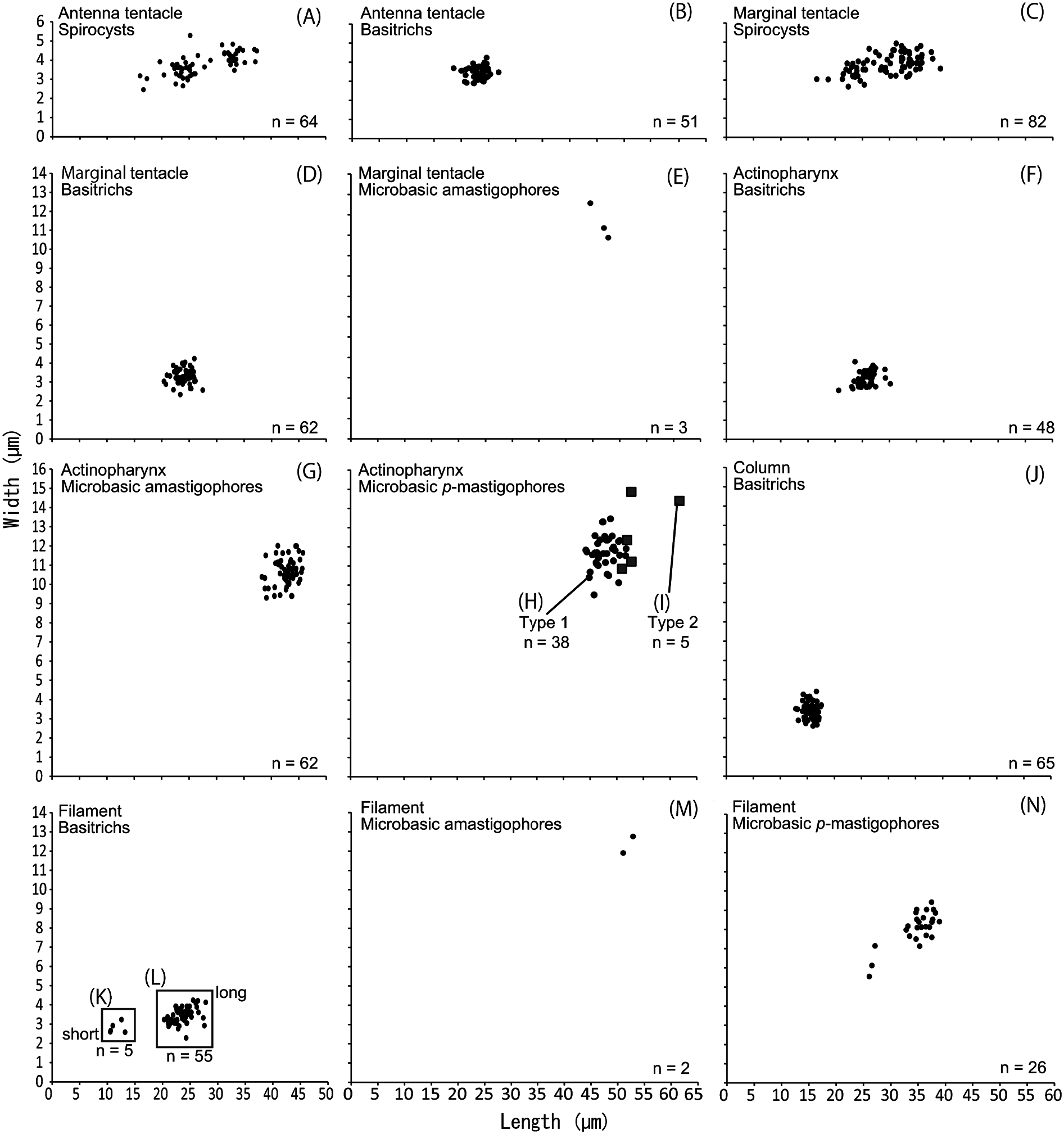
Cnidom. Spirocyst in both tentacles, basitrich in every tissue, microbasic amastigophore in actinopharynx and filament, microbasic p -mastigophore in actinopharynx and filament ( Table 1, Fig. 4 View Fig ). Basitrichs in filament distinguished as two types by their size. Microbasic p -mastigophore in actinopharynx morphologically distinguished as two types (“ Type 1” and “ Type 2”; Fig. 4H, I View Fig ): Type 1 comparatively shorter than Type 2 ( Figs 4 View Fig , 5 View Fig ); ratio of shaft length for capsule length of Type 1 smaller than that of Type 2 (0.65–0.76 [n =10] vs. 0.92–1.01 [n =5]); and slight middle expansion on shaft present in Type 1 but not in Type 2.
Remarks. This species resembles A. setouchi in having antenna tentacles, forming a siphonoglyph (which is connected to conchula) beside their actinopharynx and having wrincled orange color column. Antennapeachia jambio , however, has many different features from A. setouchi ; antenna tentacles are far smaller than marginal tentacles, in contrast those of A. jambio are bigger and more prominent than marginal ones ( Fig. 2 View Fig , Izumi et al. 2016); A. jambio has a different type of cnidae from A. setouchi , especially the microbasic p -mastigophores are only in actinopharynx and filament of A. jambio ( Table 1, Figs 4 View Fig , 5 View Fig ); the two pairs of macrocnemes between ventro-lateral mesenteries and ventral directives are only formed in the column of A. jambio . The last difference is most characteristic, that is why we judged that A. jambio is appropriate to be treated as new species, and we revised the diagnosis of genus (see the Discussion part).
| CMNH |
The Cleveland Museum of Natural History |
No known copyright restrictions apply. See Agosti, D., Egloff, W., 2009. Taxonomic information exchange and copyright: the Plazi approach. BMC Research Notes 2009, 2:53 for further explanation.
|
Kingdom |
|
|
Phylum |
|
|
Class |
|
|
Order |
|
|
Family |
|
|
Genus |