
Acrogonia virescens (Metcalf 1949)
|
publication ID |
https://doi.org/ 10.11646/zootaxa.3915.4.4 |
|
publication LSID |
lsid:zoobank.org:pub:0AE967DB-31CE-4BA2-AAB9-D69B54C62C26 |
|
DOI |
https://doi.org/10.5281/zenodo.6105927 |
|
persistent identifier |
https://treatment.plazi.org/id/03D14438-FFEE-FFA5-FF44-AD76FF08FE9A |
|
treatment provided by |
Plazi |
|
scientific name |
Acrogonia virescens (Metcalf 1949) |
| status |
|
Acrogonia virescens (Metcalf 1949) View in CoL
Diagnosis. General coloration greenish, with dark V-shaped spot at apex of crown; clypellus with dark spot of variable shape and size. Male: connective Y-shaped; styles elongated and curved toward the midline; aedeagus long and thin, apex sharp, and basal portion Y-shaped.
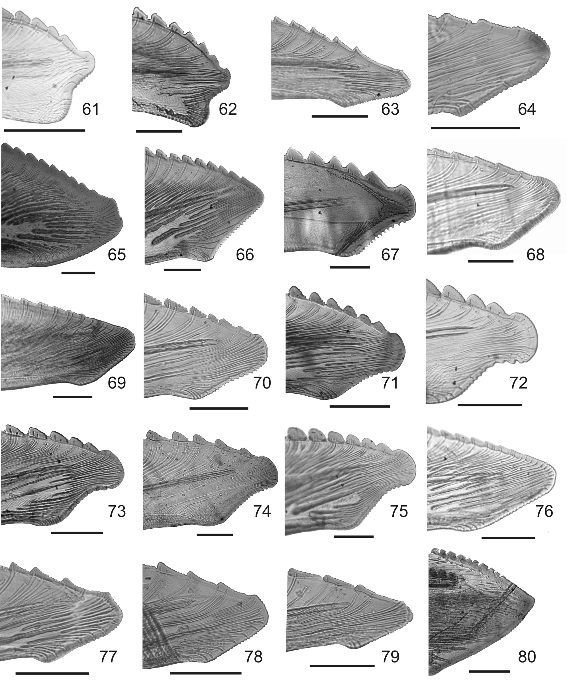
Female terminalia. Abdominal sternite VII, in ventral view ( Fig. 2 View FIGURES 1 – 20 ), with lateral margins slightly parallel, lateroposterior margins broadly rounded, and posterior margin deeply emarginate bearing conspicuous median lobe inside emargination; with a few macrosetae at edges of emargination. Pygofer, in lateral view, slightly produced posteriorly; surface with scattered microsetae and macrosetae on posterior one-third of disc. First valvifers, in lateral view, longer than tall; with small spiniform processes in posterior one-third. First ovipositor valvulae, in lateral view, short and slightly rectilinear, basal portion expanded; dorsal sculptured area formed by scale-like processes arranged in oblique lines; ventral sculptured area formed by scale-like processes arranged irregularly; apex truncate with acute median projection ( Fig. 22 View FIGURES 21 – 40 ). Second valvulae, in lateral view, short and slightly rectilinear; dorsal margin of blade bearing 18 to 20 noncontiguous teeth, each tooth subtriangular, declivous posteriorly, with denticles along entire dorsal margin ( Fig. 42 View FIGURES 41 – 60 ); size of teeth increases from proximal portion to middle region, and then decreases towards apex, ducts extending toward teeth and toward apical blade portion; apex obtuse, forming a concavity with preapical ventral prominence ( Fig. 62 View FIGURES 61 – 80 ). Gonoplacs, in lateral view, with basal half narrow, and apical half short and distinctly expanded; apex acute.
Material examined. ARGENTINA. Salta: Abra Grande, Orán, 2♂ 1♀, III/1967; 3♂ 1♀, 10/I-28/II/1967, R. Golbach ( IMLA). Misiones: Puerto Iguazú, 25°37´19´´S 54°32´52´´W, 1♀, 7/I/2008, hand collection, C.H.
Dietrich ( INHS); Eldorado, 1♂ 2♀, 31/X/2008, Logarzo & Palottini; Parque Nacional Iguazú, 2♂, 11/XII/2008, water trap, Zamudio & Colleselli Gomez de Olivera; Eldorado, Cueva Miní, 26°22,29´S 54°39,65´W, 5♂, 14/II/ 2012, hand collection, G. Dellapé ( MLP).
Distribution. Guyana, Brazil, Peru, Paraguay ( Young 1968) and Argentina: Misiones and Salta ( Paradell et al. 2012).


No known copyright restrictions apply. See Agosti, D., Egloff, W., 2009. Taxonomic information exchange and copyright: the Plazi approach. BMC Research Notes 2009, 2:53 for further explanation.