
Temnocephala gargantua, León & Volonterio, 2018
|
publication ID |
https://doi.org/ 10.11646/zootaxa.4378.3.2 |
|
publication LSID |
lsid:zoobank.org:pub:0FC4A657-3F63-4766-8FD7-73CFE48A54B9 |
|
DOI |
https://doi.org/10.5281/zenodo.5973313 |
|
persistent identifier |
https://treatment.plazi.org/id/494CFC91-8D86-4DC7-96D0-AE799EC0C743 |
|
taxon LSID |
lsid:zoobank.org:act:494CFC91-8D86-4DC7-96D0-AE799EC0C743 |
|
treatment provided by |
Plazi |
|
scientific name |
Temnocephala gargantua |
| status |
sp. nov. |
Temnocephala gargantua sp. nov. Ponce de León & Volonterio
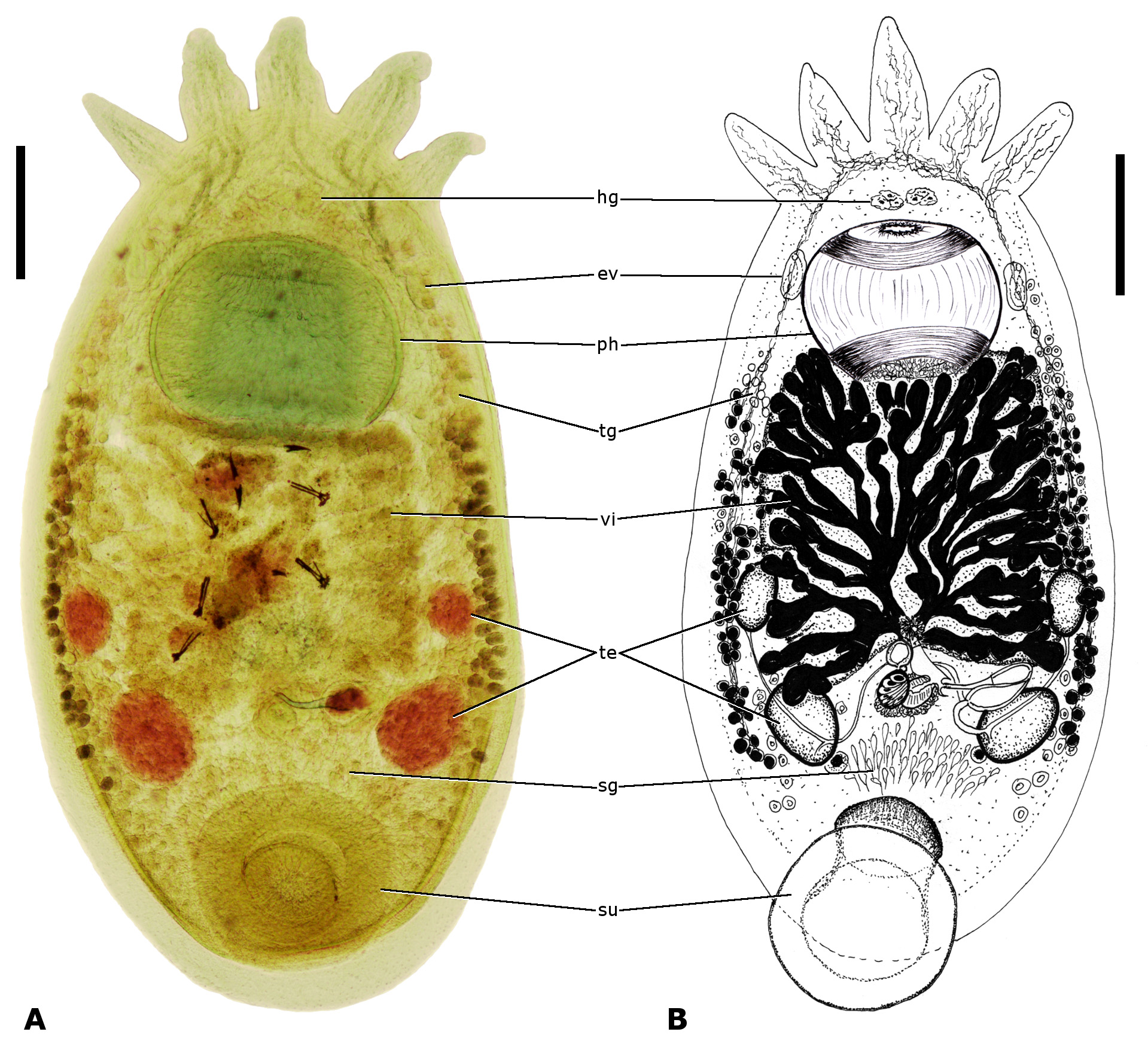
( Figs. 1–7 View FIGURE 1 View FIGURE 2 View FIGURE 3 View FIGURE 4 View FIGURE 5 View FIGURE 6 View FIGURE 7 )
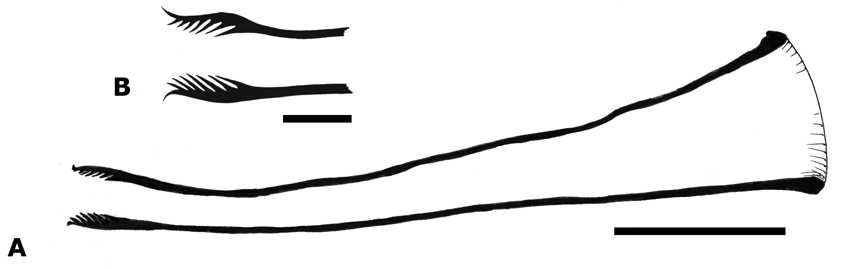
Diagnosis. Temnocephala gargantua sp. nov. is characterized by having a slightly curved penial stylet (225 µm in length) with a symmetrical introvert (27 long by 18 µm in diameter) that has at least 44 spine-like projections, each bearing 7 rows of internal spines; a seminal vesicle that opens sub-polarly into the contractile vesicle; a muscular vagina with a strong asymmetrical sphincter; preequatorial nephridiopores close to the internal borders of the elongate-oval excretory syncytia; a large pharynx with anterior and posterior sphincters of about the same diameter and a narrow pharyngeal lumen, nearly uniform in width.
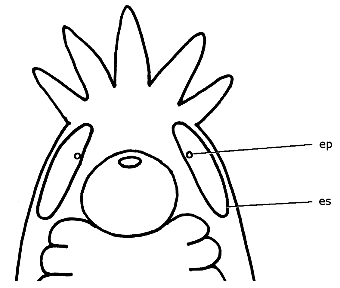
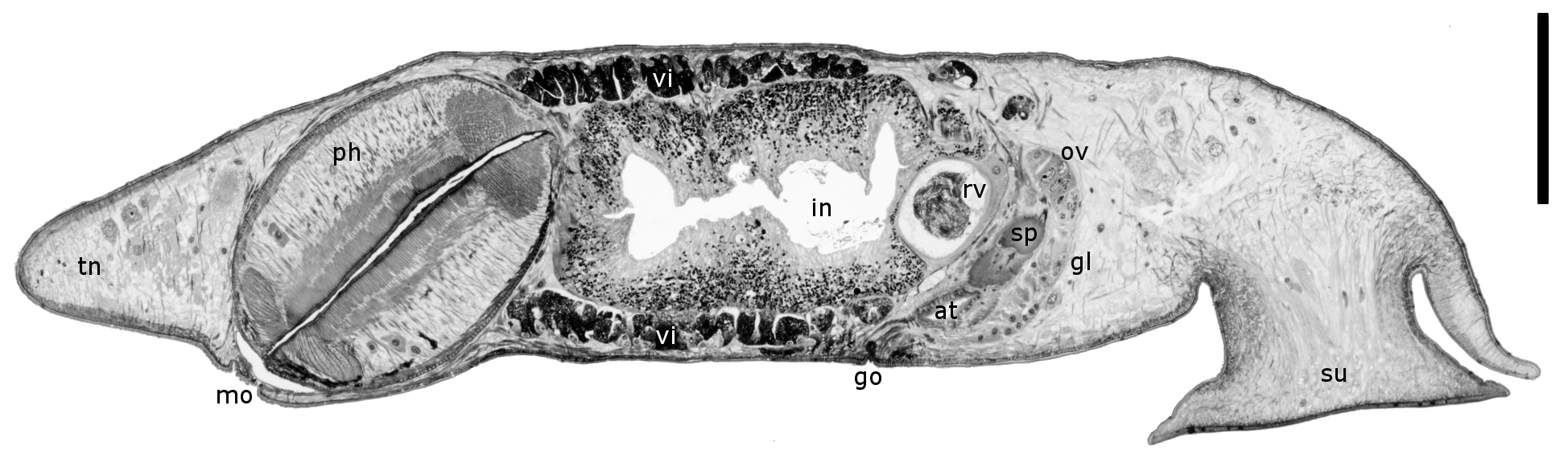
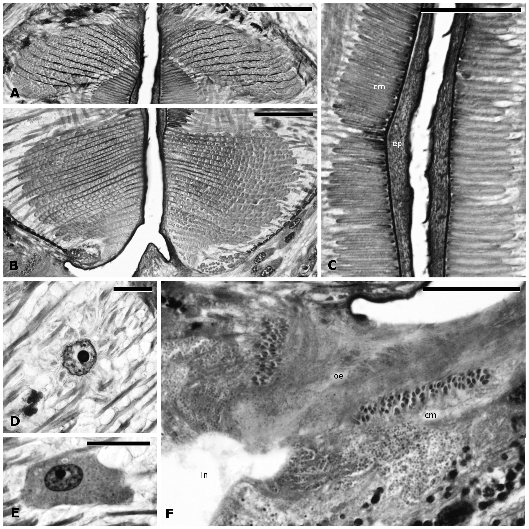
Description. Temnocephalan of large size; elliptic body, with maximum width at the level of the space between anterior and posterior testes ( Fig. 1 View FIGURE 1 ). Body without tentacles 2166.7 to 4074.1 (2925.9; 612.76; 10) long by 1148.1–1833.3 (1514.8; 259.81; 10) wide. Sucker subterminal, 518.5–907.4 (670.4; 117.70; 10) in diameter. Peduncle generally with an oblique, ventro-caudal orientation. Ratio body length without tentacles to sucker length 1:0.18–0.35 (mean 1:0.24). Sucker glands anterior to sucker peduncle, scattered between, and posterior to, posterior testes ( Fig. 1 View FIGURE 1 ). Epidermis with elongate-oval excretory syncytia, extending from the base of the outermost tentacles to the level of the anterior portion of the intestine ( Fig. 2 View FIGURE 2 ). Excretory vesicles lateral to pharynx; nephridiopores dorsal, close to internal border of excretory syncytia and preequatorial respect to them ( Figs. 1 View FIGURE 1 , 2 View FIGURE 2 ). Haswell glands conspicuous, anterior to pharynx ( Fig. 1 View FIGURE 1 ). Abundant tentacular glands, situated laterally to the intestine ( Fig. 1 View FIGURE 1 ).
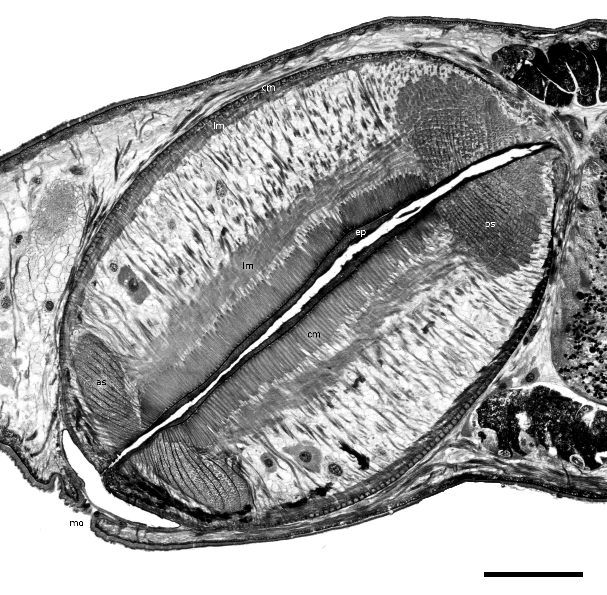
Large, elliptical pharynx, of about the same size as the intestine ( Fig. 3 View FIGURE 3 ), 444.4–759.3 (606.3; 84.31; 10) long by 481.5–851.9 (690.7; 120.68; 10) wide. Pharynx wall 192.9–202.1 (197.5; 2) maximum width. Anterior sphincter of about the same diameter as, but more flattened than, the posterior one ( Figs. 4 View FIGURE 4 ; 5A,B); section of the muscle bundle 107.1 measured radially and 44.3 measured anteroposteriorly. Section of the muscle bundle of the posterior sphincter 115.7 measured radially and 101.4 measured anteroposteriorly. The external circular musculature is a monolayer of fibers with a height of 0.7 at the level of the anterior sphincter. Posteriorly to this level the layer abruptly acquires a height of 5 and at the level of the posterior sphincter it decreases gradually to 1.8. This monolayer seems to be confluent with a small bundle of circular muscle fibers that is situated at the base of the pharynx, seemingly surrounding the entrance to the esophagus ( Fig. 5B View FIGURE 5 ). External longitudinal muscle layer roughly homogeneous in height (14.3). The internal longitudinal fibers arise and end in the pharyngeal sphincters, giving a layer of about 28.6 in height. The internal layer of circular muscles extends between both sphincters without overlapping with them; it is composed by a monolayer of high fibers ( Fig. 5C View FIGURE 5 ) whose sections measured radially are 9.3 in the anterior region, 32.5 in the median region and 6.4 in the posterior region of the pharynx. Besides the circular and longitudinal muscle fibers, abundant radial fibers were seen ( Fig. 4 View FIGURE 4 ). Among the radial muscle fibers between the sphincters, at least 2 different types of cells were observed, the first type with clear cytoplasm and abundant extensions toward the surrounding tissues, and the second type with abundant dark granules in the more dense cytoplasm, and fewer extensions ( Figs. 4 View FIGURE 4 , 5D,E View FIGURE 5 ). The pharyngeal epithelium is a wide layer of fibrous aspect, deeply staining dark-blue with Heidenhain’s haematoxylin ( Figs. 4 View FIGURE 4 , 5C View FIGURE 5 ). No nuclei were seen at this level; these are placed outside of the pharynx as cytons. The height of the epithelial layer is 2.4 anteriorly, 6.4–11.5 (8.3; 2) medially and 3.1 posteriorly. Pharyngeal lumen narrow and nearly uniform in width ( Fig. 4 View FIGURE 4 ). Short esophagus surrounded by a layer of circular muscle fibers resembling a sphincter ( Fig. 5F View FIGURE 5 ). Intestine longer than wide, with a slight central constriction and conspicuous septa. The posterior end of the intestinal sac reaches the level of the posterior testes; ratio body length without tentacles to distance between the posterior end of the intestine and base of tentacles 1:0.58–0.72 (mean 1:0.65).
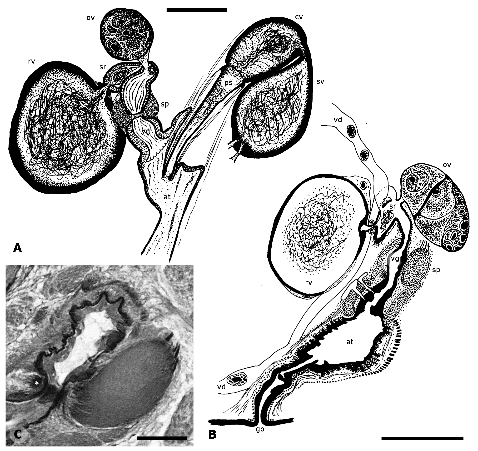
Ovoid ovary, 91.0–145.5 (119.5; 17.96; 9) long by 78.2–134.5 (103.6; 17.70; 9) wide. The short oviduct opens into the ootype just opposite the entrance of the seminal receptacle ( Fig. 6A,B View FIGURE 6 ). Abundant glandular cells surround the ootype, with ducts that open into it. Resorbiens vesicle in the space left by the posterior constriction of the intestine and generally posterior to the level of the gonopore ( Figs. 1 View FIGURE 1 , 3 View FIGURE 3 ), ovoid in shape, 149.1–236.4 (186.2; 35.76; 5) long by 118.2–207.3 (162.6; 36.07; 5) wide. The posterior part of the resorbiens vesicle is more flattened and has a thicker wall than the anterior one; this thick-walled portion gives rise to the short duct that leads to the seminal receptacle, which is surrounded by a sphincter ( Fig. 6A, B View FIGURE 6 ). The seminal receptacle is an expansion of the wall of the duct that connects the ootype with the resorbiens vesicle ( Fig. 6A,B View FIGURE 6 ). Vagina 109.9–147.2 (128.6; 26.38; 2) long by 81.1–108.1 (94.6; 19.09; 2) maximum width; distal portion with an asymmetrical sphincter, 68.5– 108.1 (87.1; 19.91; 3) in diameter ( Fig. 6C View FIGURE 6 ). Female portion of atrium surrounded by several gland cells with ducts that open into its lumen ( Fig. 6B View FIGURE 6 ). Vitelline glands branched ( Fig. 1B View FIGURE 1 ), surrounding intestine completely ( Fig. 3 View FIGURE 3 ). Eggs not observed.
Testes ovoid; two lateral to the posterior portion of the intestine and two larger, posterior to the same organ and more central ( Fig. 1 View FIGURE 1 ). Anterior testes 166.7–296.3 (224.1; 39.47; 10) long by 74.1–185.2 (138.9; 34.10; 10) wide. Posterior testes 203.7–351.9 (290.7; 50.18; 10) long by 129.6–240.7 (198.1; 32.71; 10) wide. The thick vasa efferentia enter the seminal vesicle adjacent to each other ( Fig. 6A View FIGURE 6 ). The seminal vesicle is pyriform, 191.0–298.2 (257.9; 33.59; 9) long by 95.0–173.0 (128.5; 29.22; 8) maximum width; wall 5.4–11.7 (7.8; 1.88; 11) thick; it opens into the contractile vesicle in a sub-polar position. Contractile vesicle 109.1–163.6 (143.7; 17.39; 10) long by 64.0–109.1 (92.5; 13.38; 10) maximum width; wall 2.7–7.2 (5.6; 1.75; 11) thick. No prostatic glands were seen externally to the contractile vesicle. Penial stylet slightly curved ( Fig. 7A View FIGURE 7 ); 200.0–258.2 (225.4; 18.59; 9) long by 45.5–63.6 (52.3; 6.64; 9) proximal width. Symmetrical introvert 27.3 long by 14.5–22.0 (17.8; 2.42; 9) maximum width, bearing at least 44 spine-like projections, each bearing 7 rows of internal spines ( Fig. 7B View FIGURE 7 ). Gonopore at the level of the space between the anterior and posterior testes, surrounded by glands; ratio body length without tentacles to distance between gonopore and base of tentacles 1:0.54–0.65 (mean 1:0.57).
No known copyright restrictions apply. See Agosti, D., Egloff, W., 2009. Taxonomic information exchange and copyright: the Plazi approach. BMC Research Notes 2009, 2:53 for further explanation.
|
Kingdom |
|
|
Phylum |
|
|
Class |
|
|
Order |
|
|
Family |
|
|
Genus |