
Laccobius ( Laccobius ) kunashiricus Shatrovskiy, 1984
|
publication ID |
https://doi.org/10.1515/aemnp-2017-0060 |
|
publication LSID |
lsid:zoobank.org:pub:C105A783-646F-498F-9585-D50D0D3B8333 |
|
DOI |
https://doi.org/10.5281/zenodo.5335349 |
|
persistent identifier |
https://treatment.plazi.org/id/03D18D3C-FFDA-191D-BE2E-92F1FE7FFD28 |
|
treatment provided by |
Marcus |
|
scientific name |
Laccobius ( Laccobius ) kunashiricus Shatrovskiy, 1984 |
| status |
|
Laccobius ( Laccobius) kunashiricus Shatrovskiy, 1984 View in CoL
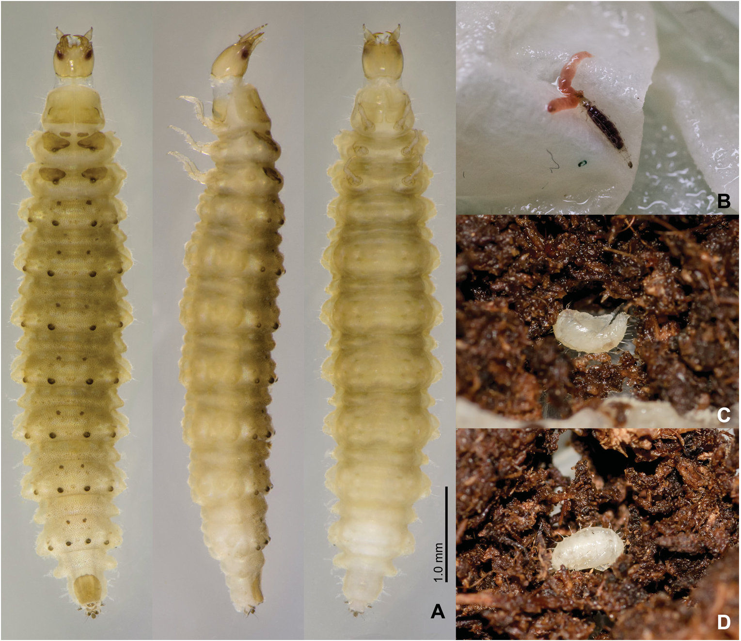
( Figs 1–12 View Fig View Fig View Fig View Fig View Fig View Fig View Fig View Fig View Fig View Fig View Fig View Fig )
Material examined. 4 EC, 13 L1, 5 L2, 4 L3, 3 P, JAPAN: NIIGATA PREF.: Jôganji, Nagaoka-shi, 27.v.2012 (adults collected in the field), Y. Iwata leg., Y. Minoshima rearing: R21.
Description of egg-case. Egg-case white in colour, constructed on substrate in water or at water’s surface; spherical with narrow, long filiform mast of variable length, often extremely long ( Fig. 1 View Fig ).
Description of larva. General morphology. Third instar. Body ( Fig. 2A View Fig ) rather thick with weak lateral projections, widest between abdominal segments 2–4. Colour pale brownish white with sclerotised areas darker, tubercles on abdomen dark brown.
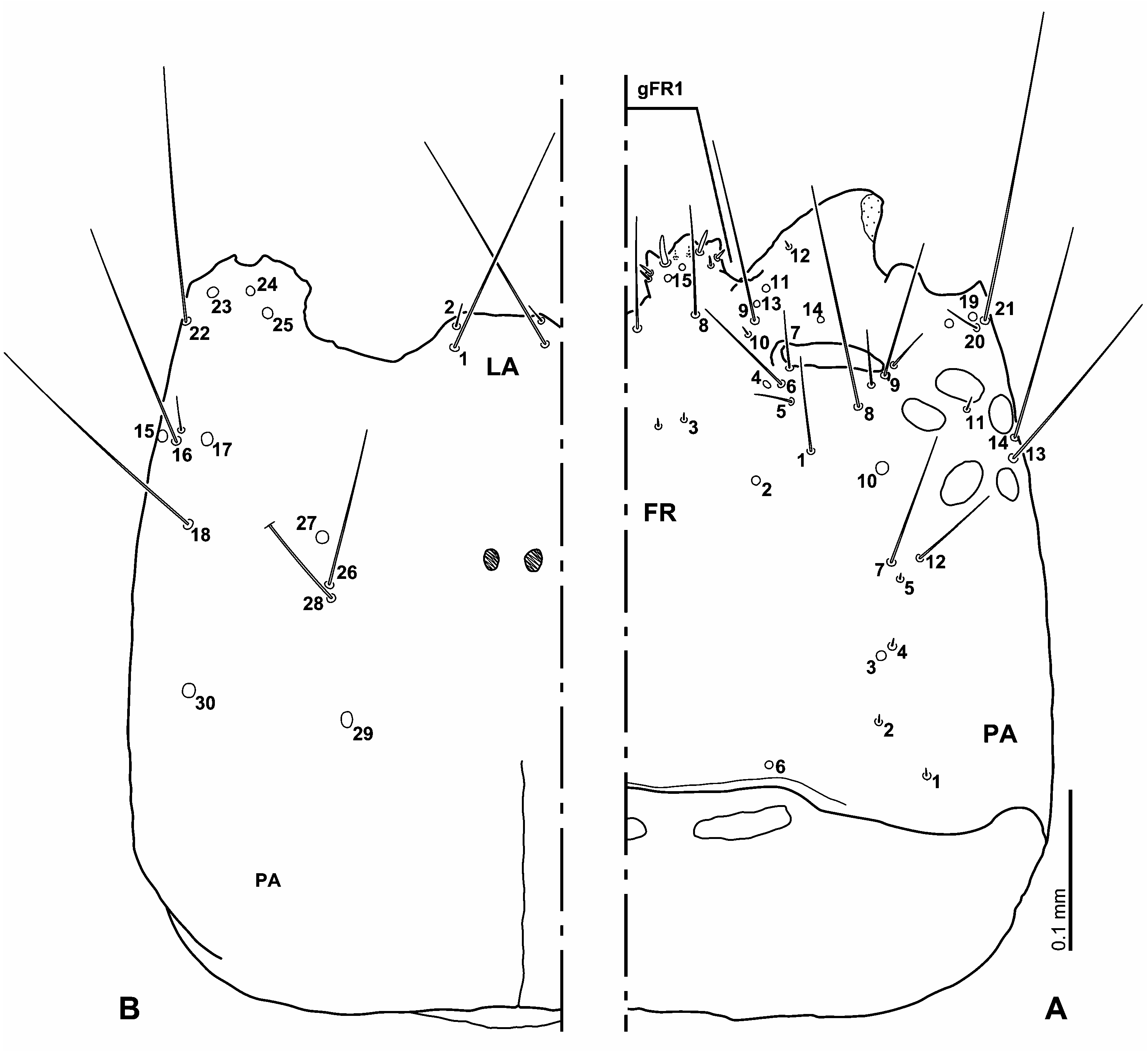
Head. Head capsule ( Figs 7A View Fig , 8–9 View Fig View Fig ) subquadrate; cervical sclerites small. Frontal line almost straight, parallel, almost invisible in third instar; coronal line absent; gular sulcus visible in basal part only. Surface of head capsule smooth. Six stemmata on each anterolateral corner of head capsule. Clypeolabrum ( Fig. 9A View Fig ) asymmetrical. Nasale with median projection bearing three teeth. Lateral lobes of epistome present; left lobe very strongly projecting anteriorly; right lobe strongly projecting anteriorly with membranous area laterally; both lobes projecting further than nasale, left lobe projecting further than right lobe. Left epistomal lobe bearing a group of stout, strongly bent ventrally, seta-like cuticular projections consisting of two rows (dorsal and ventral row) on inner margin, mesally to setae of gFR1. Ventral anterior margin of head capsule slightly asymmetrical. Dorsal and ventral mandibular articulation of left side projecting anteriorly further than right one ( Fig. 9 View Fig ).
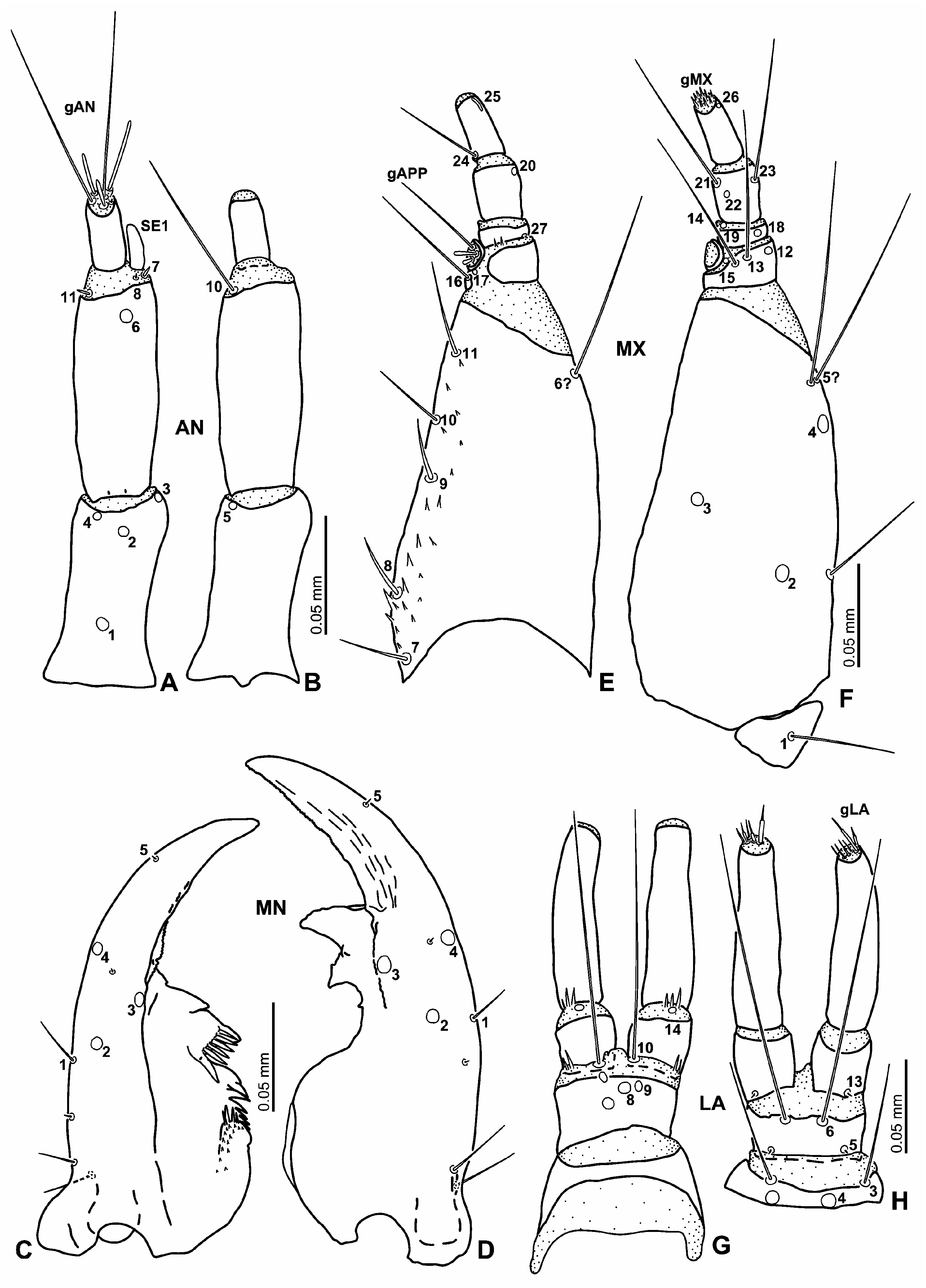
Antenna ( Figs 10A–B View Fig ) 3-segmented, short, rather slender. Antennomere 1 straight, shorter than antennomere 2; antennomere 2 longest and slightly narrower than antennomere 1; antennomere 3 shortest and narrowest. Mandibles ( Figs 10C–D View Fig ) strongly asymmetrical; left mandible shorter than right one. Right mandible with two inner teeth closely aggregated on median part. Left mandible with one inner tooth; inner tooth bearing a small subbasal tooth anteriorly and a comb composed by five to six spine-like projections posteriorly. Subbasal part of inner face of mandible with several short cuticular spines and minute cuticular teeth. Longitudinal mandibular groove present dorsally on midline of left mandibular base to incisor area. Maxilla ( Figs 10E–F View Fig ) 6-segmented, longer than antenna. Cardo moderate in size, subtriangular. Stipes the longest and widest segment, longer than palpomeres 1–4 combined; inner face with cuticular spines dorsally along inner face. Maxillary palpus 4-segmented; palpomere 1 widest, longer than palpomere 2 and shorter than palpomeres 3 and 4, bearing about two minute cuticular spines on dorsal surface of intersegmental membrane between palpomeres 1 and 2; palpomere 2 the shortest, wider than palpomere 3; palpomere 3 about as long as and wider than palpomere 4; palpomere 4 narrowest; dorsal surface of palpomere 1 incompletely sclerotised; inner process sclerotised. Labium ( Figs 10G–H View Fig ) developed, but small. Submentum fused to head capsule, large, subpentagonal, wider than mentum; submental sulcus hardly visible. Mentum transverse, narrowly cylindrically sclerotised, slightly wider than prementum; dorsal surface bare. Prementum subquadrate, slightly transverse; anterior membranous area with a few short cuticular spines dorsally on lateral face. Ligula reduced as small protuberance. Labial palpus long; palpomere 1 slightly wider than palpomere 2 and distinctly shorter than palpomere 2; intersegmental membrane between palpomeres 1 and 2 with a few short cuticular spines dorsally on median to inner part; palpomere 2 almost straight or slightly curved inwards in apical half.
Thorax. Thoracic membrane covered with fine cuticular pubescence. Prothorax wider than head capsule. Proscutum formed by one large plate subdivided by fine sagittal line, anterior part rather weakly sclerotised; whole sclerite bearing fine cuticular pubescence. Prosternal sclerite ( Fig. 7B View Fig ) divided into two closely aggregated plates thus can also be considered a subdivided single plate, bearing setae along anterior margin. Mesonotum with three sclerites on each side ( Figs 2A View Fig , 7C View Fig ); anterior two small, inner one larger than lateral one; posterior one large, subtriangular, bearing fine cuticular spines on lateral to posterior margin and setae of variable length. Two small tubercles present dorsally on each side; mesal one sclerotised, behind each posterior mesonotal sclerite; lateral one membranous, on laterodorsal surface, behind mesonotal spiracle. Metanotum with one pair of subtriangular to oval metanotal sclerites. Three tubercles on each side; mesal one sclerotised, situated behind each sclerite; remaining two membranous, one posterolaterally to sclerite, one on laterodorsal face. Legs ( Fig. 11B View Fig ) moderate in length, slightly visible in dorsal view, 5-segmented; all three pairs similar in shape.
Abdomen ( Fig. 2A View Fig ) 10-segmented, tapering posteriad, covered with fine cuticular pubescence; segments 1 to 7 similar in shape and size. Segment 1 with one pair of small dorsal sclerites bearing one short seta, medially on anterior part; four partially sclerotised tubercles behind dorsal sclerites; two membranous tubercles situated on each lateral face, one laterally to sclerotised tubercles, one on posterior part. Segments 2 to 7 similar to segment 1, dorsal sclerites on segment 2–7 similar or slightly smaller than those on first segment. Spiracular atrium ( Fig. 7D View Fig ) small. Segment 8 with large oval dorsal plate covered with fine cuticular projections and setae of variable length; dorsal surface of posterior edge of segment 8 densely covered with fine pubescence; procercus incompletely sclerotised, with one long and two short setae. Segment 9 trilobed, partially sclerotised; each lateral lobe without distinct acrocercus; urogomphi short, one-segmented; prostyli absent.
Second instar. Closely similar to third instar larva; sclerites on meso- and metathorax and abdominal segments more weakly sclerotised than in third instar.
Head. Frontal lines clearly visible; frontal line nearly straight, connecting lateral margin of antennal socket and posterior margin of head capsule; posterior end of frontal line unclear, possibly, frontal lines curved mesally at the base of head capsule.
Antenna and maxilla ( Fig. 6 View Fig ) proportionally stouter than in third instar. Thorax and abdomen. Arrangements of cuticular projections and pubescence on thorax and abdomen similar to third instar but projections and pubescence finer than in third instar.
First instar. Similar to second instar larva; sclerites on meso- and metathorax and abdominal segments more weakly sclerotised than in second instar.
Head. Antenna ( Figs 5A–B View Fig ) proportionally stouter than that of second and third instar larvae. Mandible ( Figs 5C–D View Fig ). Distal inner tooth of left mandible with four to five seta-like projections basally on posterior margin. Maxilla ( Figs 5E–F View Fig ). Intersegmental membrane between palpomeres 2 and 3 bearing a few small cuticular spines dorsally. Labium ( Figs 5G–H View Fig ) without small cuticular spines on anterior membranous area of prementum and intersegmental membrane between palpomeres 1 and 2.
Thorax and abdomen. Arrangements of cuticular projections and pubescence on thorax and abdomen similar to third instar but projections and pubescence finer than in second instar.
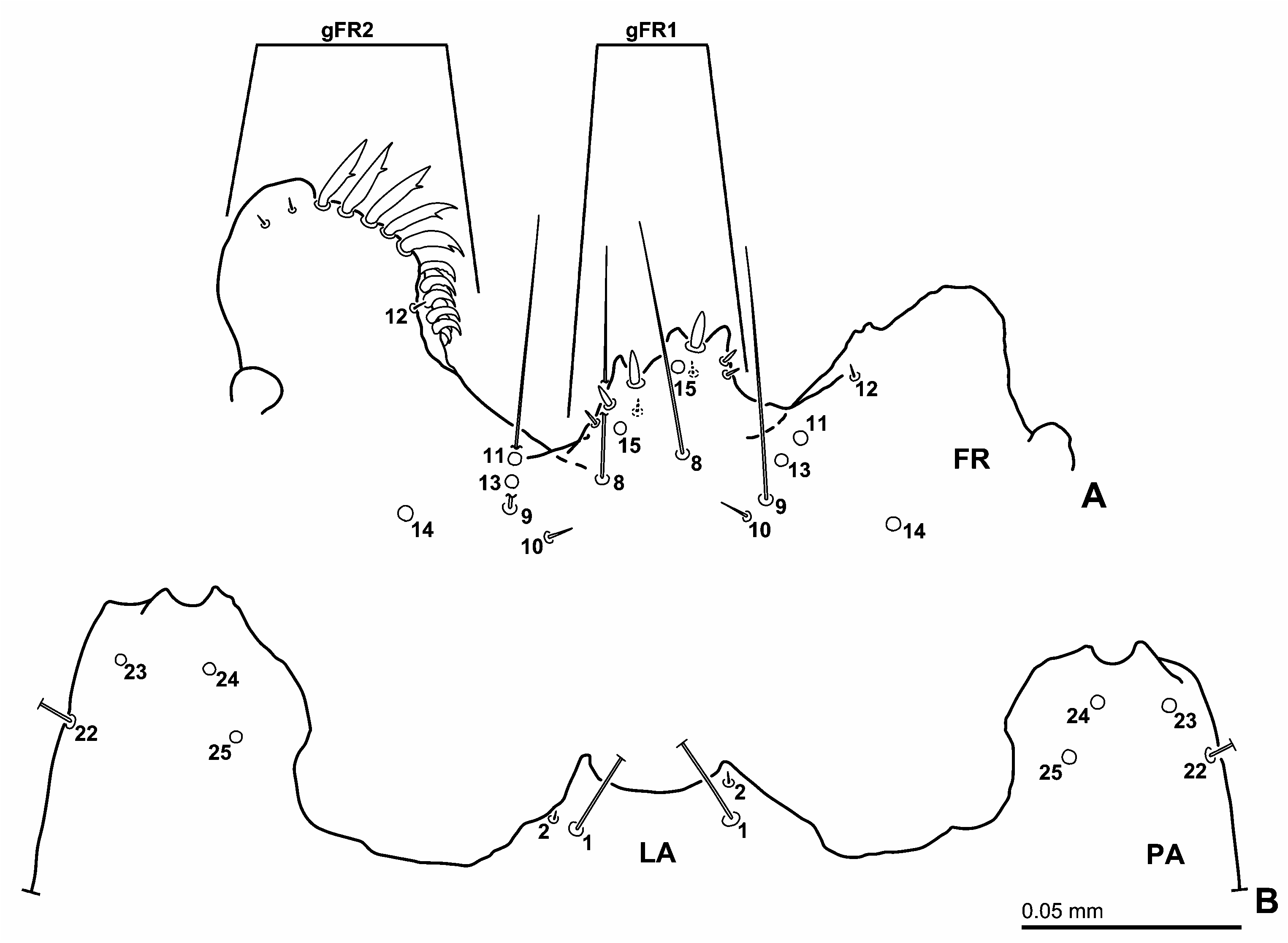
Chaetotaxy of head. Primary chaetotaxy. Frontale ( Figs 3A View Fig , 4A View Fig ). Median part with three pairs of sensilla (FR1–3); long seta FR1 situated close to frontal line, minute seta FR3 on mesal part, pore-like sensillum FR2 between FR1 and FR3, posteriorly to line connecting FR1 and FR3. Three setae FR5–7 and pore-like sensillum FR4 situated behind antennal socket, close to each other; FR5 and FR7 rather short, FR6 long; FR7 on inner margin of antennal socket; FR4 and FR6 very close to each other, slightly posteromesally to FR7; FR4 mesally to FR6; FR5 situated posteriorly to FR6. Long seta FR8 situated on mesal part of clypeolabrum, posteriorly to nasale; pore-like sensillum FR15 on median part of nasale; pore-like sensillum FR14 anteriorly to antennal socket. Three setae (FR9–10 and FR12) and two pore-like sensilla (FR11 and FR13) situated mesally on epistome; FR9 long, FR10 short, FR12 very short. FR9–FR11 and FR13 forming a slightly irregular oblique row, FR10 situated posteriorly, FR11 anteriorly, FR9 and FR13 between FR10 and FR11, FR9 posteriorly to FR13; FR12 on inner margin of epistomal lobe, distance between FR12 and FR11 on left side longer than on right. Nasale with group of six short setae, and with (at least) two short ventral setae (gFR1); left one and right two narrow, remaining ones stout. Left epistomal lobe with group of setae (gFR2); lateral two short, remaining ones stout, with small apical tooth; within stout setae, lateral five are true setae but remaining ones could possibly not be true setae (i.e., seta-like cuticular spines); therefore left epistomal lobe may bear two short setae and five stout setae; gFR2 on right lobe absent.
Parietale ( Figs 3 View Fig , 4B View Fig ). Dorsal surface with a group of five sensilla (PA1–5) forming an irregularly longitudinal row close to frontal line in posterior part of parietale; PA1–2 and 4–5 short setae, PA3 pore-like; a very small pore-like and sensilla-like structure situated posteriorly to PA1. PA6 pore-like, situated behind frontale, on posteromesal margin of head capsule. Long setae PA7 and PA12 situated laterally and close to midlength of frontal line, anteriorly and close to PA5; PA12 laterally to PA7. Very long setae PA8–9 situated behind antennal socket, PA9 on outer margin of antennal socket, PA8 posteromesally to PA9 and close to frontal line. Pore-like sensillum PA10 situated at midlength of line connecting PA8 and PA12. Four setae (PA13–14, PA16 and PA18) and two pore-like sensilla (PA15 and PA17) situated on about anterior third of lateral surface of parietale; PA13–14 and PA18 very long, PA16 long. PA13–17 forming irregular transverse row; PA13 dorsally to remaining ones, posteromesally to PA14; PA14–16 closely aggregated, PA14 dorsally to PA15–16, PA16 ventrally to PA14–15, PA15 between PA14 and PA16; PA 17–18 situated ventrally to PA13–16, PA17 mesally to PA16, and PA18 posteriorly to PA17. Short seta PA11 situated between PA9 and PA13. PA19–22 closely aggregated on anterior corner of head capsule, PA19 pore-like, PA20 rather short seta, PA21–22 very long setae; PA19–20 situated dorsally to PA21–22, PA19 anteriorly to PA20, and PA22 anteroventrally to PA22. Pore-like sensilla PA23–25 situated close to ventral mandibular articulation; PA23 laterally to PA24–25; PA25 posteromesally to PA25. Two very long setae (PA26 and PA28) and pore-like sensillum PA27 situated ventrally on median part of parietale; PA27 situated anteriorly to PA26 and PA28, PA26 between PA27 and PA28, very close to PA28. Two pore-like sensilla (PA29–30) on posterior third of ventral parietale; PA29 mesally to PA30, posteromesally to PA26–28; PA30 on lateral part.
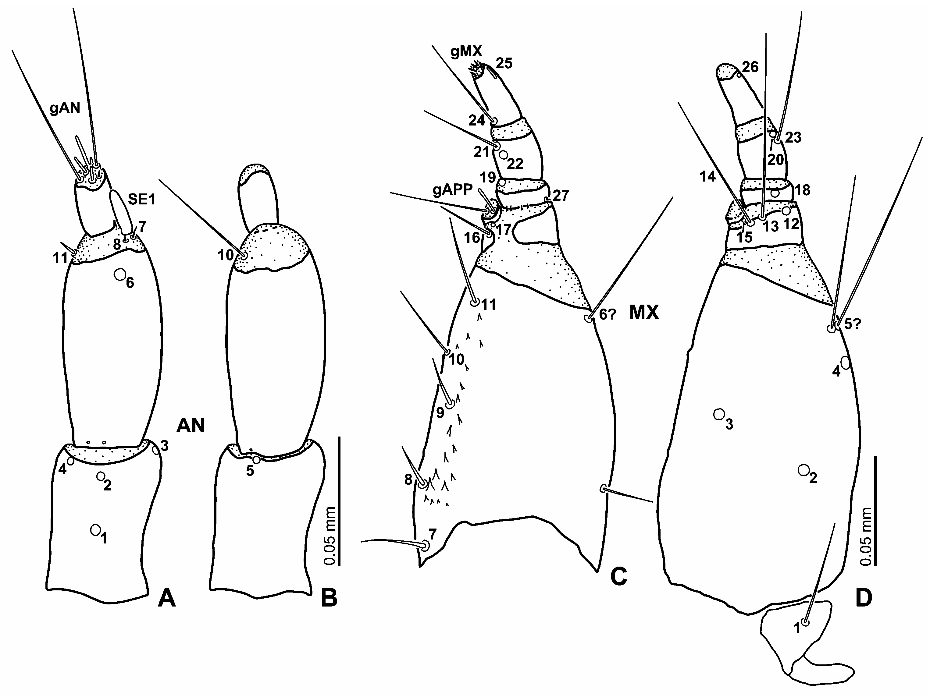
Antenna ( Figs 5A–B View Fig ). Antennomere 1 with five pore-like sensilla (AN1–5); AN1 situated dorsally on about posterior third; AN2–5 on anterior margin of sclerite, AN2 dorsally on median part, AN3 on outer face, AN4 inner face, AN5 ventrally on median portion. Antennomere 2 with one pore-like sensillum (AN6) situated dorsally on apical portion of sclerite; minute setae AN7–8 and sensorium SE1 closely aggregated on outer face of intersegmental membrane between antennomeres 2 and 3, AN9 absent; SE1 slender, about as long as to slightly shorter than antennomere 3; setae AN10–11 situated on inner face of intersegmental membrane between antennomeres 2 and 3, AN10 very long, AN11 short, both setae close to each other. Antennomere 3 with apical sensilla (gAN) in apical membranous area; gAN with two rather long setae and a few short setae of variable shape.
Mandibles ( Figs 5C–D View Fig ). Mandible with two setae (MN1 and MN5) and three pore-like sensilla (MN2–4); MN1 short, MN5 minute. Pore-like sensillum MX 6 undetectable. MN1 situated on posterior third of lateral face of mandible. MN2–4 situated on median part of mandible; MN3 situated mesally to MN1–4, MN2 between MN1 and MN3; MN4 and MN5 situated on lateral face anteriorly to MN1; MN5 subapically, MN4 at midlength between MN1 and MN5 on left mandible, closer to MN1 on right.
Maxilla ( Figs 5E–F View Fig ). Cardo with one rather long ventral seta ( MX 1). Stipes with a slightly irregular row of five setae ( MX 7–11) situated dorsally along inner face; MX 7–9 and MX 11 somewhat stout, rather long, MX 10 trichoid, long; MX 7–9 and MX 11 almost equidistant from each other; MX 10 located between MX 9 and MX 11, close to MX 9. Two very long setae ( MX 5–6) present apically on outer face of sclerite; MX 5 longer than MX 6; MX 5 situated posteriorly and very close to MX 6; pore-like sensillum MX 4 posteriorly to MX 5. Pore-like sensilla MX 2–3 situated ventrally on median part of sclerite; MX 2 on outer part, posterolaterally to MX 3, MX 3 on inner part. Dorsal surface of palpomere 1 with one long trichoid seta ( MX 16) situated on inner face. Ventral surface of sclerite with three sensilla ( MX 12–14) along distal margin of sclerite; MX 12 pore-like on lateral part, MX 13 very long seta between MX 12 and MX 14, MX 14 long seta, very close to MX 13. Pore-like sensilla ( MX 15 and MX 17) situated on membrane behind inner appendage; MX 17 dorsally, MX 15 ventrally. Inner appendage with one long seta and a few short setae (gAPP). Palpomere 2 with two pore-like sensilla ( MX 18 and MX 19) and one minute seta ( MX 27); MX 18 situated ventrally on median to lateral portion of sclerite; MX 19 on inner face of intersegmental membrane between palpomeres 2 and 3; MX 27 basal on outer face of sclerite. Palpomere 3 with two setae ( MX 21 and MX 23) and two pore-like sensilla ( MX 20 and MX 22); MX 21 long, MX 23 very long; MX 20 situated on distal margin of outer surface of sclerite; MX 23 posterodorsal and close to MX 20; MX 21–22 on inner face of sclerite, MX 21 on distal margin, MX 22 posteroventrally to MX 21. Palpomere 4 with one long seta ( MX 24) situated basally on inner face, and with digitiform sensillum ( MX 25) and pore-like sensillum ( MX 26) apically on outer face of sclerite; MX 25 dorsally, MX 26 ventrally. Apical membranous area of palpomere 4 with several minute setae (gMX).
Labium ( Figs 4B View Fig , 5G–H View Fig ). Submentum with two pairs of setae (LA1–2); LA1 very long, LA2 very short, both situated on anterolateral portion; LA1 very close and posteriorly to LA2. Ventral surface of mentum with one pair of rather long setae (LA3) and pore-like sensilla (LA4); LA4 situated behind LA3, LA3 close to distal margin, LA4 close to proximal margin. Prementum with three pairs of sensilla (LA8–9 and LA10) on dorsal surface and with two pairs of sensilla (LA5–6) on ventral surface. Pore-like sensillum LA8 situated medially, very long seta LA10 on anterior membranous area, anteriorly to LA8; LA9 close to LA8; LA11–12 absent. Minute seta LA5 situated ventrally at base of outer face; very long seta LA6 on median part, on borderline between sclerite and membrane of prementum. One minute seta (LA13) situated lateroventrally on basal margin of palpomere 1; pore-like sensillum LA14 on dorsal surface of intersegmental membrane between palpomeres 1 and 2. LA15 absent. Apical membranous area of palpomere 2 with several setae of variable length and shape (gLA).
Second instar. Primary sensilla on second instar similar to those of first instar, and secondary chaetotaxy on second instar similar to that of third instar. Frontale (e.g., Figs 8A View Fig , 9A View Fig ). Median two setae of gFR1 proportionally longer and stouter than in first instar, rounded apically. Parietale with three secondary setae and one pore-like secondary sensillum (e.g., Fig. 8 View Fig ); two short setae close to lateral edge of antennal socket and PA9, one situate between PA8 and PA9, one on anterolaterally to PA9; one pore-like sensillum very close to PA20–21; one short seta close to PA16.
Antenna ( Figs 6A–B View Fig ). Three minute sensilla-like structures present at base of antennomere 2, two dorsal and one ventral; the presence or absence of these is variable. SE1 stout, slightly shorter than antennomere 3. Mandible (e.g., Figs 10C–D View Fig ) with four secondary sensilla on each mandible; one minute sensillum situated dorsally on lateral part, close to MN4; one minute to short seta posteriorly to MN1; two rather short setae on lateral face of mandibular base. Maxilla ( Figs 6C–D View Fig ). Stipes with two secondary setae; one rather short seta situated on about posterior third of outer face of sclerite, one long seta close to MX 5–6.
Third instar. Similar to second instar. Antennal sensorium SE1 stout, about two-third as long as antennomere 3 ( Figs 10A–B View Fig ).
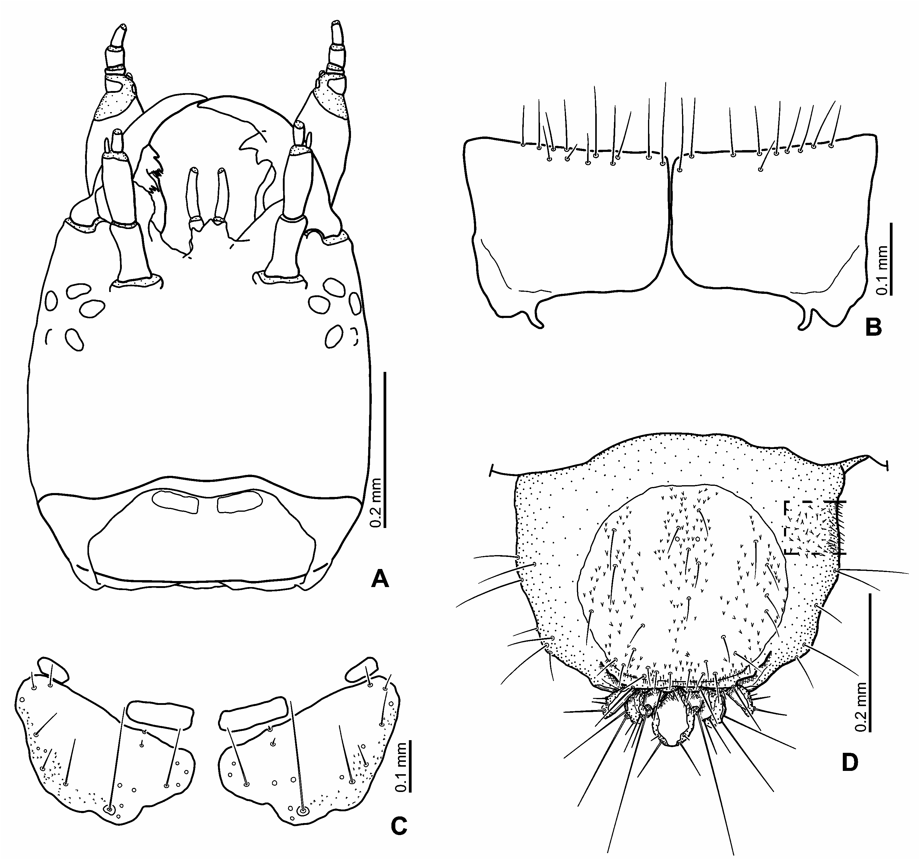
Description of pupa. Body ( Figs 2C–D View Fig , 12 View Fig ) moderately broad. Head, thorax and abdomen with styli. Colour. Whitish when alive ( Figs 2C–D View Fig ); eyes yellowish white to reddish brown.
Head. Deflexed ventrally, covered by pronotum in dorsal view. Frontoclypeal sulcus partly visible.Antennae completely covered by head and pronotum in dorsal view; mouthparts visible in ventral view; maxillary palpi long. Styli on head: Two pairs of rather short supraorbital styli close to inner margin of each eye.
Thorax. Pro-, meso- and metathoracic legs visible in ventral view; metathoracic legs partially covered by wingpads, only tibiae to tarsi visible; meso- and metathoracic legs strongly bent ventrally. Apex of tarsi ending with one minute projection. One pair of small projections mesal to metanotal styli. Styli on thorax: Pronotum with 11 pairs of rather short styli ( Figs 12D, E View Fig ). Five pairs of styli on anterior margin of pronotum, three pairs situated laterally, two pairs mesally; mesal two pairs slightly longer than others. Two pairs of styli on median part, one posterior to anterior three pairs, one on mesal part. Four pairs of styli on posterior margin. Meso- and metanotum with one pair of styli on median part.
Abdomen. Abdomen with nine segments, attenuate towards apex. Posterior corner of segment 8 with triangular projection ( Figs 12B, F View Fig ). Segment 9 with well-developed, non-articulated urogomphi; surface of urogomphi finely denticulated ( Fig. 12F View Fig ). Styli on abdomen: Abdominal styli rather short, almost equal in size and shape excluding segment 8. Abdominal segment 1 with two pairs of rather short styli, one on median part, one lateral. Segments 2 to 7 with a transverse row of three pairs of styli; one mesal, two on lateral face close to spiracle. Segment 8 with one pair of styli without apical hair on posterior margin, hook-shaped, finely denticulate ( Fig. 12F View Fig ).
Biology. Both adults and larvae of L. kunashiricus live in standing water. Two larvae likely belonging to this species were found at the edge of a pond in Hokkaido Prefecture (Y. N. Minoshima, personal observation). Larvae are sluggish, and do not swallow atmospheric air into the alimentary canal (see MINOSHIMA and HAYASHI (2015) for a more general discussion about this behaviour). We placed a living larva into water and confirmed that it sank in the water as it was not buoyed by air bubbles in the alimentary canal. WILSON (1923) mentioned that the larva of L. ( Laccobius) agilis ( Randall, 1838) sometimes hangs from the water surface supported by its spiracular atrium.


No known copyright restrictions apply. See Agosti, D., Egloff, W., 2009. Taxonomic information exchange and copyright: the Plazi approach. BMC Research Notes 2009, 2:53 for further explanation.