
Sphaeriodiscus mirabilis A.M. Clark, 1976
|
publication ID |
https://doi.org/ 10.5281/zenodo.276783 |
|
DOI |
https://doi.org/10.5281/zenodo.6184379 |
|
persistent identifier |
https://treatment.plazi.org/id/03D28792-FFEF-FFB0-84E4-13A768DE8494 |
|
treatment provided by |
Plazi |
|
scientific name |
Sphaeriodiscus mirabilis A.M. Clark, 1976 |
| status |
|
Sphaeriodiscus mirabilis A.M. Clark, 1976 View in CoL
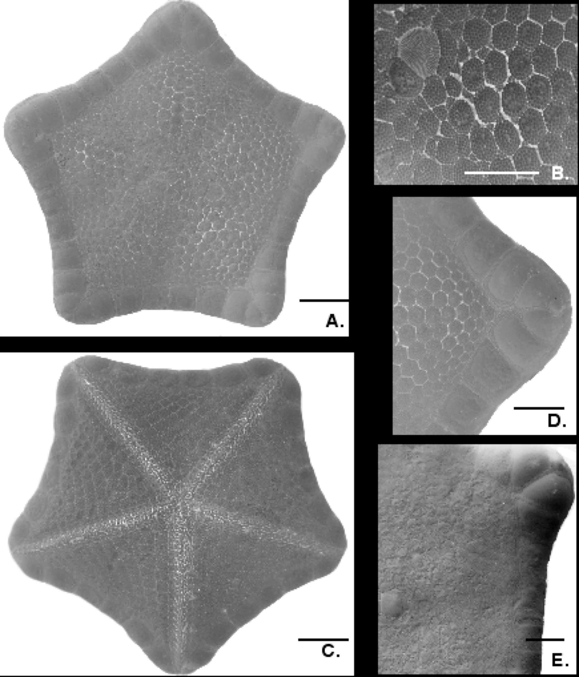
( Figures 18 View FIGURE 18 A–E)
A.M. Clark, 1976: 248
Distribution. South Indian Ocean (St. Paul and Amsterdam Islands) to the South Atlantic ( Bouvetoya Island and Tristan de Cunha). 195– 462 m.
Material examined. South Atlantic. USNM 1116242 Tristan de Cunha 37˚19’29”S, 12˚29’48”W, 195 m. Coll. Moss Landing Marine Laboratories. (1 dry spec. R=2.9, r=2.2). CASIZ 174584 Bouvetoya Island 54˚21’36”S, 3˚10.3’E, 455–462 m, Coll. S. Lockhart (2 wet specs. R=3.5, r=2.5, R=4.2, r=3.3).
Description. Body pentagonal (R/r=1.3 to 1.4), stout, thick. Interradial arcs weakly concave ( Fig. 18 View FIGURE 18 A). No pedicellariae observed on body surface.
Abactinal plates hexagonal to round in outline, closely abutted forming a crowded pavement ( Fig. 18 View FIGURE 18 B). Plates become more oblong and oval-shaped and dense on abactinal surface at contact with superomarginal plates. Larger specimens (R>3.0 cm) with more rounded abactinal plates whereas smaller specimens (R= 2.9 cm) with more hexagonal plates (e.g., Fig. 18 View FIGURE 18 B). Plates most tightly articulated interradially, adjacent to superomarginal plate contact, where papulae are absent but are more widely separated radially along arms where papulae are exposed. Abactinal plates in well-ordered rows. Mid-radial carinal plate row extending to contact point between penultimate superomarginals in contact along midradius. Carinal plate row flanked by multiple adjacent rows (approximately four to six) before becoming distributed in a more irregular pattern interradially. Primary circlet is clearly present with large primary plate present in each interradial region. Each abactinal plate covered by 70–90 flat-topped granules, round to quadrate in cross-section. Collectively granules form a dense cover over abactinal surface, apparently extending onto fringes of marginal plates. Approximately 25–30 granules form a discrete border around each plate. The remaining granules cover the center of the abactinal plate surface forming crowded rows within the border formed by the peripheral granules. Smaller secondary plates are present and are only a fraction of the size of the larger plates with only minimal granular coverings (7–12 granules present). Abactinal plates surrounded by approximately 12 papulae per plate, with 2 per side. Fasciolar grooves present, moderately deep to shallow around radial regions but becoming absent interradially. Madreporite triangular in smaller specimens with well-developed sulcae, flanked by six to seven abactinal plates ( Fig. 18 View FIGURE 18 B). In largest specimen, madreporite is seven-sided and flanked by seven to eight abactinal plates.
Marginal plates eight in each interradius (from armtip to armtip), each slightly convex ( Fig. 18 View FIGURE 18 A). Granules, rounded, blunt forming dense but evenly distributed covering which almost seems continuous with abactinal granule cover. When large bare spot is present, granules form two to four rows forming periphery on marginal plate surface. Granules cover most of marginal plate surface with the exception of a small poorly defined bare spot on the center of interradial superomarginal or inferomarginal. Infrequently, bare spot is absent. On superomarginal plates, bare spot on surface with 10–13 widely spaced granules ( Fig. 18 View FIGURE 18 A, D). Distalmost marginal plates (2–3 from terminal) are largely bare with granular periphery. Granular periphery at distalmost marginal plates thickened and confluent on some specimens.
Superomarginals and inferomarginals in 1:1 correspondence with same number of plates in each series, although plate morphology is not identically mirrored in each series. Distalmost superomarginals in full contact along, at least, one side. Penultimate superomarginals barely in contact at one edge but granular covering of two penultimate superomarginals are confluent (fig. 18D). Distalmost and penultimate superomarginals enlarged, swollen, strongly convex ( Fig. 18 View FIGURE 18 E), becoming more flattened and similar to other superomarginals interradially. Degree of swelling and enlargement is more pronounced for individuals larger then R= 3.5 cm ( Fig. 18 View FIGURE 18 E) with the smallest individual (R= 2.9 cm) showing nearly no difference from other superomarginal plates ( Fig. 18 View FIGURE 18 D). Distalmost inferomarginal plate is smallest and triangular in outline relative to terminal plate. Penultimate inferomarginal plate is swollen but not enlarged to the degree observed in superomarginal.
Actinal plates in four to six chevron rows. Actinal plates proximally, sharply quadrate in outline, becoming more hexagonal distally adjacent to contact with inferomarginal plate series ( Fig. 18 View FIGURE 18 C). Each plate covered with granules, rounded to polygonal in outline. Granules, 40–60 cover each plate, blunt distally becoming more pointed proximally; distributed more widely distally an becoming more crowded adjacent to mouth. Peripheral granules, 15–30 in discrete rows with central granules crowded, evenly distributed on the surface of each.
Adambulacral plates elongate. Furrrow spines blunt and rounded, five on each adambulacral plate, quadrate in cross-section. Four of those spines identical in length. Fifth spine, proximally directed, was about 75–80% of the length, of other furrow spines, triangular in cross-section and set off from the other four identical furrow spines. Subambulacral spine rows, each set off from the other furrow spines by discrete spaces. Subambulacral spines three, approximately 50–60% of the height of the furrow spine, each spine differs in size with smallest proximally with largest distally, quadrate in cross-section, thickened (twice the thickness of the furrow spines). Subambulacrals backed by three short granules, round to quadrate to triangular in cross section. Smallest of adambulacral spination, becoming similar to identical in size to actinal granule coverings. Oral plate furrow spines seven on each side, 10–20% thicker then other furrow spines, quadrate to triangular in cross-section. Surface of oral plates covered by 20–30 granules, triangular to angular quadrate in cross-section and well-spaced from one another relative to the crowded granules on actinal surface.
Comparisons with other taxa. The Atlantic specimens described herein of S. mirabilis are largely identical to the specimen described from the Indian Ocean by A.M. Clark (1976). However, all of the Atlantic specimens are substantially smaller then the holotype (R=7.0 cm) and show some variation. The penultimate superomarginals in the Indian Ocean specimen are substantially more swollen then those of the smaller Atlantic specimens, especially in the smallest (R= 2.9 cm) specimen, which showed almost no superomarginal plate enlargement. The description included herein also elaborates on morphological characters, such as the adambulacral spination which are not closely detailed in the holotype description.
Further comparisons of other species of Southern Hemisphere Sphaeriodiscus from H.E.S. Clark (2001) that other Pacific species are closely related, further adding strength to a notion of close affinities between S. mirabilis and S. bourgeti from north-west Africa as suggested by A.M. Clark (1976).
Morphological data from Mah (2007 and unpublished data) do not support affinities between Pentagonaster and Sphaeriodiscus mirabilis as implied by A.M. Clark (1976), suggesting that the enlarged marginal plates are convergent. The presence of enlarged superomarginal plates near the armtips is present in several unrelated Southern Hemisphere goniasterids and goniasterid-like taxa. This includes both species in the genus Pentagonaster ( Mah 2007) as well as the goniasterids Calliaster acanthodes and Toraster tuberculatus (Mortensen, 1933) . One species of New Zealand odontasterid, Diplodontias dilatatus also shows this character ( McKnight 2001).
| USNM |
Smithsonian Institution, National Museum of Natural History |
No known copyright restrictions apply. See Agosti, D., Egloff, W., 2009. Taxonomic information exchange and copyright: the Plazi approach. BMC Research Notes 2009, 2:53 for further explanation.