
Peliococcus spinigerus (Goux)
|
publication ID |
https://doi.org/ 10.11646/zootaxa.3920.2.1 |
|
publication LSID |
lsid:zoobank.org:pub:57A4B8A3-C5A5-45FB-96E6-B26123271F66 |
|
DOI |
https://doi.org/10.5281/zenodo.6102199 |
|
persistent identifier |
https://treatment.plazi.org/id/03D2879A-B377-FFDA-DFDE-FADF9CF5FB69 |
|
treatment provided by |
Plazi |
|
scientific name |
Peliococcus spinigerus (Goux) |
| status |
|
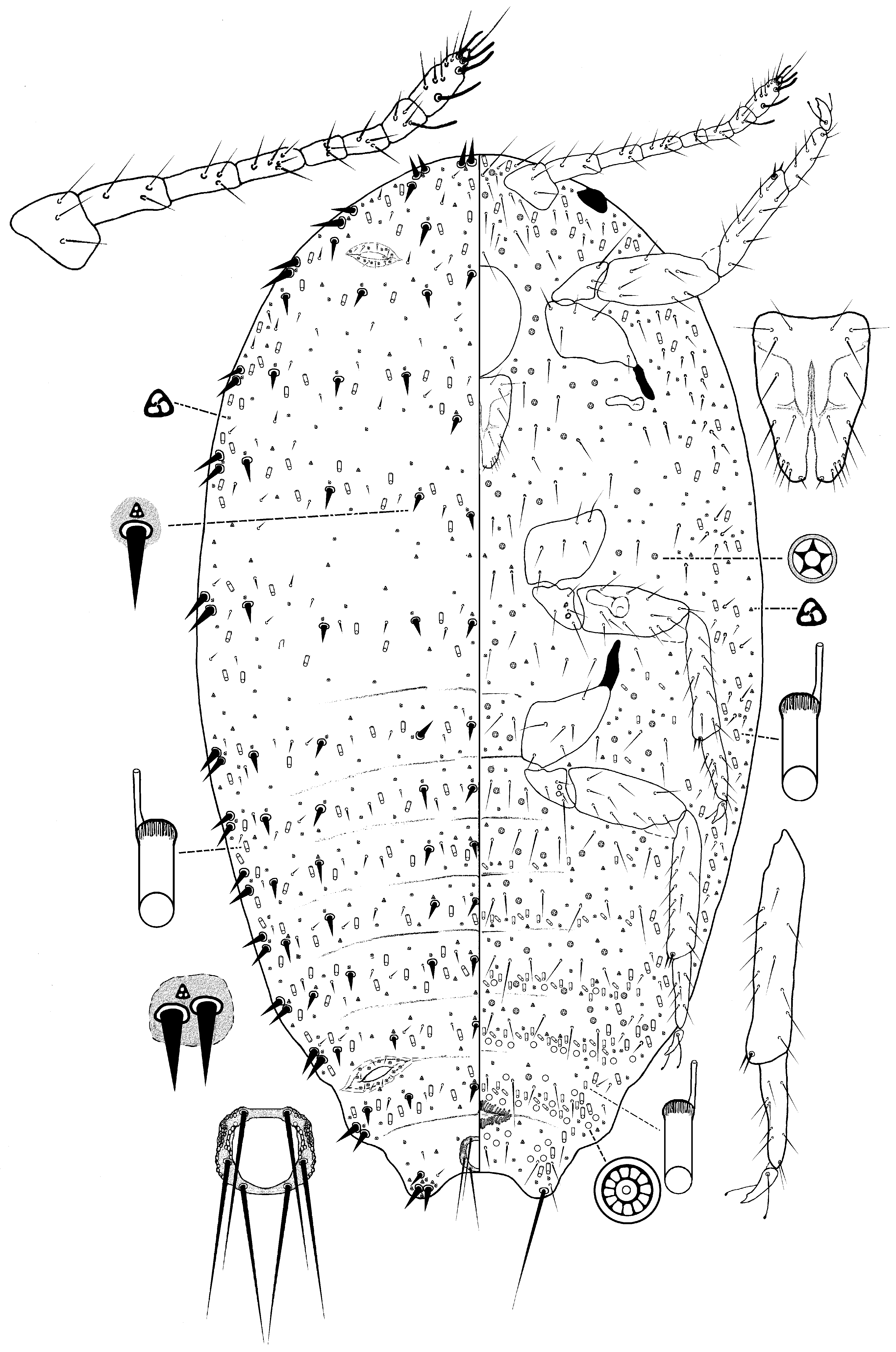
( Fig. 9 View FIGURE 9 )
Pedronia spinigera Goux, 1938: 455 View in CoL .
Parapedronia spinigera (Goux) View in CoL : Balachowsky, 1953: 283 (change of combination).
Spinococcus spinigerus (Goux) : Danzig, 1960: 178 (change of combination in connection with synonymy of Parapedronia View in CoL with Spinococcus View in CoL ).
Peliococcus spinigerus (Goux) : Danzig, 1980: 118 (change of combination in connection with synonymy of Spinococcus View in CoL with Peliococcus View in CoL ), 2001: 123.
Material studied. Paratypes: 6 adult females on 3 slides: France, Rhône, Bessenay, on Thymus serpyllum (Lamiaceae) , 8.viii.1935, coll: L. Goux ( MNHN: 3958-19, 20, 22).
Description. Adult female. Body elongate oval, 1.08–1.43 mm long, 0.54–0.78 mm wide. Eye marginal, 37.5–45 µm wide. Antenna 8 segmented, 315–350 µm long; apical segment 60 µm long, 20 µm wide; apical setae 25 µm long, plus 3 fleshy setae 25–30 µm long. Tentorium 135–140 µm long, 120–140 µm wide. Labium 85–100 µm long, 110 µm wide. Anterior spiracles 55–60 µm long, 20 µm wide across atrium; posterior spiracles 60–63 µm long, 27 µm wide. Legs well developed; posterior legs: trochanter + femur 190–225 µm; tibia + tarsus 240–270 µm; claw 22–30 µm long. Ratio of lengths of tibia + tarsus to trochanter + femur 1.20–1.32:1; ratio of lengths of tibia to tarsus 2.00–2.35:1; ratio of length of trochanter + femur to greatest width of femur 2.85–3.23:1. Tarsal digitules hair-like, each 20–25 µm long. Claw digitules knobbed, each 20–23 µm long. Anterior ostioles with a total for both lips of 4–8 trilocular pores and 2 or 3 setae; posterior ostioles with a total for both lips of 7–14 trilocular pores and 0–2 setae. Anal ring 50–70 µm wide, with 6 setae, each seta 130–158 µm long. Cerarii numbering 15 marginal pairs, each on an elevated area, most with 2 cerarian setae and 1 trilocular pore; anal lobe cerarii each with 2 enlarged cerarian setae, each 25 µm long, plus 1 smaller enlarged seta, 17–20 µm long, 7–9 trilocular pores, and 2 or 3 spine-like auxiliary setae; 8–10 dorsal cerarii in a longitudinal line medially.
Dorsum. Setae spine-like, of 2 sizes: (i) enlarged setae, each slightly smaller than cerarian setae, each 15–20 µm long, on an elevated area with a trilocular pore near basal socket; present in 6 longitudinal rows, and (ii) smaller setae, each 5.0–7.5 µm long, not on an elevated area and lacking an associated trilocular pore, scattered in a single row across all body segments. Oral collar tubular ducts, each 8–10 µm long, 6–7 µm wide, present in a single row on each segment, with a total of 65–81 ducts on head and thorax, and also on abdominal segments as follows on: I 12–18, II 17–19, III 17– 19, IV 16–31, V 23–32, VI 8– 14, VII 13–19, VIII + IX 0. Trilocular pores, each 4–5 µm in diameter, scattered over entire surface.
Venter. Setae of 2 types: (i) slender hair-like setae, each 12–100 µm long, longest setae medially on head; and (ii) spine-like setae, each 5–10 µm long, in submarginal rows. Apical setae of anal lobes each 175 µm long. Multilocular disc pores, each 8–10 µm in diameter, in single rows on posterior abdominal segments as follows: IV 0–2, V 4–7, VI 16–20, VII 24–34, VIII+ IX 15–23. Quinquelocular pores, each 4–6 µm in diameter, present medially on head, thorax and abdominal segments II-VII. Trilocular pores, each 3–4 µm in diameter, scattered over entire surface. Oral collar tubular ducts of 2 sizes: larger ducts similar to those on dorsum, in a marginal and submarginal band; and smaller ducts, each 7.5–10 µm long, 3–5 µm wide, concentrated on body margins and in single rows on abdominal segments as follows; II+ III 21–65, IV 53 –76, V 66 –94, VI 66 –75, VII 41 –63, VIII + IX 15–17.
Comments. Peliococcus spinigerus is most similar to P. marrubii in lacking: (i) multilocular disc pores on dorsum, and (ii) a circulus, but differs in having: (i) 12–16 dorsal oral collar tubular ducts on each abdominal segment, in an almost continuous row ( P. marrubii has more than 18 dorsal oral collar tubular ducts on each abdominal segment), and (ii) 15 pairs of marginal cerarii ( P. marrubii has 18 pairs).
Danzig & Gavrilov-Zimin (2014) regarded P. spinigerus as a junior synonym of P. marrubii , but in this study these two species are regarded separate because of the clear differences mentioned above.
Host plants. On Thymus serpyllum (Lamiaceae) .
Distribution. France (Bessenay, Rhone).
| MNHN |
Museum National d'Histoire Naturelle |
No known copyright restrictions apply. See Agosti, D., Egloff, W., 2009. Taxonomic information exchange and copyright: the Plazi approach. BMC Research Notes 2009, 2:53 for further explanation.
|
Kingdom |
|
|
Phylum |
|
|
Class |
|
|
Order |
|
|
Family |
|
|
Genus |
Peliococcus spinigerus (Goux)
| Kaydan, Mehmet Bora 2015 |
Peliococcus spinigerus
| Danzig 1980: 118 |
Spinococcus spinigerus
| Danzig 1960: 178 |
Parapedronia spinigera
| Balachowsky 1953: 283 |
Pedronia spinigera
| Goux 1938: 455 |