
Cladorhiza moniqueae, Ekins & Erpenbeck & Hooper, 2020
|
publication ID |
https://doi.org/10.11646/zootaxa.4774.1.1 |
|
publication LSID |
lsid:zoobank.org:pub:B0C4A2F8-F2AB-4147-BB12-63720EEF2516 |
|
DOI |
https://doi.org/10.5281/zenodo.3846413 |
|
persistent identifier |
https://treatment.plazi.org/id/E4131AC2-95FD-4E25-8CBF-6A1ED16566D5 |
|
taxon LSID |
lsid:zoobank.org:act:E4131AC2-95FD-4E25-8CBF-6A1ED16566D5 |
|
treatment provided by |
Plazi |
|
scientific name |
Cladorhiza moniqueae |
| status |
sp. nov. |
Cladorhiza moniqueae View in CoL sp. nov.
Figures 14 View FIGURE 14 & 15 View FIGURE 15 , Table 5 View TABLE 5
urn:lsid:zoobank.org:act:E4131AC2-95FD-4E25-8CBF-6A1ED16566D5
Material Examined. Holotype: QM G337496 off Newcastle , New South Wales, Australia, Station 65, 33° 26’ 27.6”– 33° 26’ 6.0” S, 152° 42’ 7.2”– 152° 39’ 54.0” E, 4280– 4173 m, Beam Trawl, Coll. Merrick Ekins on RV Investigator, Cruise IN2017_ V03 , Sample 65-162, 30/v/2017. GoogleMaps
Etymology: Named after Monique Grol for her enthusiasm for SCUBA diving and the reef.
Distribution: This species is presently known only from the type locality off Newcastle, New South Wales, Australia, at abyssal depth.
Description:
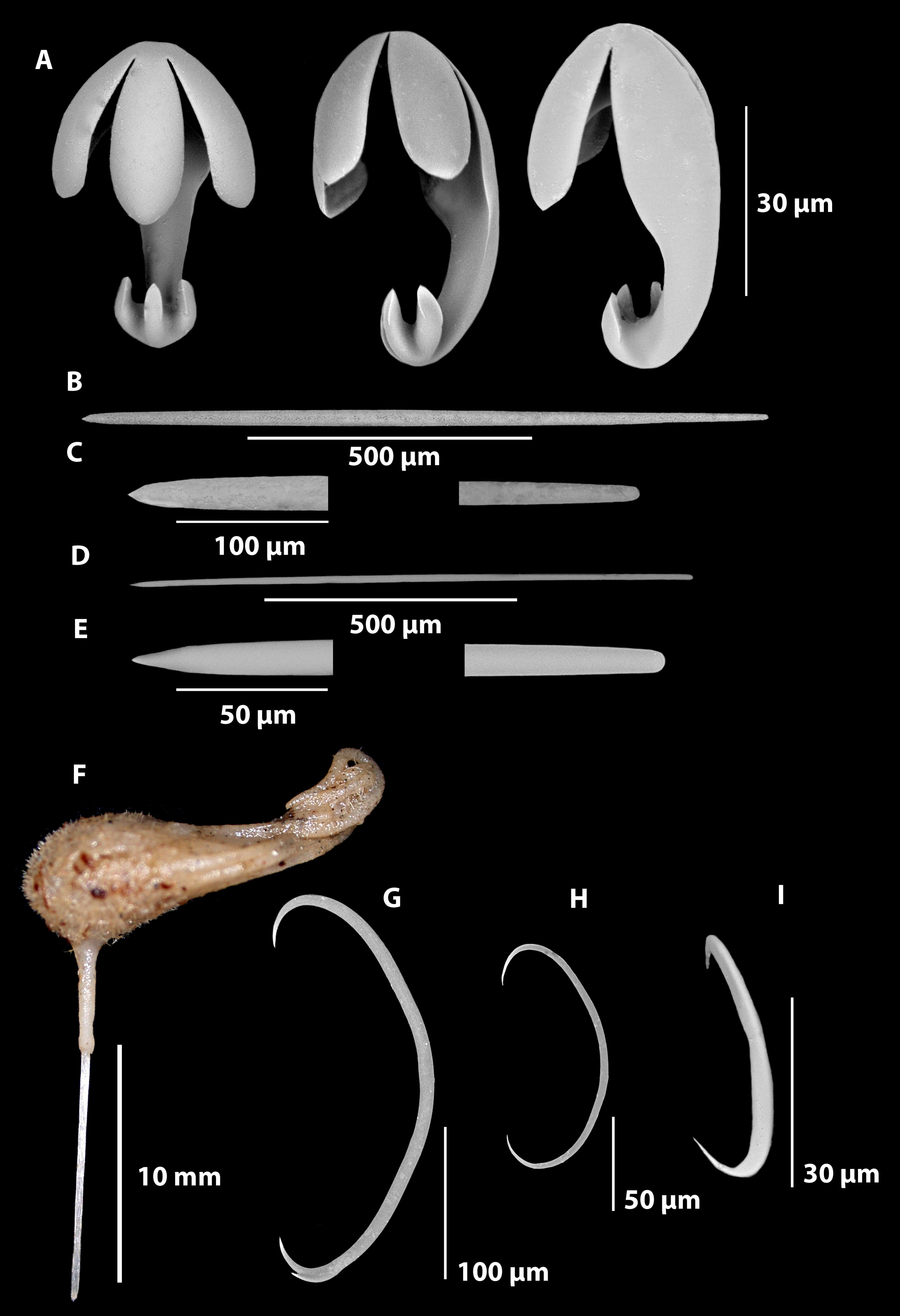
Growth form: An erect pedunculate unbranched growth form with an aerodynamically shaped-body ( Figure 14 F View FIGURE 14 ). The broken stem is 15 mm long and 0.7 mm wide, with basal attachment not collected. The main body is a horizontally aligned teardrop-shaped cushion 7 mm in length and width, accentuated by the medusoid-like tentacular filaments that trail in one direction only, as if the sponge was positioned in a strong unidirectional current. The six tentacular filaments are 15 mm long and 0.4 mm wide. The specimen did not have a basal attachment intact.
Colour: Tan in colour on deck and in ethanol.
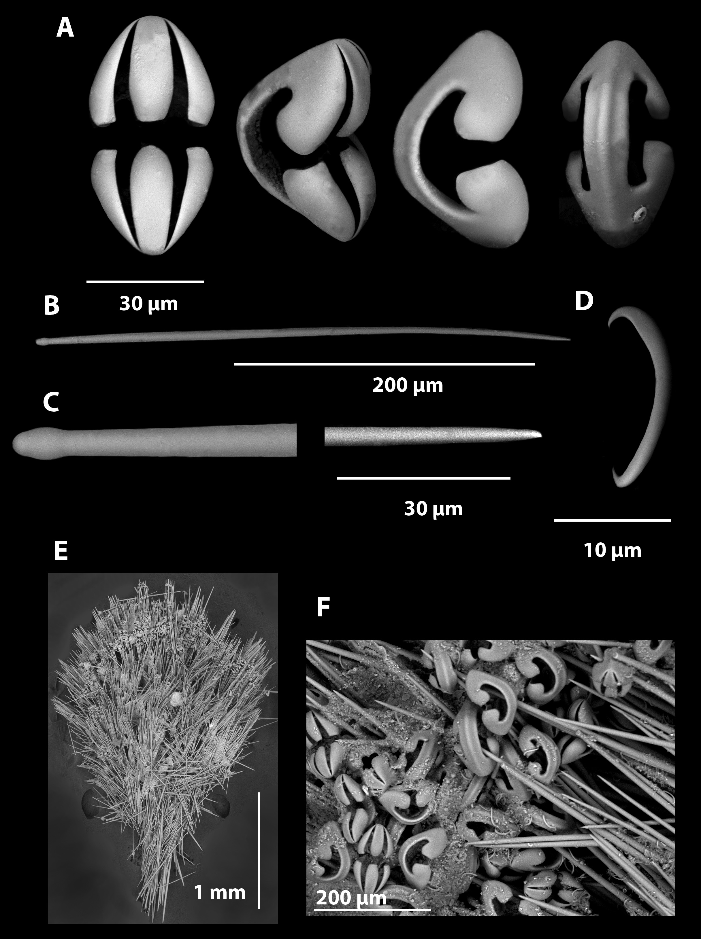
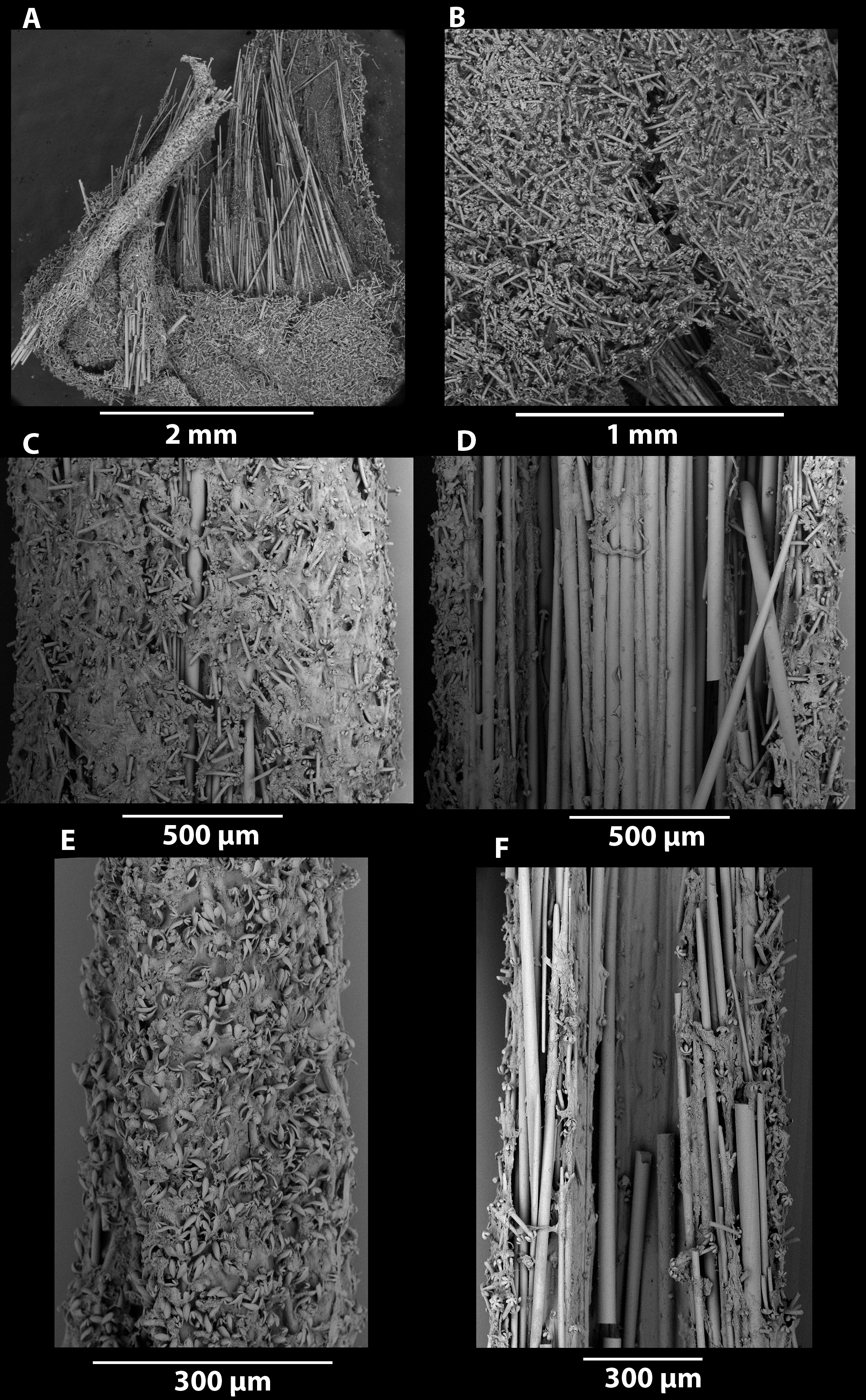
Ectosomal skeleton: The ectosome of the pedunculated body consists of soft tissue encrusted with anisochelae and sigmas, and penetrated by both sizes of the mycalostyles producing a hispid surface ( Figure 15 B View FIGURE 15 ). The ectosome of the stem is a very thick layer of anisochelae well packed perpendicular to the surface with only the large alae showing ( Figure 15 C View FIGURE 15 ). This ectosome also contains sigmas. The ectosome of the tentacular filaments consists of the perpendicular orientated anisochelae and sigmas ( Figure 15 E View FIGURE 15 ).
Endosomal skeleton: The endosomal skeleton of the stem consists of bundles of the large mycalostyles. The large mycalostyles from the stem extend into the main body along with the smaller mycalostyles where they form a thick radiating mass. Both types of mycalostyles are radially orientated that pierce the ectosome producing the hispid surface. Bundles of mycalostyles within the body also terminate into spherical bodies which may contain retracted filaments ( Figure 15A View FIGURE 15 ). The endosomal skeleton of the tentacular filaments originates in the body with both type of mycalostyles, but apically becomes dominated by the smaller mycalostyles.
Megascleres: Mycalostyles are of two different types, large with very obvious central thickening (860-(1527)- 2440 x 17.9-(27.8)-40.1 μm, n=89), and a thinner and usually much shorter mycalostyle, (318-(898)- 1120 x 8.2- (13.1)-17.3 μm, n=43).
Microscleres: Tridentate unguiferate arcuate anisochelae with spatulate alae on the head and canine-like alae on the foot (49.0-(55.0)-60.0 x 25.1-(31.5)-36.2 μm, n=88). Large sigmas (165-(241)-289 x 6.6-(10.3)-15.3 μm, n=89), medium sized sigmas with slightly more contortion (71-(109)-166 x 2.4-(4.2)-6.3 μm, n=67), and small sigmancis- tras (36.3-(43.7)-49.0 x 1.2-(2.5)-3.6 μm, n=46).
Molecular data: The 28S sequence of QM G337496 is provided in the Sponge Barcoding Database under accession number SBD#2309 and the molecular difference to other congenerics displayed in Figure 3 View FIGURE 3 .
Remarks: The external morphology of C. moniqueae sp. nov. is superficially similar to Chondrocladia (Chondrocladia) albatrossi Tendal, 1973 , from NE Brazil and Antarctica, as illustrated in Dressler-Allame et al. (2017, Fig. 4 View FIGURE 4 ). However, the spicule composition is very different between both species, with the present species having unguiferate anisochelae and belonging to Cladorhiza . As appears to be the case with many carnivorous sponges, the growth form shows convergent evolution to adapt to these abyssal environments, even if their spiculation is divergent.
The spatuliferous upper alae seen on the anisochelae of C. moniqueae sp. nov. have a similar morphology to those of C. diminuta Lopes & Hajdu, 2014 , from the mesophotic off central Brazil, although all of the spicules in the latter species, and indeed the sponge itself as the name suggests, are very diminutive compared to those of C. moniqueae sp. nov. ( Table 5 View TABLE 5 ).
Similarly, the shape of the alae on the anisochelae also resemble those of Cladorhiza tridentata Ridley & Dendy, 1887 , from the Antarctic and comprehensively illustrated by Dressler-Allame et al. (2017, Fig. 10 View FIGURE 10 ), but the lower alae of the anisochelae in the latter species are bifurcate (compared to canine-like in our new species). Other differences between the new species and C. tridentata include their very different growth forms (pedunculate versus encrusting, respectively), two size classes of sigmas, and presence of sigmancistras in C. moniqueae sp. nov. Cladorhiza schistochela Lévi, 1993 , from New Caledonia, which also has similar-shaped anisochelae, is cylindricallyshaped pedunculate, has much larger styles and anisochelae, smaller sigmas, and lacks sigmancistras. Cladorhiza nicoleae Castello-Branco et al., 2016 , from the mesophotic off central Brazil, also has similar anisochelae but they are much smaller than those of the new species, as are the sigmas and sigmancistras, and it has a spherical clubshaped body.


| QM |
Queensland Museum |
| RV |
Collection of Leptospira Strains |
No known copyright restrictions apply. See Agosti, D., Egloff, W., 2009. Taxonomic information exchange and copyright: the Plazi approach. BMC Research Notes 2009, 2:53 for further explanation.
|
Kingdom |
|
|
Phylum |
|
|
Class |
|
|
Order |
|
|
Family |
|
|
Genus |