
Microplana grazalemica, Vila-Farré, Miquel, Mateos, Eduardo, Sluys, Ronald & Romero, Rafael, 2008
|
publication ID |
https://doi.org/ 10.5281/zenodo.181468 |
|
DOI |
https://doi.org/10.5281/zenodo.5695778 |
|
persistent identifier |
https://treatment.plazi.org/id/03D287BA-FFFA-D313-85C7-F8AEFBD5FDDF |
|
treatment provided by |
Plazi |
|
scientific name |
Microplana grazalemica |
| status |
sp. nov. |
Microplana grazalemica sp. nov.
Material. Holotype, CRBA 438, CRBA 439, Llano del Berral (lat. 36.75428, long. -5.45399; alt. approx. 657 m) in the central part of the Sierra de Grazalema, Cádiz ( Spain), 5 December 2004, sagittal sections on 2 slides.
Other material examined: CRBA 440, CRBA 441, CRBA 442, Río Majaceite (lat. 36.77368, long. - 5.486587; alt. approx. 278 m) in the western part of the Sierra de Grazalema, Cádiz, 6 December 2004, sagittal sections on 3 slides.
Diagnosis. With respect to external features, M. grazalemica sp. nov. can be distinguished from its congeners by its size (12 mm), cylindrical body tapering anteriorly to a blunt point, bluntly pointed tail, brown dorsal surface with dark spots, and anterior end without conspicuous eyes. Anatomically the species is characterized by the presence of a large and rounded penis bulb, obliquely oriented conical penis papilla, a copulatory bursa not connected to the intestine and communicating with the atrium through a slightly obliquely orientated bursal canal.
Ecology and distribution. The species is known only from two localities in the Sierra de Grazalema.
Etymology. The specific epithet is based on the name of the mountain system from where the specimens were collected, i.e. the Sierra de Grazalema in Southern Spain.
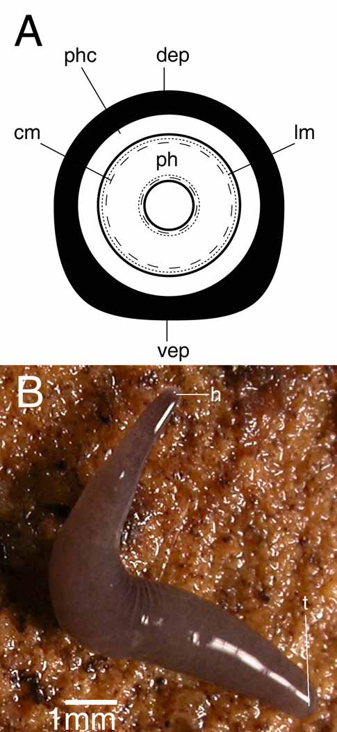
Description. In elongated state the living, sexually mature specimens measured 10–12 mm in length, with a width of about 1 mm ( Fig. 3 View FIGURE 3. A B). The preserved holotype specimen measured 3.45 x 0.68 mm. The cylindrical body tapers anteriorly and posteriorly to form blunt points. The dorsal surface is brown with darker spots all over the surface; the anterior end is dark brown. The anterior end of the body is slightly invaginated. The creeping sole is less than one-third of the body width.
The two small eyes (eye cup diameter 13–16 Μm in sections) are located at a short distance anterior to the brain and are only clearly visible under observation through a dissecting microscope. In CRBA440-442 a third reduced eye is present close to the anterior body margin.
The subepidermical longitudinal fibres of the body musculature are weak. In the ventral body region numerous longitudinal fibres are especially present over and under the ventral nerve cords. The scarce dorsal longitudinal parenchymal fibres are very weak and apparently discontinuous.
In specimen CRBA438-439, the cylindrical pharynx is about one-eight of the total body length (0.3 mm) and placed in a posterior horizontal position, while in specimen CRBA440-442 the pharynx represents about one-twelfth of the body length (0.4 mm) and is situated slightly posterior to the central region. The outer epithelium is ciliated and underlain by a layer of longitudinal muscles followed by a layer of circular muscles. Underneath the inner pharynx epithelium lies an outer layer of circular muscles, followed by an inner layer of longitudinal muscles fibres. The mouth is located in the middle of the pharyngeal pouch in specimen CRBA438-439 at 2.2 cm from the tip of the body, but in specimen CRBA440-442 it is situated in the posterior portion of the pharyngeal pocket, somewhat anterior to the hind wall of the pharyngeal pouch at 2.7 cm from the tip of the body. In specimens CRBA438-439 and CRBA440-442 the gonopore is situated at 0.23 and 0.6 mm from the mouth, respectively.
There are 13 to 15 testes situated on either side of the body (CRBA438-439 and CRBA440-442, respectively). The rounded or oval-shaped, irregularly sized follicles occupy approximately one-eight of the dorsoventral diameter and are arranged in ventral longitudinal rows, extending anteriorly from about the root of the pharynx up to two-fifth of the distance between the root of the pharynx and the ovaries.
The vasa deferentia open separately into the bulbar lumen, which communicates with an ejaculatory duct that opens at the tip of the penis papilla ( Fig. 4 View FIGURE 4 A). The bulbar lumen and the ejaculatory duct are lined with a nucleated epithelium that is underlain with a layer of circular muscle fibres.
The conical penis papilla has a relatively broad base. It has an oblique ventro-caudal orientation and in both specimens the tip projects into the bursal canal. The outer wall of the penis papilla is covered with a thin epithelium, which is underlain with a layer of circular muscles at its distal section; on the middle and more proximal parts of the papilla the circular muscle layer is considerably thicker and bounded by a layer of longitudinal muscles.
The genital atrium is lined with a nucleated epithelium that is underlain with a subepithelial circular muscle layer, followed by a longitudinal one.
The small ovaries are situated close to the ventral nerve cord, occupying about one-fifth of the dorso-ventral diameter. The ovaries are located at approximately one-third of the distance between the first testis and the brain. Somewhat posterior to the gonopore, the oviducts turn towards the middle of the body and open separately into the bursal canal.
The copulatory bursa is rounded and slightly flattened; it is lined with tall, vacuolated cells, with the nuclei mainly in peripheral position ( Fig. 4 View FIGURE 4 B). The bursa is connected with the atrium by means of a somewhat obliquely running bursal canal ( Fig. 5 View FIGURE 5 A, B). The lining epithelium of the canal bears long cilia and consists of nucleated cells surrounded by a subepithelial layer of circular muscle fibres. Shell glands could not be discerned.
Discussion. This species can be distinguished from the other European native land planarians by a combination of external features and anatomical characteristics of the genital apparatus.
The dorsal colouration pattern of M. grazalemica resembles that of M. nana and M. gadesensis sp. nov, the latter described below. However, in both M. nana and M. gadesensis a copulatory bursa is absent.
There are three European species that are provided with a copulatory bursa and lack a genito-intestinal duct, viz. M. mahnerti , M. styriaca , M. aixandrei . In M. mahnerti the bursal canal runs parallel to the body surface and the oviducts open at its most distal end. In contrast, in M. grazalemica the bursal canal is an obliquely running structure with the opening of the oviducts at its central region. ( Minelli, 1977). In M. styriaca the penis bulb is very large and elongated, while an expanded bulbar lumen is absent, in contrast to the rounded penis bulb from M. grazalemica that is provided with a distinct bulbar lumen. In M. styriaca the oviducts form a short common oviduct that enters the bursal canal ( Freisling, 1935), contrasting with the oviducts of M. grazalemica , which open separately into the bursal canal.
Differences between M. aixandrei and M. grazalemica were detailed above.


No known copyright restrictions apply. See Agosti, D., Egloff, W., 2009. Taxonomic information exchange and copyright: the Plazi approach. BMC Research Notes 2009, 2:53 for further explanation.
|
Kingdom |
|
|
Phylum |
|
|
Class |
|
|
Order |
|
|
Family |
|
|
Genus |