
Paratrizygia alvesi, Oliveira, Sarah Siqueira & Amorim, Dalton De Souza, 2010
|
publication ID |
https://doi.org/ 10.5281/zenodo.198297 |
|
DOI |
https://doi.org/10.5281/zenodo.6204454 |
|
persistent identifier |
https://treatment.plazi.org/id/03D287BE-B73E-FFB6-FF3B-FAD1F2874A1F |
|
treatment provided by |
Plazi |
|
scientific name |
Paratrizygia alvesi |
| status |
sp. nov. |
Paratrizygia alvesi View in CoL , sp. nov.
( Figs. 2 View FIGURE 2 , 4 View FIGURES 3 – 6 , 8 View FIGURE 8 , 9 View FIGURE 9 )
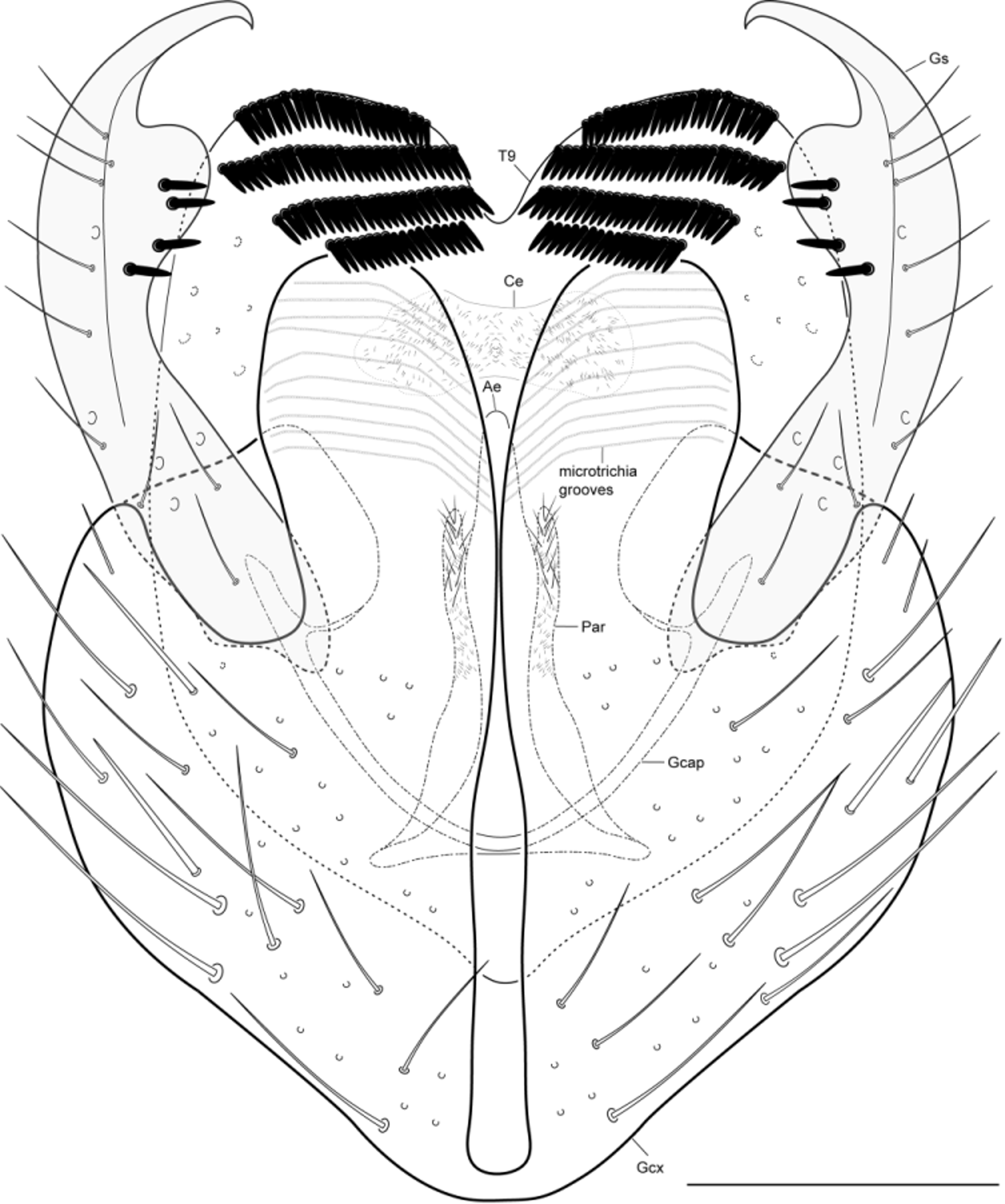
Diagnosis. Projections of gonocoxite rounded apically, with regular rows of microtrichia in grooves ( Fig. 8 View FIGURE 8 ). Gonostyle hook-shaped, with a rounded inner projection before apex, four dark spines apically. Aedeagus without a medial projection directed ventrally at base.
Material examined. Holotype ɗ, BRAZIL, State of Rio de Janeiro, Rio de Janeiro, Parque Estadual do Desengano, Malaise trap, 21°50’ S, 41°40’ W, 20–23.iv.2002 (BIOTA-FAPESP). Paratypes, 2 ɗ, 1 Ψ same data as holotype; 2 ɗ, 2 Ψ, same data as holotype, except 17–20.iv.2002; 5 ɗ, 5 Ψ, State of Minas Gerais, Presidente Olegário, Fazenda Gigante, 1,000 m, Malaise trap, 18°31’ S, 46°18’ W, 02–09.i.2010, Ribeiro, Amorim, Silva & Berbert cols. (BIOTA-FAPESP); 2 ɗ, 2 Ψ, Botelhos, Córrego da Onça, 21°40’90” S, 46°22’05” W, Malaise trap, 09.vii.2008, João Basso col. (BIOTA-FAPESP); 2 ɗ, 3 Ψ, same data, but 07.xii.2008 – 06.i.2009; 1 Ψ, same data, but 19.xi.2008 (BIOTA-FAPESP); 1 Ψ, State of São Paulo, Jundiaí, Serra do Japi, 23º11’11” S, 46º53’03” W, 22.x.1197 – 12.iv.1998, Amorim, Martins & Urso-Guimarães cols. 1 ɗ, State of Santa Catarina, São Bento do Sul, Cepa Rugendal, 26°15’01,0” S, 49°22’43,0” W, 13–19.x.2001, A.P. Dias et al. col. (BIOTA-FAPESP).
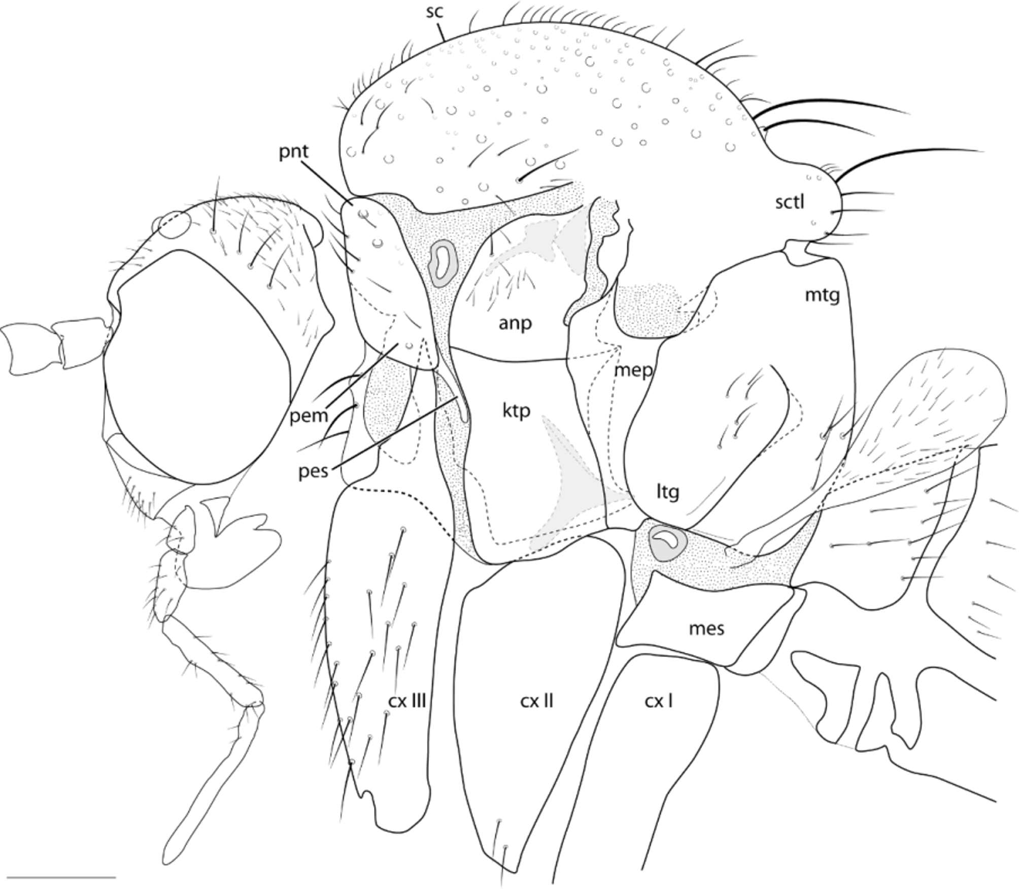
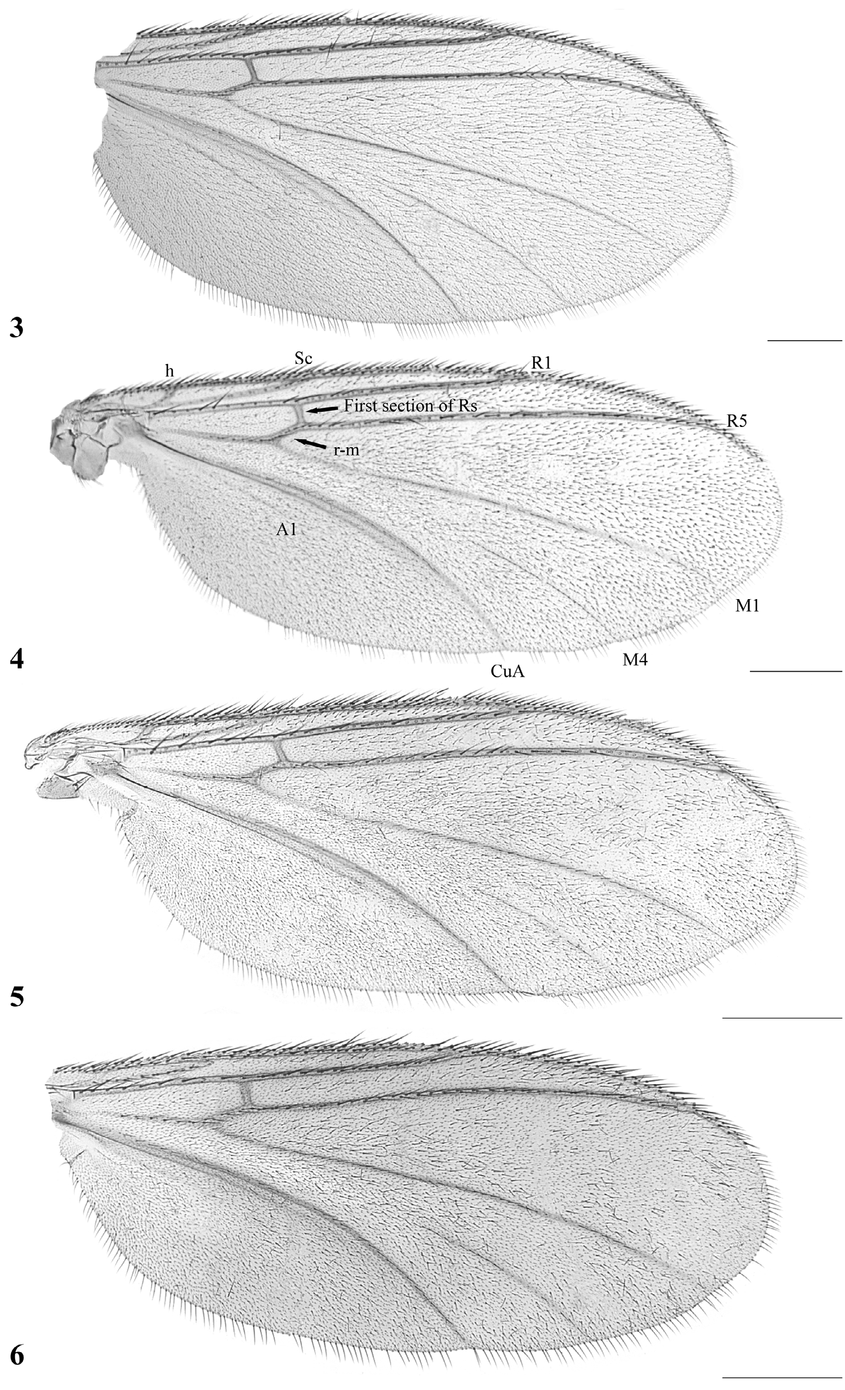
Description. Male. Head ( Fig. 2 View FIGURE 2 ). Vertex light-brown, with scattered setae. Three ocelli, mid ocellus smaller and slightly more ventral in position than lateral ones. Occiput brown. Eyes setose. Scape and pedicel yellow, with longer setae dorsally along apical margin; 14 brownish flagellomeres, almost twice longer than wide, with scattered setae and a short apical neck. Front and clypeus light-brown, covered with short setae; labella yellow; maxillary palpus yellow, five palpomeres, basal palpomere very small, apical ones increasingly longer, last palpomere almost twice penultimate. Thorax ( Fig. 2 View FIGURE 2 ). Scutum and scutellum brownish. Pleural sclerites brownish. Pleural membrane yellowish. Scutum moderately arched, covered with scattered small setae and stronger supra-alar, dorsocentral and acrostical. Scutellum with four scutellar bristles of slightly different sizes and many setulae. Pronotum densely setose, with some stronger setae. Anepisternum with some stronger setae and many setulae; katepisternum more or less squared ventrally. Mesepimeron reaching ventral margin of thorax, devoid of setae. Laterotergite only slightly projected, with 6–9 setae of different sizes, suture separating from mediotergite incomplete dorsally. Mediotergite slightly curved on profile, ventral half with a pair of patches with some longer setae and some setulae. Haltere whitish yellow, with some few setae on pedicel and more densely setose on knob. Coxae whitish yellow, femora, tibiae and tarsi yellow, darker to the apex. Mid and hind first tarsomere more than twice the length of second tarsomere; mid and hind tibiae and tarsi with erect darker short bristles along almost entirely length, those on hind tibia more or less aligned dorsally and laterally. Tibial spurs 1:2:2, about twice the length of tibial width at apex, internal spurs shorter. Tarsal claws with a larger apical tooth and a smaller, more basal one. Wing ( Fig. 4 View FIGURES 3 – 6 ). Length, 1.9 mm, width 0.8 mm. Membrane homogenously hyaline, no maculation; membrane densely covered with microtrichia on all cells, macrotrichia densely distributed, less so in the basal cells. Sc complete, ending in C just beyond base of Rs, setose, well sclerotized. C ending before wing apex, extending about a third the distance between R5 and M1. First sector of Rs almost transverse, devoid of setae, about as long as rm. R1 relatively long, reaching C on apical third of wing; R5 reaching C quite before wing margin, well sclerotized; r-m almost longitudinal, well sclerotized, setose. M1+2 unforked; a loose vein between medial and cubital inforked veins (possibly M4); CuA complete, well sclerotized. A1 incomplete, not produce on the apical half. All apical veins with dorsal macrotrichia. Abdomen. Abdomen brownish, setose. T8 short and wide, S8 slender, longer than wide, rounded apically. Terminalia brownish, conspicuous, quite elongate. Terminalia ( Fig. 8 View FIGURE 8 ). Gonocoxite setose, elongate, fused to each other only at ventral margin, with a pair of distinctive apical medial extensions, pointed outwards at apex, which on untreated specimens fold covering inner structures of the terminalia, projections of gonocoxite rounded apically, with regular rows of microtrichia on grooves; gonostyle hook shaped, with a rounded inner projection before apex, four dark spines apically; aedeagus slightly sclerotized, pointed at apex, with a pair of lateral extensions at base; parameres not strongly sclerotized, just lateral of aedeagus, with small macrotrichia apically; T9 long, setose, with four characteristic regular rows of spines at a apical fold facing ventrally; cerci medially fused, weakly sclerotized, covered with setulae.
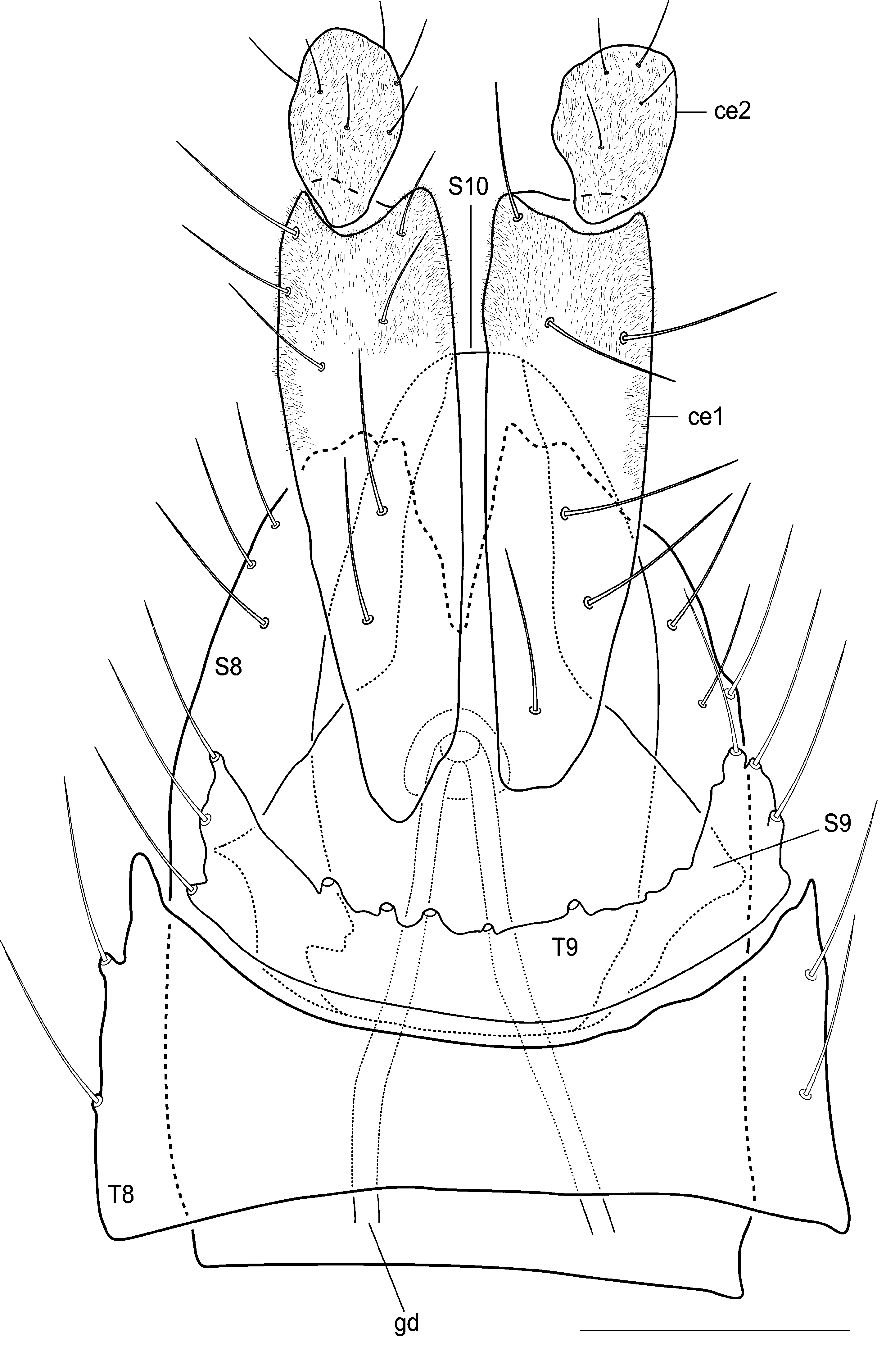
Female. As male, except for the following features. Wing length 2.1 mm, width 0.8 mm. Antennal flagellomeres not as elongate as the male, near each other. Terminalia ( Fig. 9 View FIGURE 9 ). Terminalia yellowish. A pair of weakly sclerotized spermathecae on segment 7. Sternite 8 elongated, a pair of gonapophyses apically separated by a short medial incision, covered with fine, elongated setae on apical margin; T8 wide, twice length of T9, with setae only at lateral margins; S9 (genital fork) without an anterior medial extension, a gonopore shortly connected anteriorly to a pair of ducts; T9 wide and short, setae emerging from digitiform projections along margin; S10 membranous, with microtrichia; T10 not visible, maybe fused to T9; Ce1 more than three times Ce2 length, covered with microtrichia and few scattered setae; Ce2 ovoid, covered with microtrichia and few setulae.
Etymology. The species is named after the collector, Mr. João Batista Alves, an exemplary gentleman, who has been extremely helpful with our Malaise trap collections.
Comments. There is some color variations between examined specimens. The antennal flagellomeres and thorax vary between brownish or light-brown. The specimens from Descalvado, in the state of Rio de Janeiro, are yellowish, differing from those of the state of Minas Gerais, which are much darker. Nevertheless, male and female terminalia from both localities are nearly identical, so with the data at hand we prefer to consider them a single species.
No known copyright restrictions apply. See Agosti, D., Egloff, W., 2009. Taxonomic information exchange and copyright: the Plazi approach. BMC Research Notes 2009, 2:53 for further explanation.
|
Kingdom |
|
|
Phylum |
|
|
Class |
|
|
Order |
|
|
Family |
|
|
Genus |