
Baeocera myrmidon (Achard, 1923)
|
publication ID |
https://doi.org/10.11646/zootaxa.3652.3.1 |
|
publication LSID |
lsid:zoobank.org:pub:6D53F9E2-70BC-4B69-B2B2-8C268C39FF49 |
|
DOI |
https://doi.org/10.5281/zenodo.6160719 |
|
persistent identifier |
https://treatment.plazi.org/id/03D287D3-FFB6-FFDB-808B-FA9FFDB232D5 |
|
treatment provided by |
Plazi |
|
scientific name |
Baeocera myrmidon (Achard, 1923) |
| status |
|
Baeocera myrmidon (Achard, 1923) View in CoL
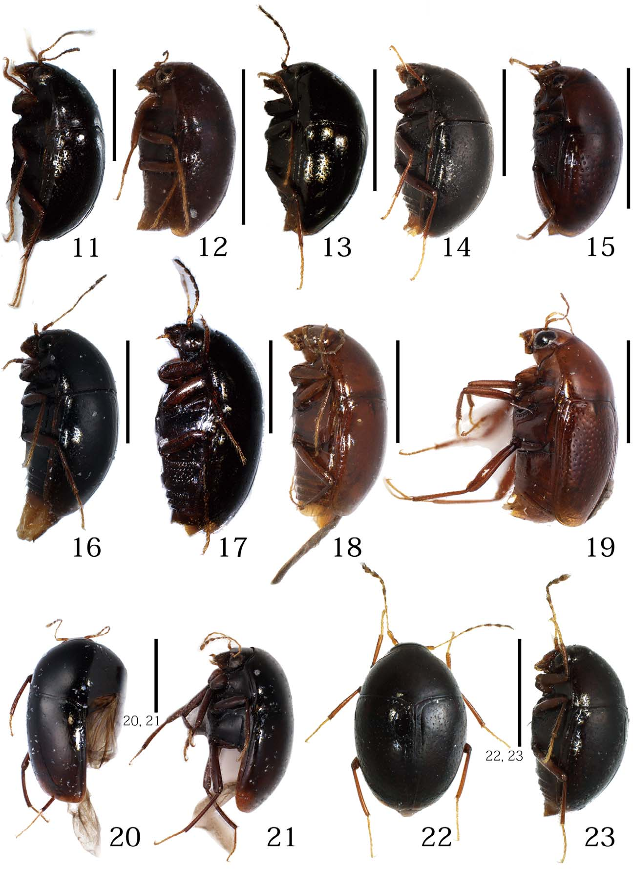
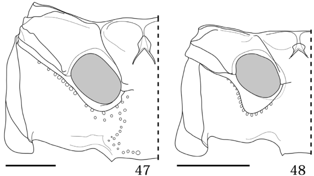
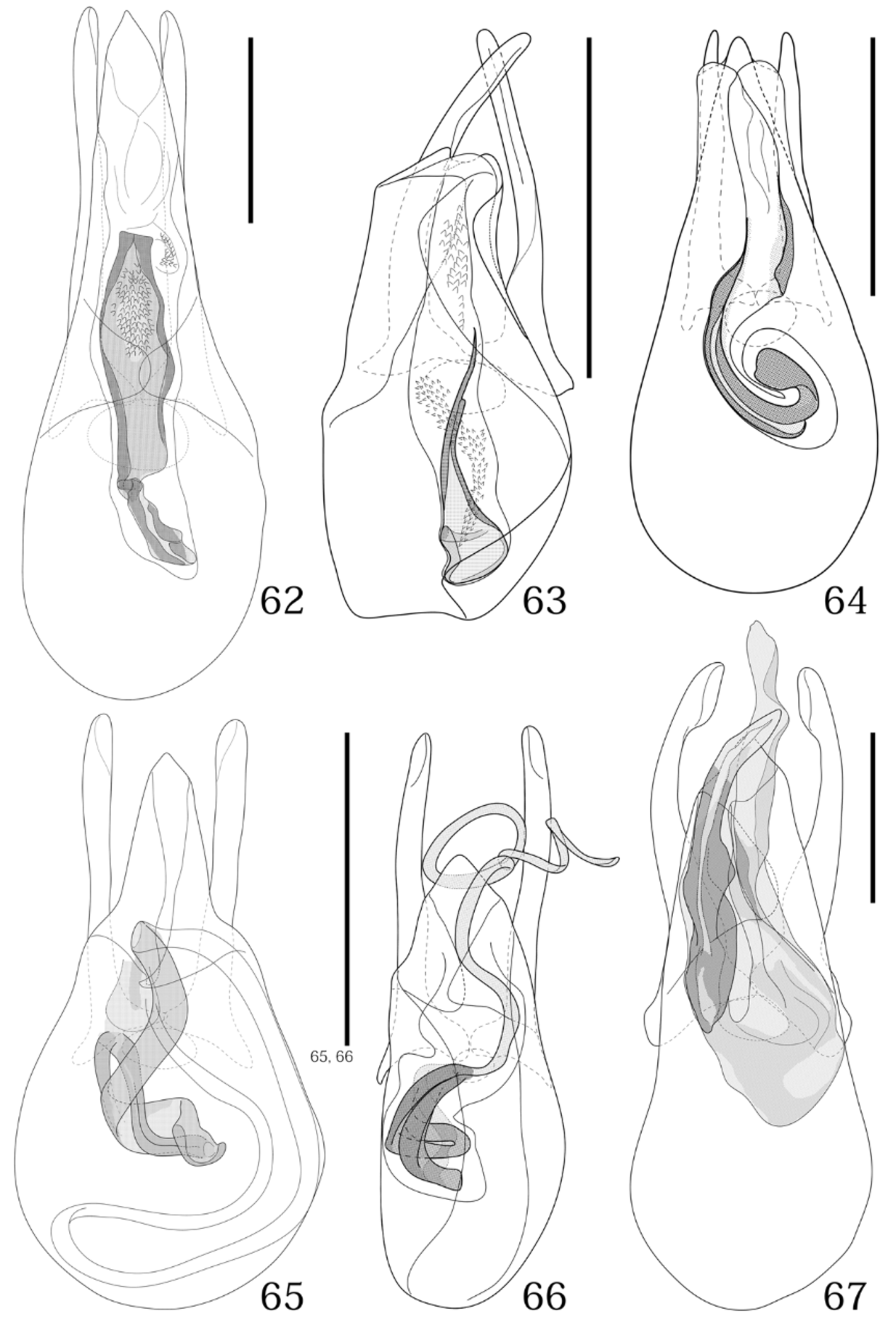
( Figs. 3 View FIGURES 2 – 10 , 12 View FIGURES 11 – 23 , 25, 48, 50, 63)
Scaphosoma myrmidon Achard, 1923: 116 ; Miwa & Mitono, 1943: 544. Scaphisoma myrmidon Nakane, 1955: 57 .
Baeocera myrmidon: Löbl, 1966: 1 ; Löbl, 1980: 94; Morimoto, 1985: 258. Eubaeocera myrmidon: Löbl, 1969: 332 .
Diagnosis. Antennomere XI about 2.5 times as long as III. Elytral punctuation sparse and coarse. Mesocoxal area broad. Lateral portion of metaventrite sparsely and finely punctuate. Ventrite I sparsely and coarsely punctuate. Parameres symmetrical; inner sac with a rod-like sclerite. Distal gonocoxite elongate and tapering apically.
Redescription. Body elongate-oval, shining. Pronotum and head brown; elytra dark brown; frons, clypeus, labrum, maxillary palpi, legs and antennomeres I–IV yellowish-brown; antennomeres V–XI dark yellowish-brown. Head, pronotum and elytra sparsely and finely pubescent.
Head with interocular distance about twice as eye width. Punctuation sparse and fine. Antennomeres I–VI with a few macrosetae, VII–XI covered with some macrosetae; each segments of IX and X about twice as long as III; XI about 2.5 times as long as III (Fig. 25).
Pronotum wider than long, lateral keel distinct except near basal angles. Punctuation sparse and fine, as on head. Scutellum wider than long, with exposed apex.
Elytra longer than wide, widest at basal fourth, lateral margins gradually narrowed apically, minutely serrate at inner part of posterior margin. Elytral disc sparsely and coarsely punctuate. Sutural striae extending outwards along basal margin to form complete basal stria joined to lateral striae.
Propygidium sparsely and coarsely punctuate; pygidium with 2 macrosetae sparsely and coarsely punctuate in basal half, finely punctuate from basal half to apex, and gradually narrower toward apex.
Hypomeron impunctate. Mesoventrite sparsely and finely punctuate, as pronotum. Lateral portion of metaventrite sparsely and finely punctuate. Mesepimeron about third as long as wide. Mesocoxa about twice as wide as interspace between mesocoxa; mesocoxal area broad ( Fig. 48 View FIGURES 47, 48 ). Metepimeron almost as long as wide. Metacoxa about four times as wide as metacoxal process. Metanepisternum twice as long as wide. Ventrite I sparsely and coarsely punctuate, with row of long basal wrinkles and 4 macrosetae; V with 8 macrosetae, sparsely and finely punctuate, and sinuate at apex.
Meso- and metafemora with microsculpture, sparsely and coarsely punctuate.
Male. Aedeagus about 0.28 mm long; parameres symmetrical; inner sac with a rod-like sclerite ( Fig. 63 View FIGURES 62 – 67 ).
Female. Distal gonocoxite elongate and tapering apically (Fig. 50); vagina membranous, without robust sclerites; bursa copulatrix not sclerotized.
Measurements (n = 2). Length (PL+EL): 1.30–1.45 mm; width (PW, EW): 0.65–0.75 mm, 0.74–0.75 mm. PL/PW: 0.80–0.88; EL/EW: 0.98–1.13. HW: 0.31–0.36 mm. ID: 0.14–0.16 mm.
Approximate ratio of each antennal length (width) as follows (n = 1); 2.0 (1.0): 1.7 (0.9): 1.0 (0.4): 1.3 (0.4): 1.4 (0.4): 1.3 (0.4): 1.5 (0.6): 1.4 (0.5): 2.0 (0.9): 2.0 (1.0): 2.6 (0.9).
Specimens examined. 13, Nakanoshima Is., Tokara Is., Kagoshima Pref., 1997, 13. VII. 1960, M. Sato leg.; 1Ƥ., Nakanoshima Is., Tokara Is., Kagoshima Pref., 17. VII. 1969, M. Sakai leg. (All preserved in EUMJ).
Distribution. Japan: Kyushu; Taiwan.
Comments. This species was not previously assigned to any species group. The male genitalia were illustrated by Löbl (1969). Löbl (1980) provided illustration of the male genitalia of a specimen from Taiwan that differs in the more elongate apical process of the median lobe.
FIGURES 49–57. Ovipositors. 49, B. frater ; 50, B. myrmidon ; 51, B. sordida ; 52, B. abnormalis ; 53, B. caliginosa ; 54, B. curtula ; 55, B. nakanei ; 56, B. satana ; 57 Baeoceroxidium micros . Abbreviations: DG, distal gonocoxites; GS, gonostylus; PG, proximal gonocoxites. Scales: 0.2 mm.

No known copyright restrictions apply. See Agosti, D., Egloff, W., 2009. Taxonomic information exchange and copyright: the Plazi approach. BMC Research Notes 2009, 2:53 for further explanation.
|
Kingdom |
|
|
Phylum |
|
|
Class |
|
|
Order |
|
|
Family |
|
|
SubFamily |
Scaphidiinae |
|
Tribe |
Scaphisomatini |
|
Genus |