
Emmenomma Simon
|
publication ID |
https://doi.org/ 10.11646/zootaxa.3931.3.3 |
|
publication LSID |
lsid:zoobank.org:pub:13BA1A35-C31A-4910-8EAC-1D840C5CAD18 |
|
DOI |
https://doi.org/10.5281/zenodo.6115507 |
|
persistent identifier |
https://treatment.plazi.org/id/03D287E3-FFD3-7F5D-FF28-FAE14847FB3B |
|
treatment provided by |
Plazi |
|
scientific name |
Emmenomma Simon |
| status |
|
Emmenomma Simon, 1884: 126 View in CoL , pl. 3, figs. 8–11 (type-species by monotypy Emmenomma oculatum View in CoL ); Lehtinen, 1967: 232; World Spider Catalog, 2014.
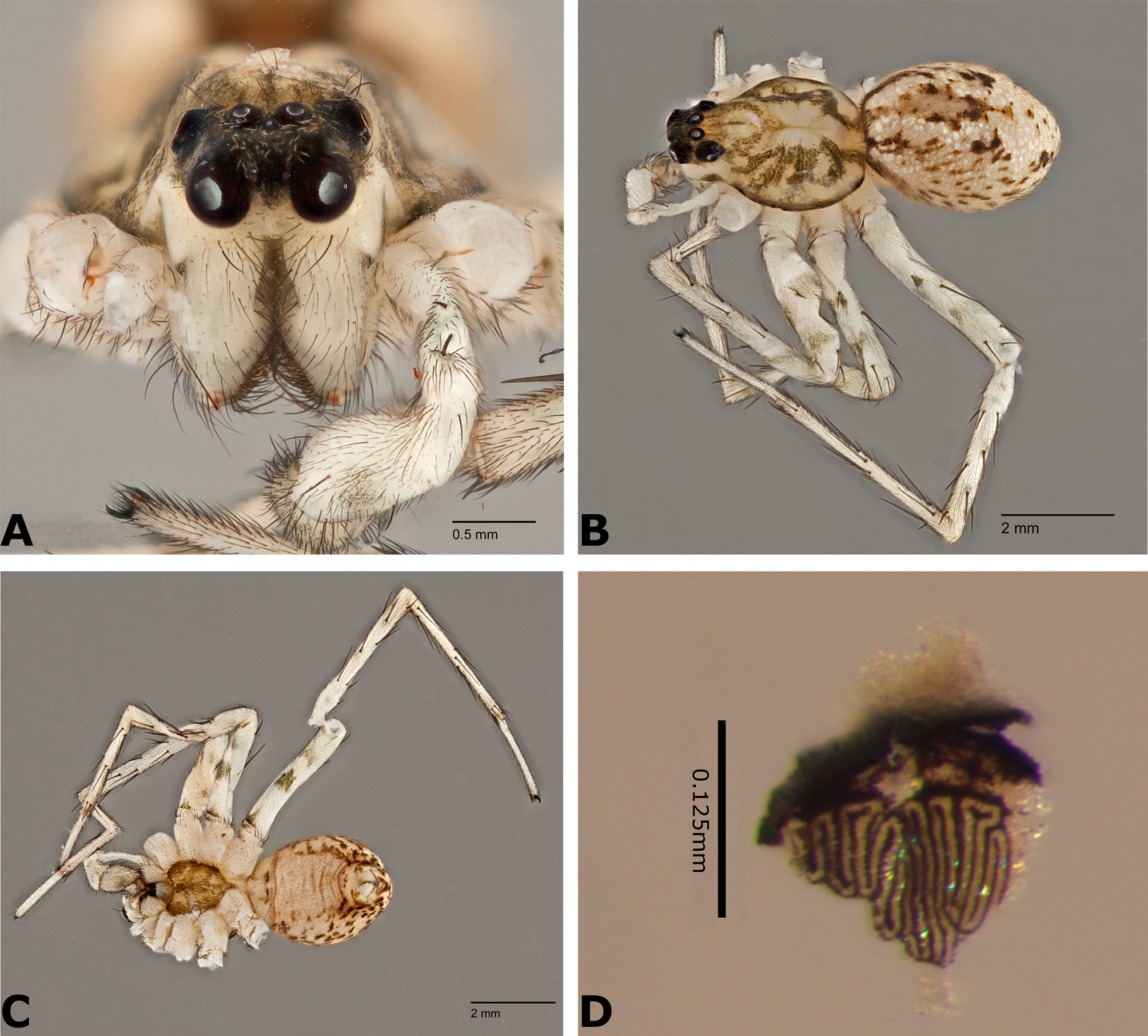
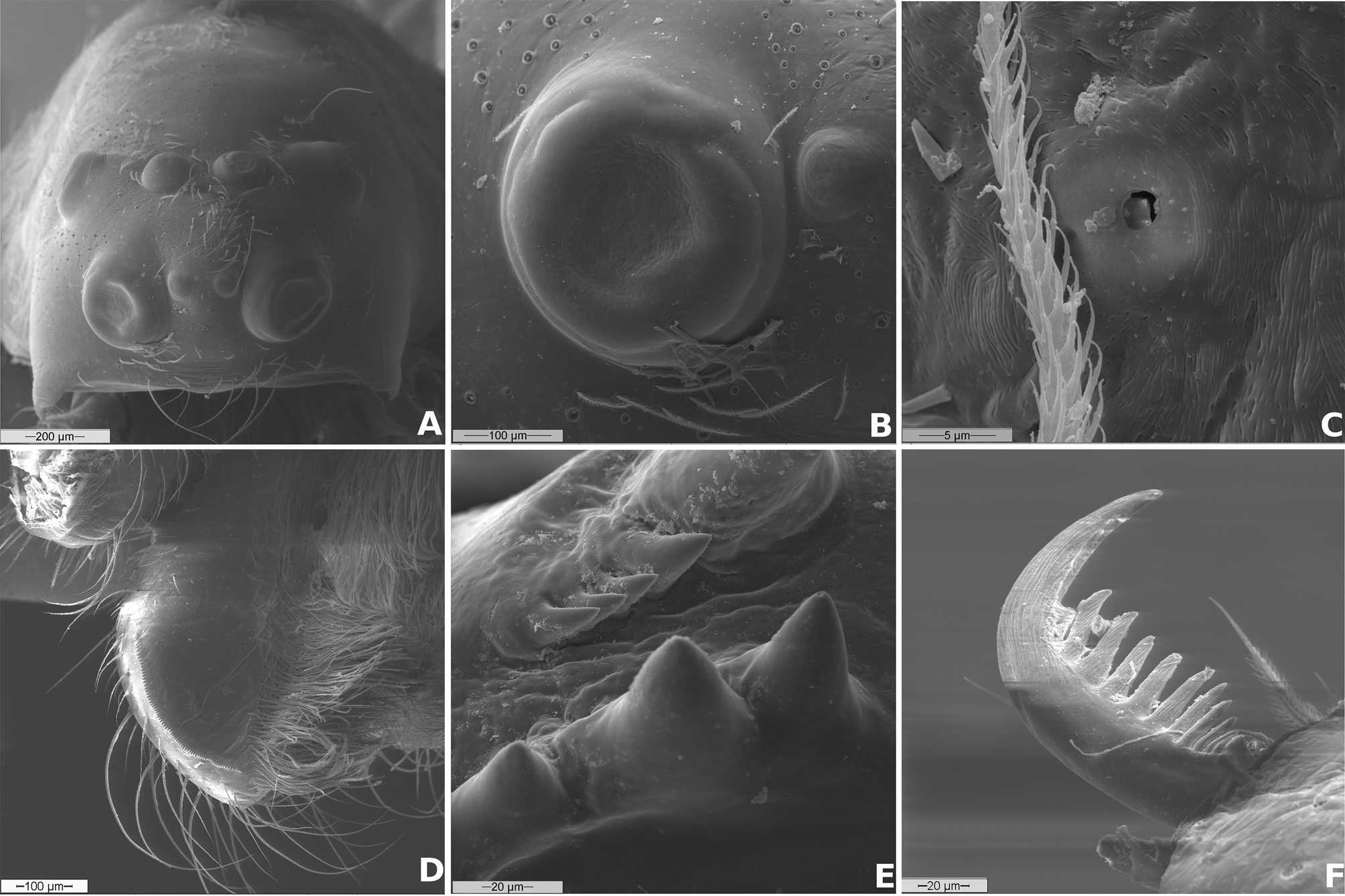
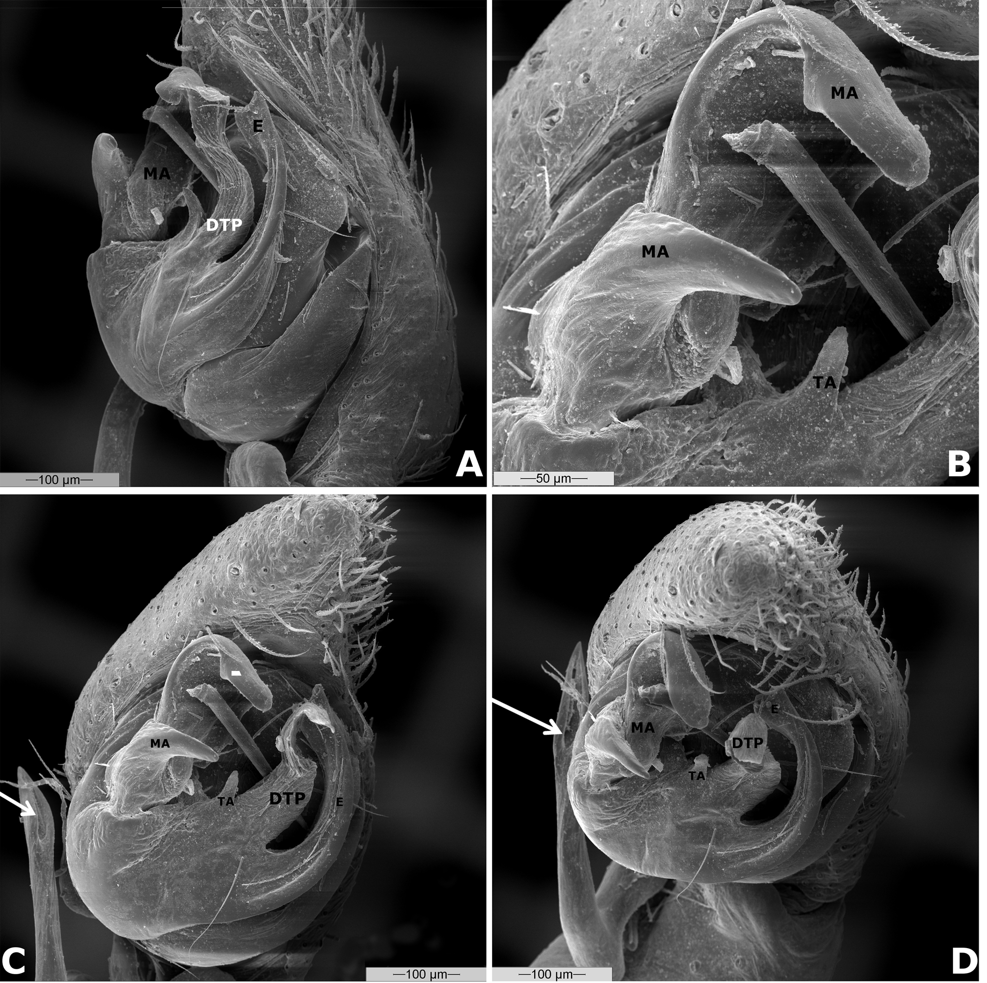
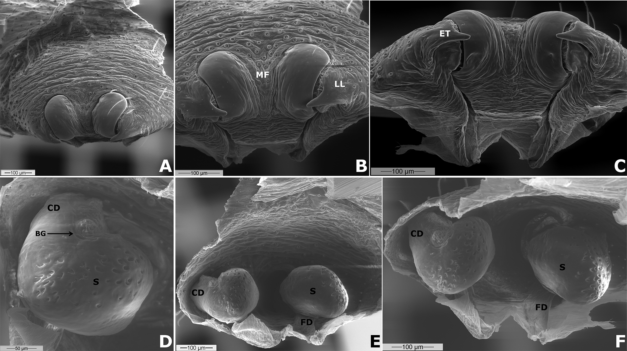
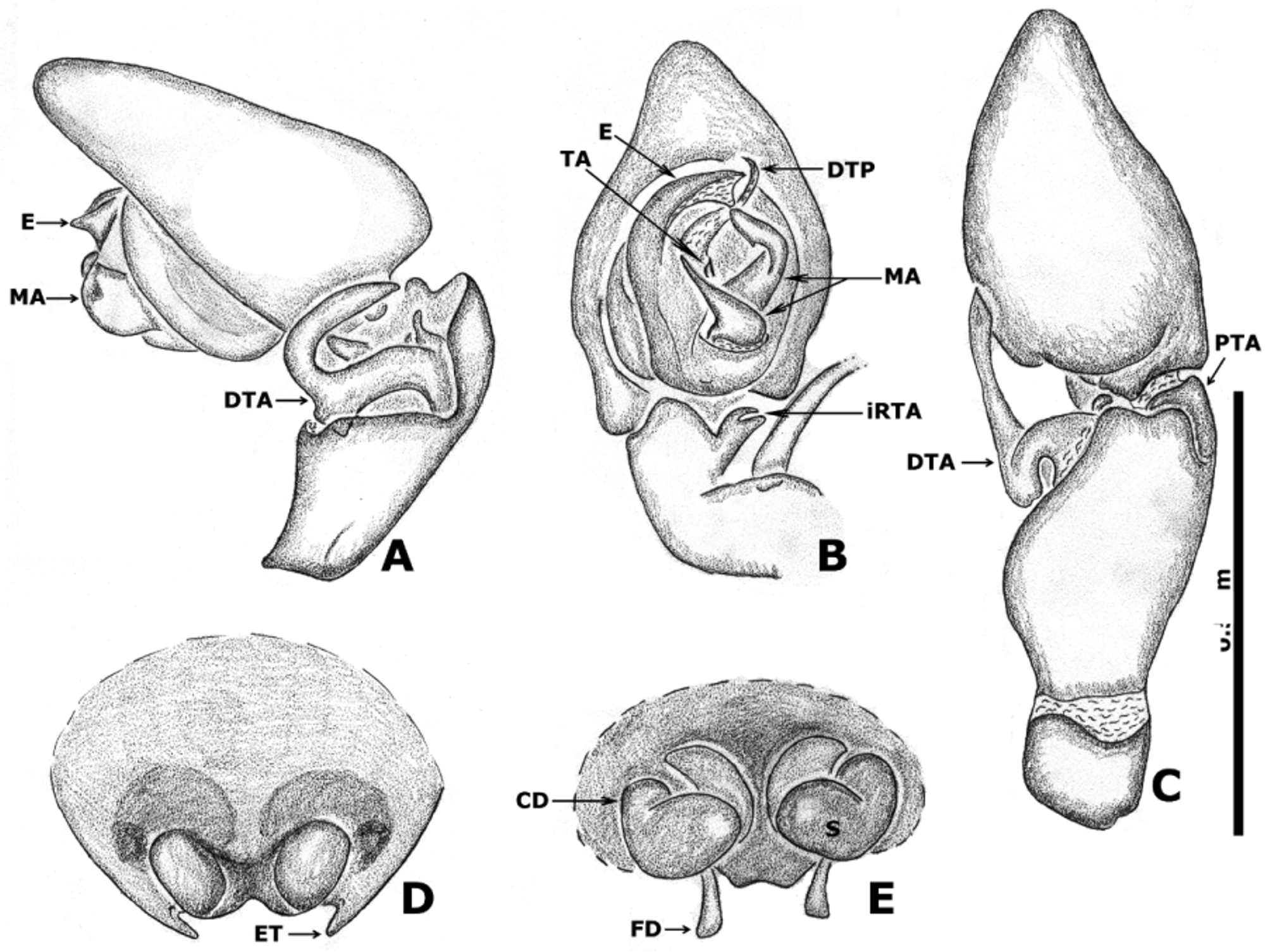
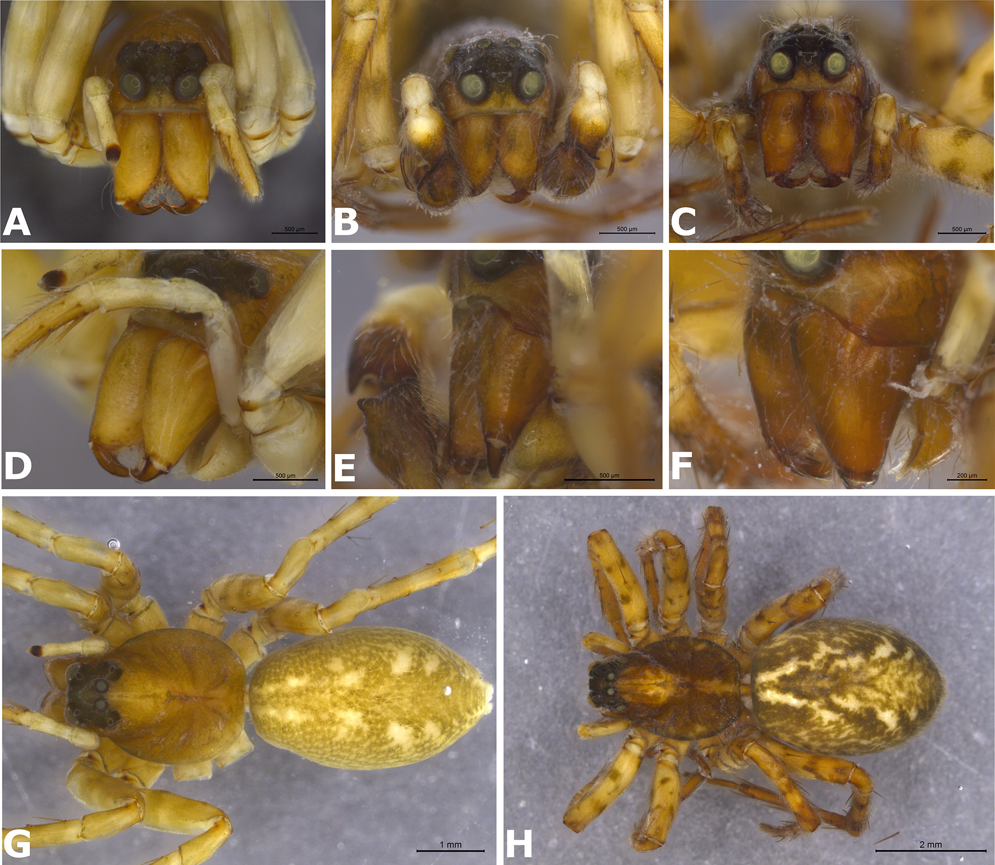
Diagnosis. Emmenomma differs from the non-macrobunine amaurobiids by the presence of denticles on the proximal end of the retromarginal row of teeth of the chelicerae ( Fig. 2 View FIGURE 2 E). Emmenomma can be recognized among other species of Macrobuninae by the recurved posterior eye row ( Fig. 1 View FIGURE 1 A), enlarged ALE and PLE ( Figs. 1 View FIGURE 1 A; 2A–B), and grate-shaped tapetum in all indirect eyes ( Fig. 1 View FIGURE 1 D). The males resemble Anisacate and Naevius by the presence of a gland in the male palpal tibia which discharges through an opening on the DTA ( Figs. 3 View FIGURE 3 C–D), but differ from Anisacate by having the DTA flexibly attached and mobile ( Figs. 4 View FIGURE 4 A–B) and from Naevius by the presence of a bifid MA ( Fig. 3 View FIGURE 3 B), by the elbowed DTA and RTA not ventrally displaced ( Fig. 4 View FIGURE 4 B). Females differ from all Macrobuninae by having an ET at the posterior end of lateral lobe of the epigynum ( Figs. 5 View FIGURE 5 B; 8A; 9D).
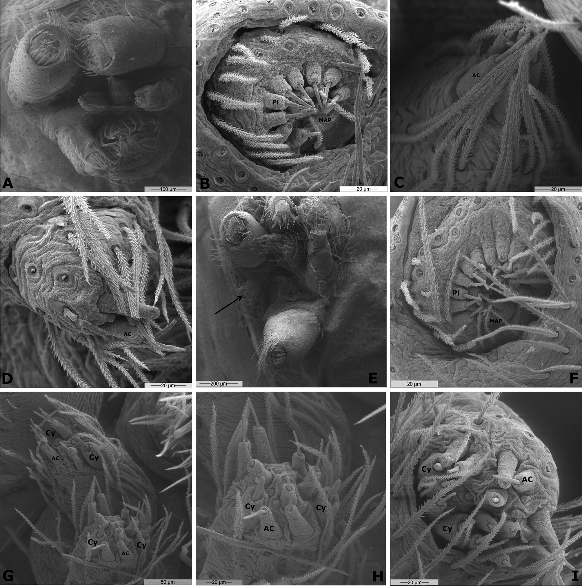
Description. Total length: 4.90–15.00. Anterior eye row slightly procurved and posterior eye row recurved; ALE enlarged, approximately twice the size of PME; AME reduced, 1/2 size of PME; PLE and PME about same size. All indirect eyes with grate-shaped tapetum. Thoracic fovea long and shallow in E. obscurum and E. joshuabelli sp. nov. and deep in E. oculatum . Coloration variable between pale and dark brown, with scattered guanine deposits in the abdomen, legs usually maculate ( Figs. 1 View FIGURE 1 A–C). Chilum absent except in females of E. oculatum where it is divided. Clypeal hood absent in E. oculatum , present in E. joshuabelli sp. nov. and E. obscurum ( Figs. 10 View FIGURE 10 A–F). Chelicerae geniculate only in E. obscurum ( Fig. 10 View FIGURE 10 A, D) and E. oculatum . Cheliceral promargin with four teeth, retromargin with variable number of teeth and denticles ( Fig. 2 View FIGURE 2 E), see details in species description. Labium slightly wider than long. Sternum longer than wide, projected between legs IV or not. Trochanter shallowly notched. Tracheal spiracle approximately as wide as colulus. Median tracheae branched and laterals simple. Ecribellate, colulus entire, wider than long, width approximately 1/4 of the distance between the outer margin of ALS ( Fig. 7 View FIGURE 7 E). PLS slightly smaller than ALS but longer than PMS. Male palp with triangular cymbium, longer than wide, cymbial apophysis present only in E. joshuabelli sp. nov. MA bifid with a triangular branch and a flat, almost rectangular, concave branch. E rising at prolateral side of bulb. TA present, located between DTP and MA. DTP membranous. DTA elbowed and retrolaterally projected with an opening for the tibial gland. RTA reduced, laminar, longer than wide and strongly sclerotized. iRTA bifid, with an external triangular and an internal laminate branch ( Figs. 6 View FIGURE 6 B; 9B–C). VTA distally rounded. PTA small and subtriangular, interacting with a small cymbial, prolateral projection ( Figs. 4 View FIGURE 4 A; 8A–C). Female epigynum with teeth and well-developed posterior plate ( Figs. 5 View FIGURE 5 B–C; 6D; 8C; 9F). CD wall heavily sclerotized ( Fig. 5 View FIGURE 5 D–F; 6E; 8B; 9F). Spermathecae oval to mushroom-shaped ( Fig. 5 View FIGURE 5 D–F; 6E; 8B; 9E).
Remarks. A circular region of closely spaced pores, slightly everted, can be found on the distal end of the copulatory duct ( Fig. 5 View FIGURE 5 D) of E. oculatum and seems to be compatible with what Ramirez (2014: 270) called Bennett’s gland (BG). Although there is not enough evidence to corroborate or refute the homology between those features we decided to call this porous area a BG so it will be noted in further studies.
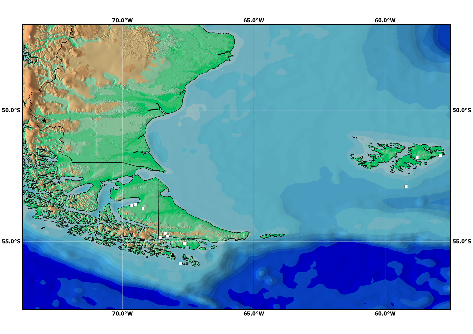
Distribution. Falkland Islands ( Malvinas), southern Chile and Argentina including islands around Tierra del Fuego—see distribution map ( Fig. 11 View FIGURE 11 ).
Composition. Emmenomma oculatum Simon, 1884 ; E. obscurum Simon, 1905 sp. reval., new rank; E. joshuabelli Almeida-Silva, Griswold & Brescovit sp. nov.

No known copyright restrictions apply. See Agosti, D., Egloff, W., 2009. Taxonomic information exchange and copyright: the Plazi approach. BMC Research Notes 2009, 2:53 for further explanation.
|
Kingdom |
|
|
Phylum |
|
|
Class |
|
|
Order |
|
|
Family |
Emmenomma Simon
| Lina, Almeida-Silva, Charles, Griswold & Antonio, Brescovit 2015 |
Emmenomma
| Lehtinen 1967: 232 |
| Simon 1884: 126 |