
Simulium (Aspathia) sandyi Coscarón, Ibáñez-Bernal & Coscarón-Arias
|
publication ID |
https://doi.org/ 10.5281/zenodo.174690 |
|
DOI |
https://doi.org/10.5281/zenodo.6258327 |
|
persistent identifier |
https://treatment.plazi.org/id/03D2C75F-7014-BC5B-FEC7-30D3FE9DFE97 |
|
treatment provided by |
Plazi |
|
scientific name |
Simulium (Aspathia) sandyi Coscarón, Ibáñez-Bernal & Coscarón-Arias |
| status |
|
Simulium (Aspathia) sandyi Coscarón, Ibáñez-Bernal & Coscarón-Arias View in CoL
Figs. 2 View FIGURES 1 – 12 P–R; 9 C, D
Simulium (Simulium) sandyi Coscarón, Ibáñez-Bernal & Coscarón-Arias, 1999: 577 View in CoL –578,
Male (based on 1 pharate male). Head holoptic. Antenna dark, with scape, pedicel, and first flagellomere longer than remaining flagellomeres. Palpomere III darker than other palpomeres, covered with black setae; palpomere V about twice as long as palpomere IV, and 1.5X longer than palpomere III; palpomere proportions: 1.0: 1.6: 3.7: 3.0: 7.1; sensory vesicle 0.33X length of its segment ( Fig.1 View FIGURES 1 – 12 ).
Foreleg: Coxa and trochanter dark; proximal and middle sections of femora pale, distal section dark; tibiae with proximal and distal sections dark and midsection pale; tarsomere dark. Midleg: similar to foreleg, except proximal section of first tarsomere pale. Hindleg: similar to midleg. All legs covered with black setae. Calcipala apically rounded, reaching proximal margin of deep pedisulcus ( Fig. 2 View FIGURES 1 – 12 ). All tarsal claws slightly curved from base and with basal tooth. Wing membrane hyaline; C and Sc with thin and stout setae along their entire length; basal sector of R bare.
Terminalia: Gonocoxite wider than long, conically produced posterolaterally. Gonostylus longer than gonocoxite, apex rounded with apical spinule, and with internal basal process. Aedeagal membrane with thin hairs at middle and marginal short and stout spines at each side ( Fig. 3 View FIGURES 1 – 12 ). Ventral plate in dorsoventral view triangular, broader than long, with posterior margin shallowly concave and anterior margin triangularly projected, forming lip that is densely covered with setae; distal portions of apodemes broad with external margins parallel ( Fig. 4 View FIGURES 1 – 12 ).
Female (based on 2 pharate females). Frons wide, with decumbent and erect setae. Antennae dark; first flagellomere broader than long, remaining flagellomeres subequal in length and width, except last conical flagellomere. Mandible with 24–28 serrations. Lacinia with 28–32 retrorse teeth. Palpus pale, except palpomere III darker; palpomere V about 2.0X longer than IV, and 1.5X longer than III, all with black setae. Palpomere proportions: 1.0: 1.75: 3.62: 3.0: 6.75 ( Fig. 5 View FIGURES 1 – 12 ). Sensory vesicle about 0.5X as long as palpomere III ( Fig. 6 View FIGURES 1 – 12 ). Basal portion of cibarium smooth and without median process.
Thorax dark; scutum, when posteriorly illuminated, with silvery pollinosity covering posterior third, and extending to front as thin lateral stripes that reach pronotal lobes, as well as pair of dorsocentral capitate stripes that reach level of posterior border of pronotal lobe, and short median pointed stripe that reaches center of scutum ( Fig. 7 View FIGURES 1 – 12 ); postnotum grayish. Leg-color pattern similar to that of male. Calcipala rounded, reaching proximal margin of deep pedisulcus ( Fig. 8 View FIGURES 1 – 12 ). All tarsal claws slightly curved from base and with basal tooth. Wing and wing veins as in male.
Terminalia: Hypogynial valve short, broad, and pointed distally ( Fig. 9 View FIGURES 1 – 12 ). Cercus nearly rectangular, hind margin truncated; anal lobe shorter than high, with posterior margin concave and ventral margin rounded, forming posteroventral triangular projection; both structures covered with setae ( Fig. 12 View FIGURES 1 – 12 ). Genital fork stem long and slender, about 1.5X as long as arms; arms forming nearly triangular space between them, with triangular sclerotized toothlike process on anterior margin ( Fig. 11 View FIGURES 1 – 12 ). Spermatheca spherical, smooth and without internal spicules, junction with spermathecal duct membranous ( Fig. 10 View FIGURES 1 – 12 ).
Pupa. Cocoon length 3.5–4.3 mm ( Fig. 14 View FIGURES 13 – 18 ). Cocoon loosely woven, with sediment particles incorporated, slipper shaped, and with variable anterodorsal projection ( Figs. 13, 14 View FIGURES 13 – 18 ).
Cephalothoracic region with many minute tubercles, giving roughened appearance ( Figs. 14, 15, 17 View FIGURES 13 – 18 ). Frontoclypeus with 2+2 frontal and 1+1 facial trichomes ( Fig. 15 View FIGURES 13 – 18 ). Gills brownish, about same length as pupa, consisting of 25–31 membranous terminal filaments arising from 3 branches originated from short trunk; filaments moderately thin, surface with striations and minute spicules, the latter more abundant near base ( Fig. 16 View FIGURES 13 – 18 ). Thorax with numerous small tubercles, each side of thorax with 16–20 dorsal trichomes, 3 on prealar lobe, 2 on supralar area, and without stigmal trichomes ( Fig. 17 View FIGURES 13 – 18 ).
Chaetotaxy of abdominal tergites as follows ( Fig. 18 View FIGURES 13 – 18 , right): I with 5+5 setae, II with 8+8 setae, III with 2+2 setae and 4+4 simple hooks, IV with 1+1 setae and 4+4 simple hooks, V with 3+3 setae, VI with 2+2 setae, VII and VIII with 1+1 setae, IX without setae but with 2 short and robust caudal spines. Chaetotaxy of abdominal sternites as follows ( Fig. 18 View FIGURES 13 – 18 , left): III and IV with 2+2 setae, V with 2+2 setae and 2+2 bifid hooks, VI and VII with 1+1 setae and 1+1 bifid hooks, VIII and IX without setae or hooks. Striated pleural membrane chaetotaxy as follows ( Fig. 18 View FIGURES 13 – 18 , midline): II with 1+1 setae, III and IV with 4+4 setae, V with 5+5 setae, VI with 3+3 setae, VII with 2+2 setae, VIII with 2+2 setae, and IX without setae; all setae simple and thin.
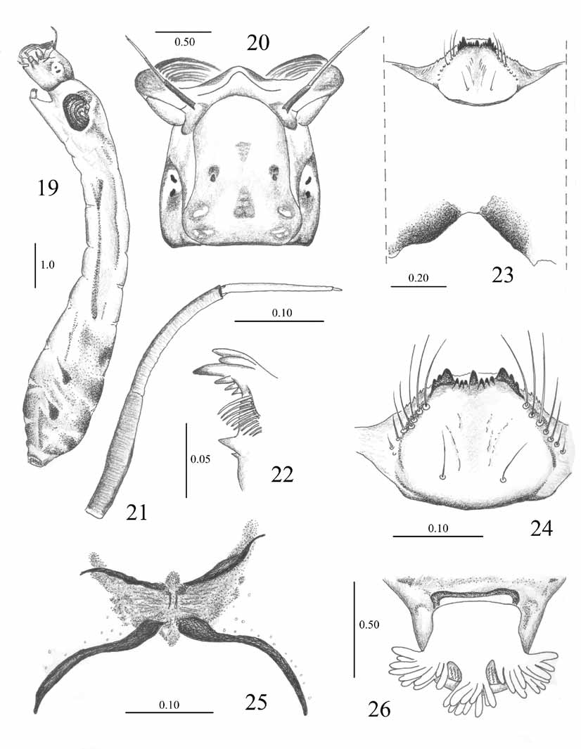
Larva. Length 8–9 mm. General body color pale grey to brown, pigmentation heaviest on dorsum of segments V–VII; abdomen gradually expanded posteriorly ( Fig. 19 View FIGURES 19 – 26 ). Head capsule pale yellowish to brown; cephalic apotome negative ( Fig. 20 View FIGURES 19 – 26 ). Antenna longer than stalk of cephalic fan, with transverse striations on proximal and median articles; proximal and distal articles pale brown and median article pale yellowish; proportions of articles (excluding apical sensillum): 1.00: 1.12: 0.87 ( Fig. 21 View FIGURES 19 – 26 ). Labral fan with 22–28 primary rays. Mandible with 1 large apical tooth, 2 outer teeth, 3 preapical teeth followed by series of 9–11 inner setalike teeth and 2 basal teeth ( Fig. 22 View FIGURES 19 – 26 ). Hypostoma with 13 teeth, median and lateral teeth subequal in length, 2+2 paralateral and 3+3 sublateral teeth; 6 or 7 lateral serrations and 4–6 small and 2 or 3 prominent setae at each side ( Fig. 24 View FIGURES 19 – 26 ). Postgenal cleft triangular to trapezoidal; hypostomal bridge distinctly longer than gular cleft ( Fig. 23 View FIGURES 19 – 26 ), gular cleft depth: hypostomal bridge length 25: 54. Cervical sclerites small and free.
Thoracic proleg with circlet of apical hooks arranged in about 17–20 rows with 11–15 hooks per row. Lateral plate of proleg trapezoidal, with thin and long spines on its distal margin.
Anal sclerite X shaped, with anterodorsal arms shorter and broader than posteroventral arms; peripheral cuticle of anal plate without setae, except for some short setae near posteroventral arms ( Fig. 25 View FIGURES 19 – 26 ). Anal papilla with 3 lobes, each giving rise to 9–12 branches ( Fig. 26 View FIGURES 19 – 26 ) Posterior circlet of hooks consisting of about 13–15 hooks in about 80–96 rows.
Material examined. Two pharate females and their pupal exuviae: Mexico, limits between Estado de Mexico and Morelos, Lagunas de Zempoala National Park (19º02’51.57” N, 99º19’1.26” W, 2841 m a.s.l.), August 11, 2006, Sandoval-Ruíz, coll. (mounted on slides except thorax of 1 pinned); 1 pharate male and its pupal exuviae: same data (mounted on slide); 3 pupae: same data (in ethanol); 3 pupae: same data, except September 19, 2005 (mounted on slides); 5 larvae (mounted on slides); 8 larvae: same data, except August 11, 2006 (1 mounted on slide, remaining material in ethanol).
Biological notes. All specimens of S. sandyi came from a small, shallow, cold, clean stream in an area partially shaded by forest and with a moderate amount of grass on the banks. The stream is about 1 m wide, with water flowing slowly. Immature stages were attached to stones and grasses in association with Simulium marquezi Vargas & Díaz- Nájera, 1957, S. capricorne De León, 1945 , and Gigantodax wrighti Vargas , Martínez- Palacios & Díaz-Nájera, 1944.
Comments. The type locality given in the original description by Coscarón et al. (1999) was Lagunas de Zempoala, “Distrito Federal (sic)”, Mexico. This location, however, is a mistake, because Lagunas de Zempoala is a National Park situated between the states of Morelos and Estado de Mexico, not in Distrito Federal. All specimens here reported come from the type locality (19º02’51.57” N, 99º19’1.26” W, 2841 m a.s.l.) of this species.
The female of S. sandyi is similar to those of S. marquezi , S. piperi Dyar & Shannon, 1927 , and S. tricorne De León, 1945 , having the same general pattern of the scutum and a smooth cibarial margin. The female of S. marquezi , however, is distinguished by the quadrangular anal lobe. The female of S. piperi has the anal lobe similar to that of S. sandyi , but can be distinguished by the distally rounded hypogynial valves. The female of S. tricorne has the sensory vesicle of the palp smaller, 0.25 the length of the basal palpomere, and the inner margins of the hypogynial valves somewhat sclerotized.
The male of S. sandyi has the ventral plate with a partially setose lip, similar to that of other species in the subgenus Aspathia , but its shape distinguishes it from that of nearly all related species. Only S. marquezi could be confused with S. sandyi by the shape of the ventral plate, but it has a broader apex of the gonostylus than does S. sandyi .
The pupa of S. sandyi is readily separated from that of other species. Despite having a cocoon with an anterodorsal projection, as in S. costalimai Vargas, Martínez-Palacios & Díaz-Nájera, 1946 , S. piperi , S. marquezi , and S. jacumbae Dyar & Shannon, 1927 , the pupa of S. sandyi can be distinguished by the granular surface of its head and thorax, number and branching pattern of the dorsal thoracic trichomes, and number of terminal branches and thickness of the gills. The gills of the aforementioned species are generally more filamentous than those of S. sandyi , with the exception of S. marquezi , which has thick gill branches, but only two secondary and eight terminal branches.
The larva of S. sandyi is similar to that of S. marquezi , S. piperi , and S. tricorne because it has the second antennal article longer than the third, the first article shorter than the second, and the gular cleft scarcely developed. It can be differentiated from that of S. marquezi because the latter species has more than 100 rows of hooks in the posterior circlet, 40–42 primary rays in the labral fan, and 13 or more mandibular inner teeth. These characteristics will not separate S. sandyi from S. piperi or S. tricorne , but the latter two species have a positive apotome ( Coscarón et al. 1999).
Simulium sandyi View in CoL corresponds with Simulium (Aspathia) sensu Adler et al. (2004) View in CoL because the female has the scutum striped and the cibarium smooth; the male has the gonostylus longer than the gonocoxite, a strong internal basal process on the gonostylus, a uniquely shaped ventral plate, and the paramere with various spines; the pupa has a slipper-shaped cocoon; and the larva has the second antennal article with transverse striations.


No known copyright restrictions apply. See Agosti, D., Egloff, W., 2009. Taxonomic information exchange and copyright: the Plazi approach. BMC Research Notes 2009, 2:53 for further explanation.
|
Kingdom |
|
|
Phylum |
|
|
Class |
|
|
Order |
|
|
Family |
|
|
Genus |
Simulium (Aspathia) sandyi Coscarón, Ibáñez-Bernal & Coscarón-Arias
| Sandoval-Ruiz, César A. & Ibáñez-Bernal, Sergio 2006 |
Simulium (Aspathia) sensu
| Adler et al. 2004 |
Simulium (Simulium) sandyi Coscarón, Ibáñez-Bernal & Coscarón-Arias, 1999: 577
| Coscaron, Ibanez-Bernal & Coscaron-Arias 1999: 577 |