
Mesenchytraeus jungsaihoi, Dózsa-Farkas & Felföldi & Nagy & Hong, 2018
|
publication ID |
https://doi.org/ 10.11646/zootaxa.4496.1.27 |
|
publication LSID |
lsid:zoobank.org:pub:7C536E1E-5D5A-4E2D-9E4F-28F3CEA9664C |
|
DOI |
https://doi.org/10.5281/zenodo.5950211 |
|
persistent identifier |
https://treatment.plazi.org/id/03D3D43A-E458-FFB9-2580-FA8BFC5EF9EB |
|
treatment provided by |
Plazi |
|
scientific name |
Mesenchytraeus jungsaihoi |
| status |
sp. nov. |
Mesenchytraeus jungsaihoi View in CoL sp. n.
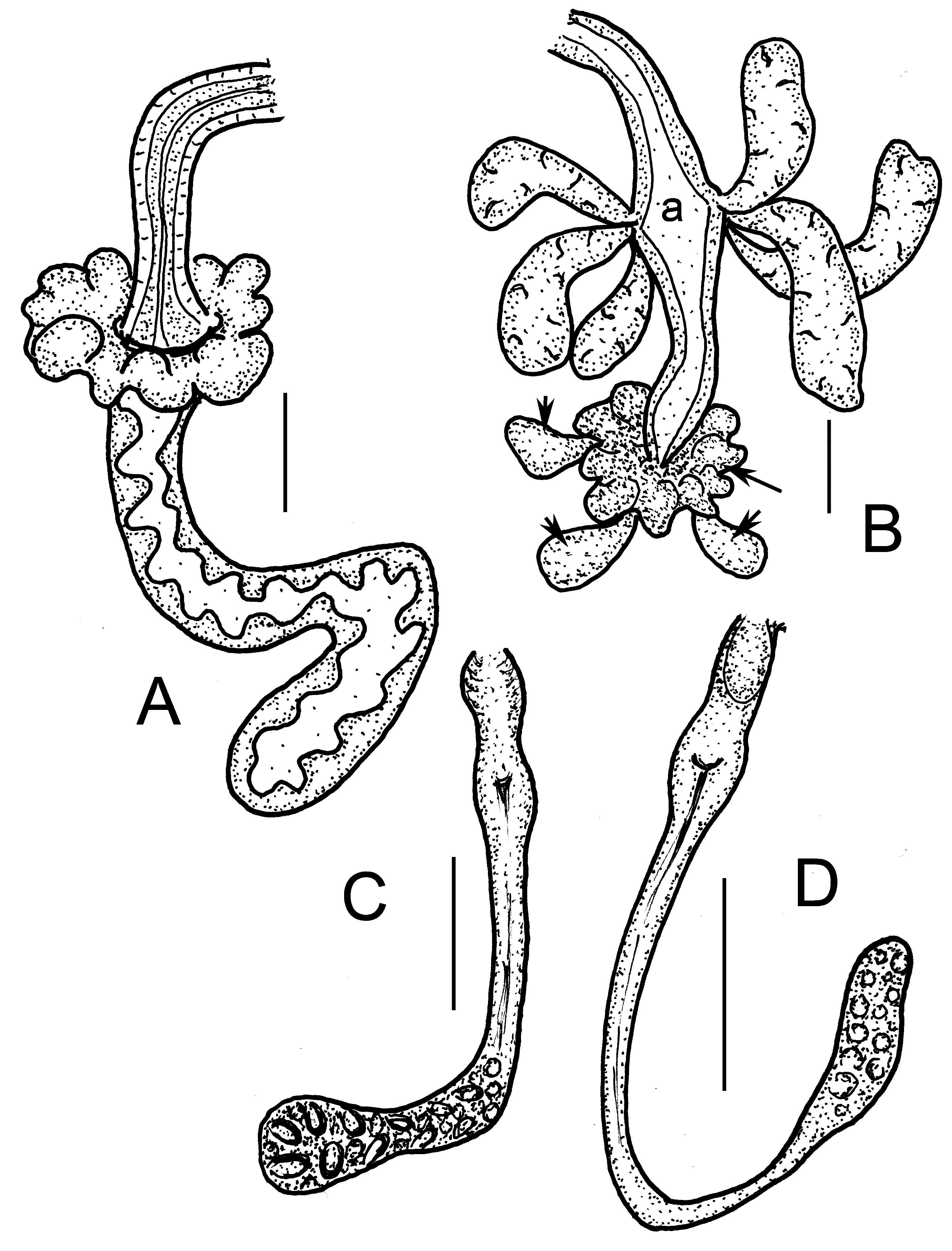
( Figures 12A–B View FIGURE 12 , 13 View FIGURE 13 , 14 View FIGURE 14 )
Type material. Holotype: NIBRIV0000810594, slide No. 2381, adult, stained specimen, anterior part of the worm opened, mounted specimen (last 12 segments, 2.45 mm; processed for DNA analysis No. 1125). Type locality: site 11. Gwaneumsa trail of Mt. Hallasan, Jeju Island, Korea, N 33˚41'75.9", E 126˚54'78.0", 634 m asl, 27.10.2016, leg. Y. Hong. Paratypes (in total 9 stained, adult specimens on slides): NIBRIV0000810595, slide No. 2278 adult, anterior 42 segments of the stained, mounted specimens. From type locality, NIBRIV0000811385, slide No. 2391, one adult, stained specimen (last 31 segments, 5.3 mm, for DNA analysis No. 1149), site 4. P.118.1.1–118.1.3. slide No. 2279 adult, stained specimen whole mounted, slide No. 2334, one adult, stained specimen (last 3 segments, 1.3 mm, for DNA analysis No. 1141), slide No. 2382, one adult, stained specimen (last 13 segments, 3 mm, for DNA analysis No. 1126) from type locality. P.118.2.1–P.118.2.2, slide No. 2390, one adult, stained specimen (last 12 segments, 2 mm for DNA analysis No. 1124), slide No. 2393a+b, anterior and posterior part of one adult, stained, mounted specimen. P.118.294 subadult, stained, whole mounted specimen anterior part opened, site 4.
Further material examined. 5 juveniles from sites 2, 5 and 12.
Etymology. The species is named after Dr. Jung Sai Ho who kindly supported collection in Mt. Hallasan.
Diagnosis. The new species can be recognized by the following combination of characters: (1) large, wide worms (15–31 mm long and about 0.9–1 mm wide in vivo), segments 47–80; head and the body forepart dorsally with brown pigment; (2) chaetae sigmoid with nodulus, maximum 5–6 (7) per bundle, in V–X ventrally only 2–3 enlarged chaetae; (3) clitellum girdle-shaped: gland cells small, in reticulate pattern; (4) five preclitellar pairs of nephridia; (5) dorsal blood vessel from XVIII–XXI, blood light pink; (6) two pairs of primary and 3 pairs of secondary pharyngeal glands, not connected dorsally; (7) small lemon-shaped coelomocytes, light yellow in aggregations in vivo; (8) sperm sacks and egg sack may extend into XXIV and XXX; (9) atrium 190 – 250 µm long with 6–7 very large atrial glands, numerous penial glands and some slightly larger accessory copulatory glands extending around male pores; (10) spermathecae free, ectal ducts of varying length, globular ampullae with a crown of diverticula-like protrusions, ampullae continuing entally into a flexible elongate bag that ends in VII or VIII, not connected to the oesophagus.
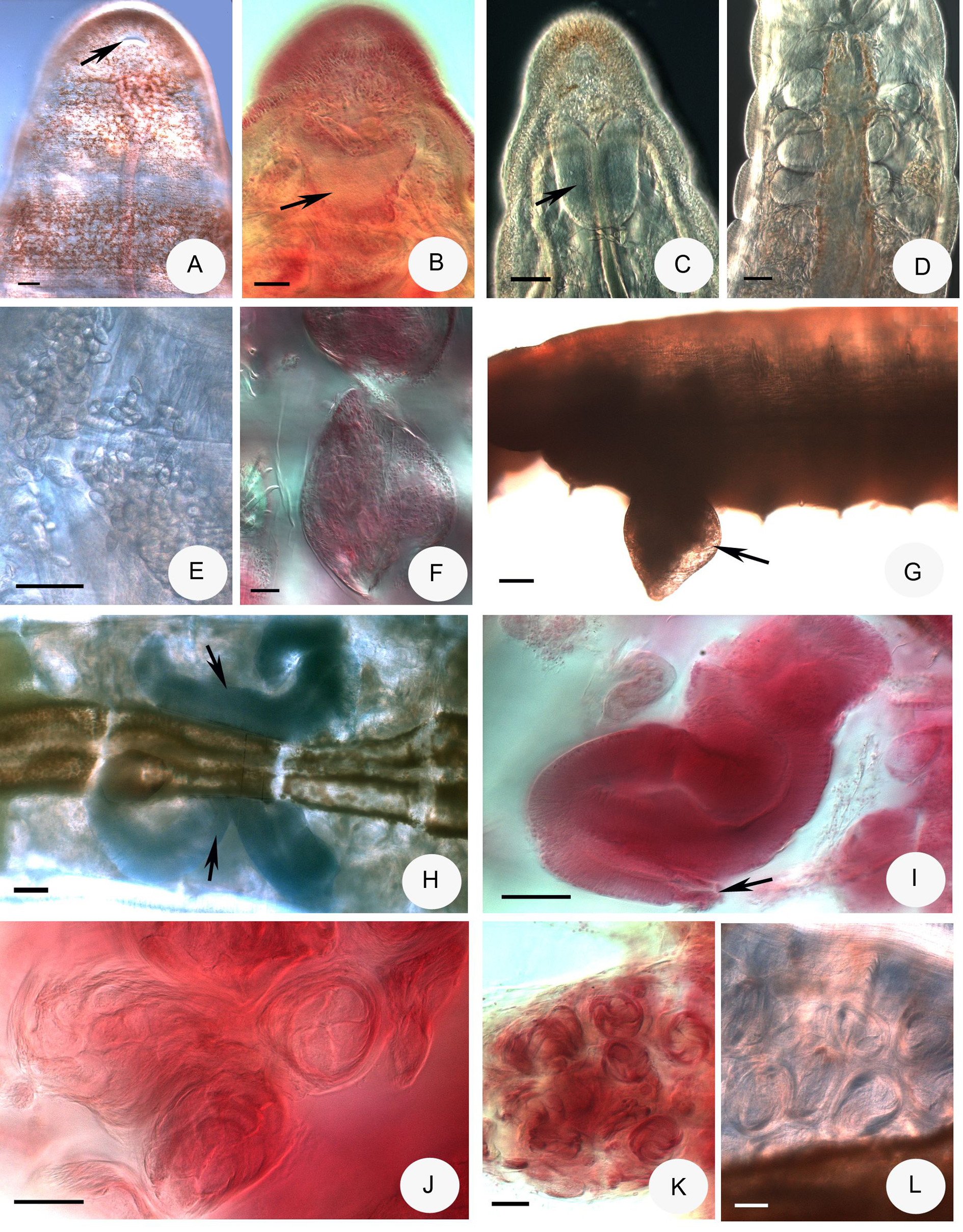
Description. Large enchytraeid worm. Colour light pink, dorsal side is light brown preclitellarly, while the prostomium is dark brown by pigmentation ( Fig. 13A, C View FIGURE 13 ). Holotype 15.9 mm long, 1 mm wide at VIII and 1.15 mm at clitellum (fixed), segment number 68. Body length of paratypes 15–31 mm, width 700–950 µm at VIII and 820–1110 µm at clitellum in vivo; length of fixed specimens 8.5–14.5 mm, width 520–1100 µm at VIII and 620– 1500 µm at clitellum. Segments 47–80. Strong external segmentation at body end. Chaetae sigmoid with nodulus. Chaetal formula: 2,3,4,5 – 4,3,2,3,4: 3, 4 ,5,– 4,5,6,(7). Chaetae mostly unequal in size within a bundle: in ventral bundles a gradual increase in length towards the ventral midline and in lateral bundles an increase in length in dorsal direction. From V to IX or X only two or three stronger chaetae per bundle ventrally (sometimes 2 one side and 3 in other side, in XI always tree or four chaetae in a bundle). The length of the longest chaetae ventrally gradually increasing from II to IV (from 100 or 115 by 8.5 µm to 150–160 by 10 µm). From V to VII–IX they are larger and stronger (195–230 by 10–15 µm). In X–XI slightly smaller (195–200 by 10–12 µm). Chaetae of lateral bundles smaller and thinner than ventrals and gradually increasing from II to XI (from 112–120 × 6–7 µm to 130– 185 x 8–10 µm). Postclitellarly longest chaetae measuring 120–105 by 7–10 µm. Chaetae in XII absent. Head pore at 0/I, a large transverse slit ( Fig. 13A View FIGURE 13 ). Clitellum girdle-shaped in XI–XII, weakly developed, gland cells small in reticulate pattern, also between bursal slits. In one case, gonadal region shifted forwards, bursal slits in X (paratype P.118.2.1 slide No. 2390). Thickness of body wall about 74–120 µm, depending on state of sexual maturity, cuticle about 2–2.5 µm, fixed.
Brain incised anteriorly and slightly convex posteriorly, slightly wider than long (e.g. 105 by 110 µm, fixed) ( Fig. 13B,C View FIGURE 13 ). Two pairs of primary pharyngeal glands (in 4/5–5/6), not united dorsally, and three or four pairs of secondary pharyngeal glands in V–VII (VIII); the secondary glands lobed ( Fig. 13D View FIGURE 13 ). Yellow-brownish chloragocytes from IV, about 10 µm long, fixed. Dorsal blood vessel from XVIII–XXIV, anterior bifurcation in I, blood pale reddish ( Fig. 13C View FIGURE 13 ). Five pairs of preclitellar nephridia from 6/7 to 10/11, anteseptale funnel only, postseptale lobed with folded canal, no interstitial tissue, efferent duct arising between the lobes. Oesophageal and intestinal appendages or diverticula absent. Coelomocytes only mucocytes, lemon-shaped, with granula, in cell aggregations light yellowish in vivo, small, size of cells 14–20 µm ( Fig. 13D,E View FIGURE 13 ). Sperm sac extending backwards to XIX–XXIV, egg sack to XXIII–XXX. In sperm sack sperm bundles in rolls ( Fig. 13J–L View FIGURE 13 ). Sperm funnel thickwalled, about 400–1250 µm long and 4–6 times longer than wide, fixed, mostly reclinate ( Fig. 13H–I View FIGURE 13 ). Sperm duct very long, reaching as far as XVI–XIX, loosely coiled, diameter 44–65 µm (fixed); diameter gradually increasing towards atrium. Atrium 190–250 µm long, maximum width 97–150 µm, here joined by 6–7 very large atrial glands (prostate glands), usually (120 – 200 µm to 420 µm long, Figs. 12B View FIGURE 12 , 14C–E View FIGURE 14 ). The atrium connects through a duct (90–110 µm long, fixed) with the male copulatory organ, which is surrounded by numerous penial glands (about 40 – 50 µm long) and some accessory copulatory glands (about 50 – 80 µm long) ( Figs. 12B View FIGURE 12 , 14F View FIGURE 14 ). The everted bursa may be conspicuous and domelike ( Fig. 13F,G View FIGURE 13 ). Bursal slits large, irregular, transversal, in XII ( Fig. 14A,B View FIGURE 14 ). Subneural glands absent. Spermathecae free ( Figs. 12A View FIGURE 12 , 14G–K View FIGURE 14 ): ectal ducts with different length, 240–260 µm long and 48–62 µm wide in vivo (85 – 180 µm long and 40–58 µm wide, fixed), devoid of glands. Spermathecal pores in 4/ 5 in lateral position. Ampullae globular with more diverticula-like protrusions, diameter 150 – 205 µm in vivo (130–190 µm, fixed). Ampullae continuing entally into a flexible elongate bag which ends in VII or VIII, not connected to the oesophagus. Eggsack with submature eggs only. It is possible that the diverticula of the spermathecae will be more distinct when the specimens are more developed sexually and with mature eggs. Unfortunately, our specimens did not reached this stadium even after half a year of culturing.
Distribution and habitat. Korea, Mt. Hallasan, Jeju Island, site 2, 4, 6, 11–12: soil, moss, grass and litter layer under Abies koreana , Quercus mongolica , Sorbus alnifolia and mixed forest.
Differential diagnosis. Up to now, seven valid species of Mesenchytraeus have been reported with enlarged ventral chaetae: M. tetrapodus Timm & Popchenko, 1978 , M. crenobius Timm, 1994 , M. kontrimavichusi Piper et al., 1982 , M. gigachaetus Xie, 2012 ( Shen et al., 2011, Xie 2012), M. longiductus Christensen & Dózsa-Farkas, 2012 , M. anisodiverticulatus Shen et al., 2012a , M. monodiverticulatus Shen et al., 2012b and M. calyx Dózsa- Farkas, Felföldi & Hong, 2015. The main differences between these species and the new species are as follows: The spermathecae of M. tetrapodus , M. crenobius , M. gigachaetus , M. anisodiverticulatus , M. monodiverticulatus , M. longiductus and M. calyx are connected with the oesophagus but in the new species they are free. M. kontrimavichusi has also free spermathecae, but the worms are smaller, 12 mm long, 53 segments (vs. 15–31 mm long and 47–80 segments in M. jungsaihoi sp. n.) and the spermathecae have one diverticulum each; moreover there is no atrium ( Timm 1994; Timm & Popchenko 1978; Piper et al. 1982; Christensen & Dózsa-Farkas 2012; Shen et al. 2011, 2012a, b; Xie 2012, Dózsa-Farkas et al. 2015). Considering all morphological characters, this species is very similar to M. calyx , by the dark-brown pigmented head, the light-pink blood, and the enlarged ventral chaetae, but apart from other differences purely on the basis of the type of spermathecae, the two species are different. It was also supported by the results of the molecular analysis.

| DNA |
Department of Natural Resources, Environment, The Arts and Sport |
No known copyright restrictions apply. See Agosti, D., Egloff, W., 2009. Taxonomic information exchange and copyright: the Plazi approach. BMC Research Notes 2009, 2:53 for further explanation.