
Portelmis guianensis, Przewoźny, Marek & Fernandes, André S., 2012
|
publication ID |
https://doi.org/ 10.5281/zenodo.280127 |
|
DOI |
https://doi.org/10.5281/zenodo.6167017 |
|
persistent identifier |
https://treatment.plazi.org/id/03D4878D-FFEE-464C-FF6B-0C2AEB8F8E7D |
|
treatment provided by |
Plazi |
|
scientific name |
Portelmis guianensis |
| status |
sp. nov. |
Portelmis guianensis View in CoL sp. n.
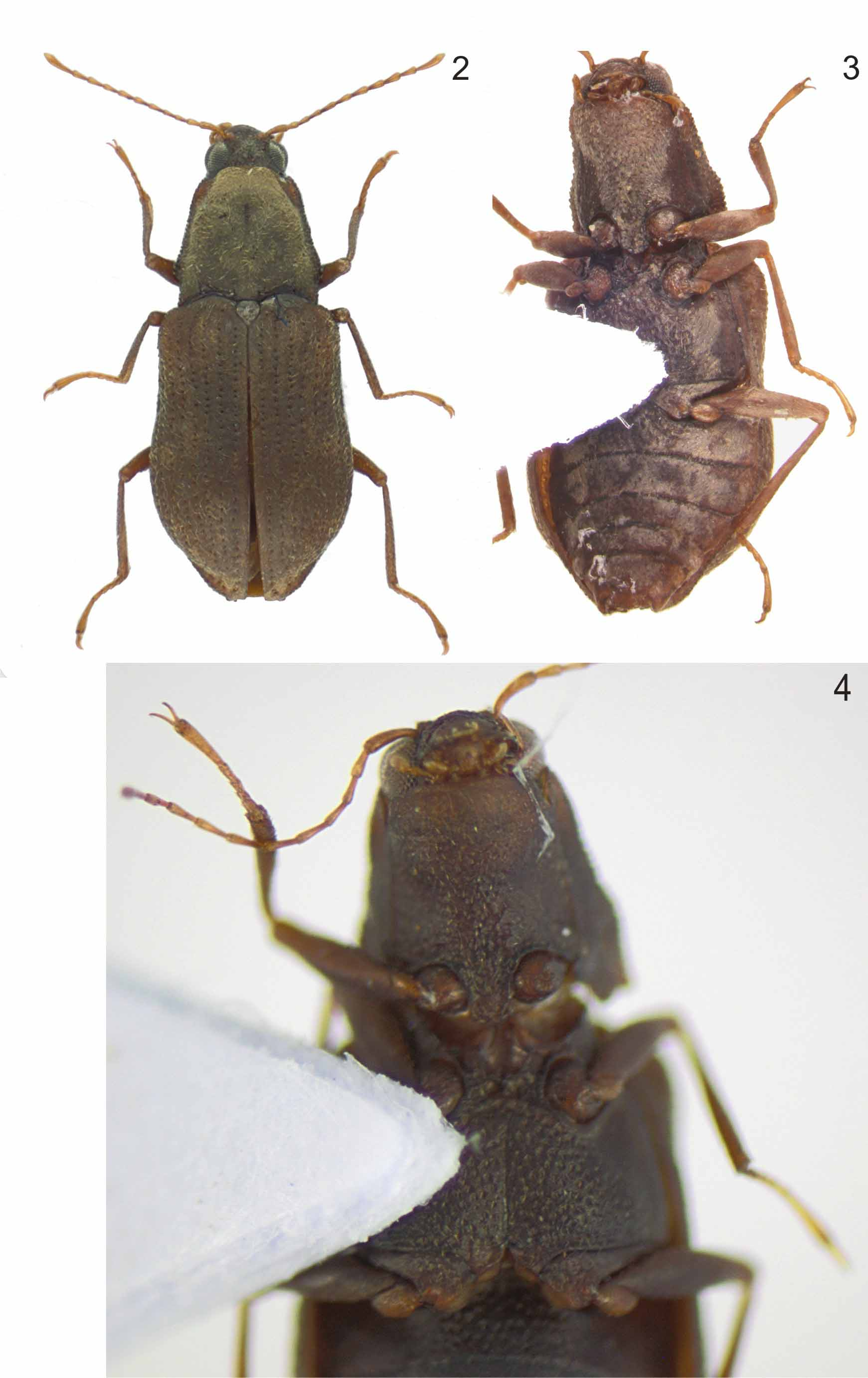
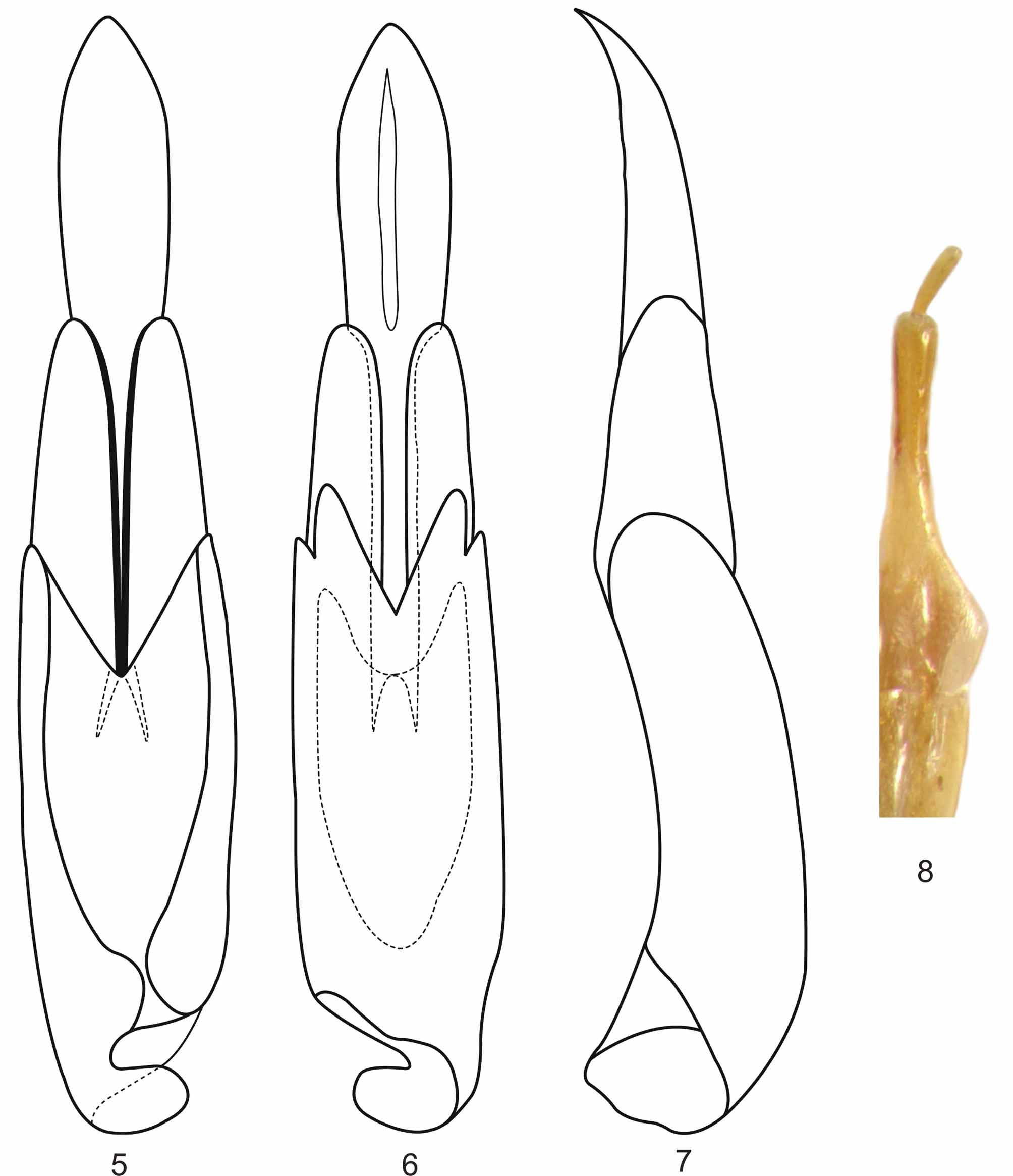
( Figs. 2–8 View FIGURES 2 – 4 View FIGURES 5 – 8 )
Type locality St. Elie Route, Sinnamary Municipality, French Guiana (ca. 5°23'3.40"N, 53°01'05"W)
Type material. Holotype: 3, French Guiana, Sinnamary, Route St. Elie, 08/xii/2006, leg. M. Snižek " Holotypus, Portelmis guianensis sp. n., Przewożny & Fernandes det. 2010" [red, printed] ( INPA). Paratypes: 3 3 and 5 Ƥ, with same label data as holotype, all paratypes with a red paratype label ( CMP, EMEC).
Diagnosis. Pronotum with sublateral carinae extending until basal 1/5; pronotal surface with distinct granules only on lateral surface; median longitudinal impression extending from basal 1/4 until apical 1/4. Prosternal process with apex wider than middle; lateral margin arcuate; apex rounded. Mesoventrite deeply depressed between pro- and metaventrite. Disc of metasternum with median longitudinal impression extending from posterior to anterior 1/8. Disc of ventrite I densely punctate, without granules. Aedeagus, in lateral view, with phallobase as wide as paramere. Ovipositor without apical setae on apical segment of styli.
Description. Holotype: male ( Fig. 2 View FIGURES 2 – 4 ). Length 2.85 mm, greatest width 1.38 mm. Body ( Fig. 2 View FIGURES 2 – 4 ) robust, subparallel; ventral and dorsal surface with irregular depressions; coarse punctures three to four times the diameter of eye facets on the prosternal process, metasternal disc and metacoxae; dorsum sparsely covered with fine, short, recumbent and pale setae, with some long setae on pronotum, and near elytral apex; scutellum glabrous; venter surface with plastron.
Head: With small irregular depressions; frontal margin emarginate. Eyes moderately protuberant, laterally rounded, separated by a distance 1.5 times wider than eye. Antenna with 11 segments, long and slender, first and last segments slightly swollen and twice as long as the remaining segments. Frontclypeal suture present between bases of antennae. Clypeus rectangular, as long as and wider than labrum; anterior margin emarginate; anterior angles rounded. Labrum rectangular; anterior margin slightly convex; anterior angles rounded, with row of short pale setae. Maxillary palpus with four segments; last segment swollen, as long as second and third segments combined. Labial palpus with three segments; last segment swollen, two times wider than second segment, as long as the remaining segments combined.
Color: Cuticle reddish-brown, varying from shiny to opaque where plastron is present. Distinctly darker on head and paler on mouth parts, antennae and tarsus.
Thorax ( Figs. 3–4 View FIGURES 2 – 4 ): Pronotum slightly longer (0.96 mm) than wide at base (0.89 mm); wider at base than at apex (0.53 mm); one sublateral carina, on each side, occupying posterior 1/5; impressions on disc (one longitudinal, beginning at anterior 1/2 and extending until anterior 3/4, deeper and wider at its anterior 1/3; one oblique on each side, on posterior 1/ 4); gibbosities (one median, on posterior 1/3; one lateral, on each side, on median 1/3); anterior angles slightly produced, subacute; anterior margin broadly convex at middle and concave behind each eye, extending over base of head; lateral margin sinuate, crenate; posterior angles produced, acute; posterior margin with three arches, two broad, one on each side in front of the elytron, and one narrow in front of scutellum. Elytra longer (1,63 mm) than wide, (maximum width, at posterior 1/6, 0,67 mm); humeral angle broadly rounded, tumid and sinuate at middle; without sublateral carinae; lateral margins crenate; disc with punctures separated by twice their diameters, half as wide as intervals between striae. Scutellum ( Fig. 2 View FIGURES 2 – 4 ) flat; anteriorly convex; posteriorly triangular; longer than wide. Prosternum ( Fig. 4 View FIGURES 2 – 4 ) with anterior margin truncate; without impressions or carinae. Prosternal process nearly as long (0.25 mm) as wide (0.24 mm), wider at base than at apex (0.14 mm); extending beyond anterior coxae; subcapitate; lateral margin feebly concave; apex rounded. Mesoventrite ( Fig. 4 View FIGURES 2 – 4 ) wider between coxae (0.26 mm) than long (0.24 mm); deeply depressed between pro- and metaventrite. Metaventrite ( Fig. 4 View FIGURES 2 – 4 ) with median, longitudinal impression extending until anterior 1/8; anterior margin between mesocoxae convex; posterior margin between metacoxae concave; posterior portion in front of metacoxae with transverse arched impression. Legs ( Fig. 2 View FIGURES 2 – 4 ) long; pro- and mesocoxae globular; tibiae with indistinct fringes of tomentum, two parallel fringes on apical 1/2 of anterior margin on the protibiae and one fringe on apical 1/2 of posterior margin on the meso- and metatibiae; tarsal claws without basal teeth.
Abdomen: Wider (maximum width, ventrite I, 1.30 mm) than long (1.13 mm). Ventrite I ( Fig. 3 View FIGURES 2 – 4 ) with intercoxal process subtriangular; without carinae or impressions; disc slightly depressed on anterior 1/3. Ventrite V ( Fig. 3 View FIGURES 2 – 4 ) with posterior angle with strong toothlike projection; posterior margin between projections truncate, with few short setae extending slightly beyond posterior margin.
Plastron: Head with plastron noticeable only on gena. Dorsum completely covered, except on pronotal impressions and carinae, edges of pronotum and scutellum. Venter completely covered. Legs completely covered, except on trochanters, basal 1/3 of tibiae and tarsus.
Aedeagus ( Figs. 5–7 View FIGURES 5 – 8 ): Parameres short, around 1/2 the length of median lobe. In dorsal view ( Fig. 5 View FIGURES 5 – 8 ): elongate; internal and external lateral margins slighly convex; internal margins divergent; apex rounded. In ventral view ( Fig. 6 View FIGURES 5 – 8 ): internal margins parallel. In lateral view ( Fig. 7 View FIGURES 5 – 8 ) as wide as apical portion of phallobase; gradually and feebly narrowed to apex; apex irregularly rounded. Median lobe 2.0 times longer than parameres. In dorsal view ( Fig. 5 View FIGURES 5 – 8 ): 1.5 times wider than paramere; lateral margins parallel until apex of parameres and then abruptly widened and ligulate; apex narrowed, subacute; basolateral apophyses slender, acute and divergent. In ventral view ( Fig. 6 View FIGURES 5 – 8 ) with fibula noticeable on apical portion, slender and as long as 1/3 of median lobe. In lateral view ( Fig. 7 View FIGURES 5 – 8 ): about the width of apical portion of paramere; ventral margin nearly straight until apical portion; dorsal margin slightly convex; apex narrow, acute and strongly curved to venter. Phallobase asymmetrical, with 0.75 times the length of median lobe. In dorsal view ( Fig. 5 View FIGURES 5 – 8 ) almost 2.0 times wider than median lobe; without setae. In lateral view ( Fig. 7 View FIGURES 5 – 8 ) moderately arched; basal portion slightly widened; apex broadly rounded.
Female. Externally similar to male.
Ovipositor ( Fig. 8 View FIGURES 5 – 8 ): Styli elongate, in dorsal view ( Fig. 8 View FIGURES 5 – 8 ) with basal segment narrowed from base until basal 1/ 3 and then feebly widened until apical portion, apical portion without setae; apex of basal segment rounded and with some short and stout setae; apical segment narrow, cylindrical and with 1/4 the length of the basal segment. Coxite trapezoidal, apically as wide as base of styli; gradually narrowed basally, with same width as baculum at base.
Etymology. The specific epithet is a reference to the country of origin of the new species.
Comparative notes. Portelmis guianensis sp. nov. can be assigned to the P. gurneyi –g roup due to having the pronotal surface without distinct granulations; the disc of ventrite I densely punctuated, without granules; and the disc of metaventrite with the median, longitudinal impression extending beyond basal 1/2. Relative to other species in the P. gurneyi –group, Portelmis guianensis sp. nov. appears to be closely related to P. gurneyi by having flat elytral intervals; the prosternal process with concave lateral margins; by the general shape of the aedeagus and the absence of setae on the phallobase.
Portelmis guianensis sp. nov. can be distinguished from all other species of the P. gurneyi –group by the mesoventrite being deeply depressed between pro- and metaventrite; the base of phallobase as wide as parameres in lateral view; and the apical segment of the ovipositor styli without apical setae.
No known copyright restrictions apply. See Agosti, D., Egloff, W., 2009. Taxonomic information exchange and copyright: the Plazi approach. BMC Research Notes 2009, 2:53 for further explanation.
|
Kingdom |
|
|
Phylum |
|
|
Class |
|
|
Order |
|
|
Family |
|
|
Genus |