
Pseudopannota pannota, Kluge, Nikita J. & Novikova, Eugenia A., 2016
|
publication ID |
https://doi.org/ 10.11646/zootaxa.4169.1.1 |
|
publication LSID |
lsid:zoobank.org:pub:F15C0038-DF14-4E4B-98F5-FE1BD7A5759F |
|
DOI |
https://doi.org/10.5281/zenodo.5616283 |
|
persistent identifier |
https://treatment.plazi.org/id/03D48C3E-E639-FFD1-76EC-B4F9FD55F96B |
|
treatment provided by |
Plazi |
|
scientific name |
Pseudopannota pannota |
| status |
sp. nov. |
Pseudopannota pannota sp. n.
( Figs 2 View FIGURES 1 – 3 , 7 View FIGURES 4 – 7 , 14 View FIGURES 8 – 15 , 17 View FIGURES 16 – 20 , 25–58 View FIGURES 21 – 25 View FIGURES 26 – 33 View FIGURES 34 – 37 View FIGURES 38 – 49 View FIGURES 50 – 53 View FIGURES 54 – 56 View FIGURES 57 – 58 )
Etymology. The adjective " pannota " refers to great dergee of fusion of protopotera with notum on mesothorax of larva.
Material examined. Holotype: L-S-IƋ {specimen [VI] (8 A) A}, ZAMBIA, river Luangwa near Luangwa Bridge, 8.VIII.2014, coll. N. Kluge & L. Sheyko . Paratypes: the same locality, 2–8.VIII.2014, coll. N. Kluge & L. Sheyko: 1 L-S-I ♂, 2 L-S-I ♀, 11 larvae ; river Zambezi near Victoria Falls, 25–31.VIII.2014, coll. N. Kluge & L. Sheyko: 1 larva.
Descriptions. Larva. CUTICULAR COLORATION. Dorsal side of head, thorax and abdomen grayish-brown with diffusive lighter blanks. Pronotum and mesonotum with several paired roundish blanks ( Figs 31, 32 View FIGURES 26 – 33 ). Metanotum and thoracic pleura mainly grayish-brown, thoracic sterna nearly colorless. Cuticle of legs at most colorless; on each leg hidden part of tibial base tinged with orange-brown; hind femur with large, orange-brown, diffusively outlined macula on posterior side close to outer margin and with smaller macula of the same color on inner margin ( Fig. 52 View FIGURES 50 – 53 ). Abdominal terga grayish-brown with diffusive unpaired blanks on median line ( Fig. 33 View FIGURES 26 – 33 ); sterna lighter, unicolor. Tergalii colorless ( Figs 31 View FIGURES 26 – 33 , 46–48 View FIGURES 38 – 49 ). Cuticle of caudalii colorless.
HYPODERMAL COLORATION. Absent; only in mature larva ready to molt to subimago, subimaginalimaginal coloration of abdominal terga can be visible through larval cuticle.
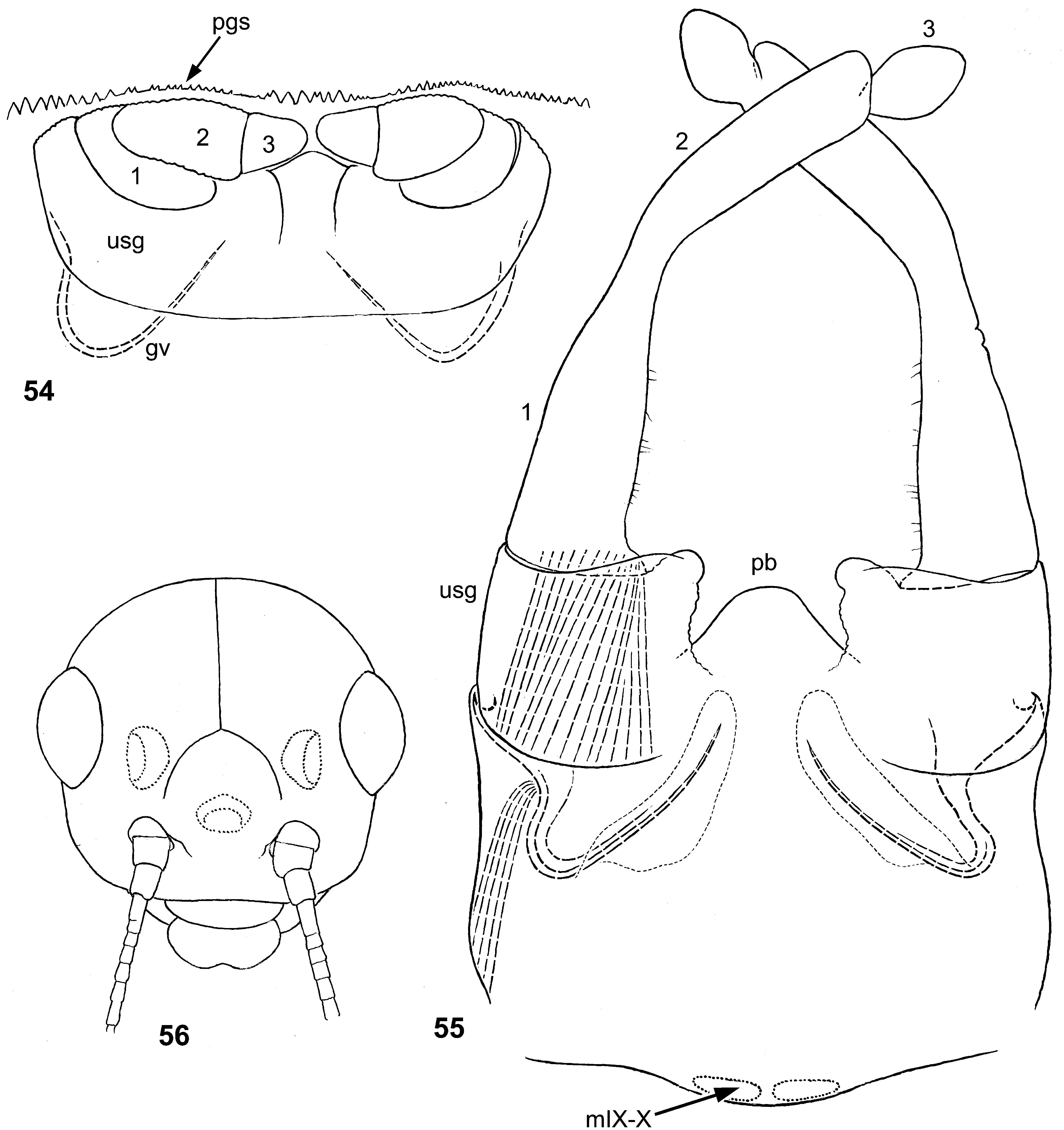
STRUCTURE. Body robust. Frons flat, bases of antennae widely separated. Frontal suture blunt-angled ( Fig. 56 View FIGURES 54 – 56 ). Labrum ( Figs 37–39 View FIGURES 34 – 37 View FIGURES 38 – 49 ) with lateral margins roundish and sclerotized, lacking setae; distal margin [soft, with numerous fine setae—see Pseudopannota (1)] with well-expressed median incision; inner side with paired row of spine-like setae, with asymmetric sclerite [see Pseudopannota (1)]. Incisor of left and right mandible represented by one small denticle [see Pseudopannota (2)]; protuberance proximad of right prostheca blunt, just after molt with fine seta-like processes ( Figs 40–42 View FIGURES 38 – 49 ) [see Pseudopannota (3)]. Maxilla as characterized for above [see Pseudopannota (4, 5)] ( Fig. 36 View FIGURES 34 – 37 ).
Maxillary palp ( Figs 14 View FIGURES 8 – 15 , 36 View FIGURES 34 – 37 ) large, with primary first segment distinctly divided into longer proximal subsegment and shorter distal subsegment, with muscle running from its base through both subsegments to base of primary second segment [see Pseudopannota (6)]; cuticle entirely colorless. Proximal subsegment of first segment on outer side with smooth and sclerotized cuticle, on inner side with membranous cuticle bearing numerous campaniform sensilla. Distal subsegment of first segment widened apically, with apex wider than next segment; on outer side with smooth and sclerotized cuticle, on inners side with membranous cuticle bearing numerous irregular soft colorless setae. Primary second segment thickest proximally; on outer side with smooth and sclerotized cuticle, on inners side with membranous cuticle bearing numerous irregular soft colorless setae; terminates by small globular portion with membranous cuticle, covered with numerous irregular soft colorless setae. Thanks to rigid outer side of each segment, palp of shed exuviae keeps its form, being dipped into maxilla only basally, because of inversion of palpiger ( Fig. 14 View FIGURES 8 – 15 ).
Paraglossae larger than glossae, both glossae and paraglossae widened distally ( Fig. 34 View FIGURES 34 – 37 ). Glossa with regular row of long setae on inner margin. Both glossae and paraglossae apically-ventrally with numerous irregularly situated long setae, apically-dorsally with row of larger setae. Each glossa and paraglossa with one long seta on distal half of dorsal side.
Distal segment of labial palp [formed by fusion of 2nd and 3rd primary segments—see Pseudopannota (8)] large, transverse, thick, widest proximally, triangular in cross section ( Fig. 35 View FIGURES 34 – 37 ). Its inner-apical margin somewhat sclerotized and bears irregular row of setae; outer and ventral surfaces soft and densely covered with numerous irregularly situated soft colorless setae ( Fig. 43 View FIGURES 38 – 49 ).
Suture between pronotum and mesonotum nearly straight, mesonotum without projected antero-lateral corners ( Figs 31, 32 View FIGURES 26 – 33 ). Fore protoptera fused nearly up to apex ( Fig. 17 View FIGURES 16 – 20 ). Vestiges of hind protoptera absent. Coxa of each leg with rounded lobe, large on fore and middle legs, smaller on hind leg ( Figs 50–52 View FIGURES 50 – 53 ). Femora stout, with strong musculature; fore femur thickest proximally; hind femur halteriform, thickest distally and thinnest medially. Outer margin of each femur with irregularly situated stout setae; besides it, distal part of anterior surface with few setae of the same structure: 2–4 on fore femur, 1–2 on middle femur, absent on hind femur. No structures resembling femoral patch of Baetofemorata. Tibiae and tarsi stout, with few stout spine-like setae on inner side. Patella-tibial suture equally developed on all legs. Each claw, besides apical hook, always with two diverging denticles, large pointed proximal one and narrow blunt distal one ( Fig. 25 View FIGURES 21 – 25 ).
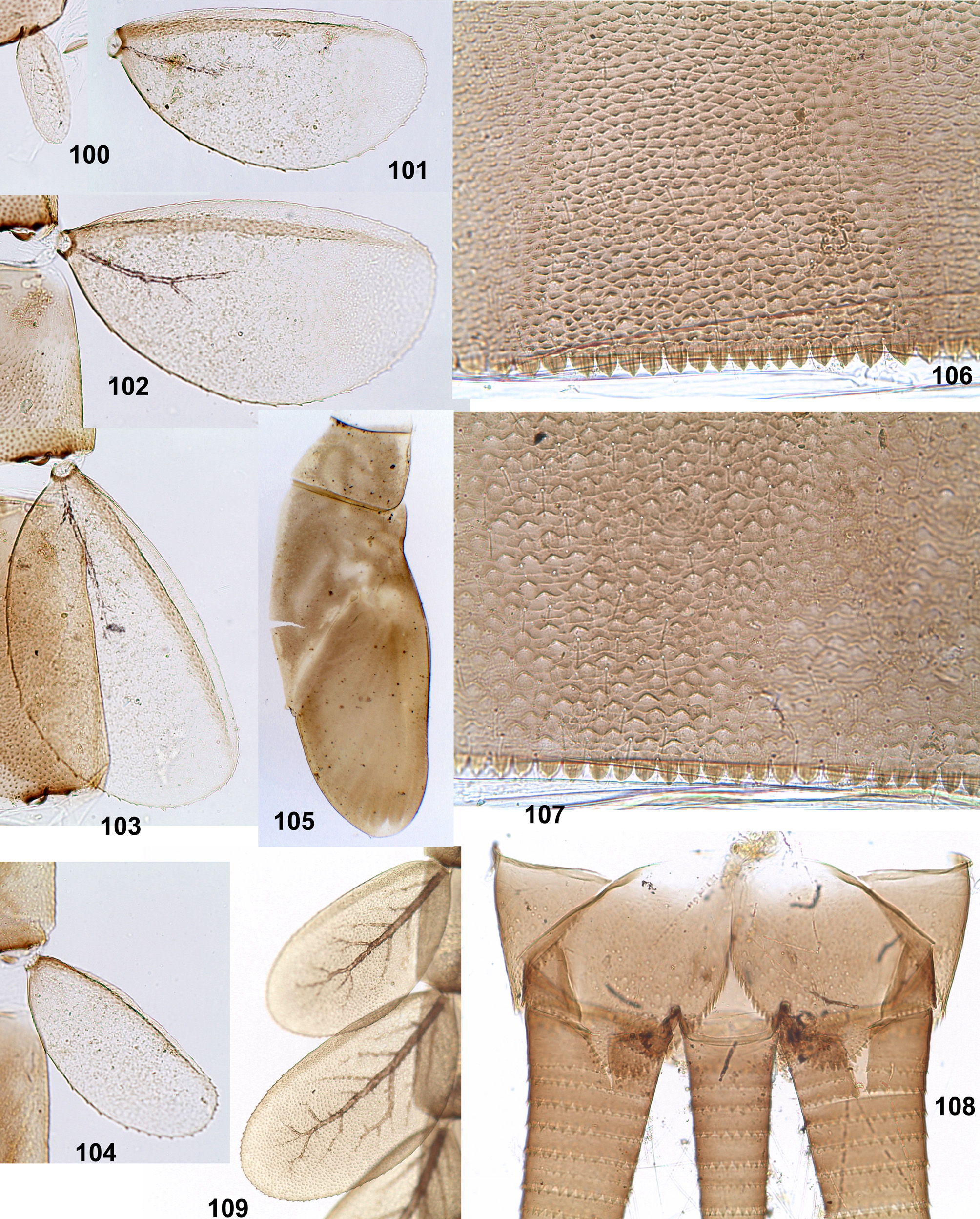
Surface of abdominal terga and sterna with pointed denticles, without scales or scale bases, with few fine setae only ( Fig. 54 View FIGURES 54 – 56 ). Posterior margin of tergim I smooth, posterior margin of each tergum II–X with regular denticles ( Fig.45 View FIGURES 38 – 49 ). Sterna I–VI with posterior margin entirely smooth, sterna VII and VIII with few small denticles on lateral parts of posterior margin, sternum IX with small denticles on posterior margin ( Fig. 54 View FIGURES 54 – 56 ). Paraprocts with many short marginal denticles ( Fig. 49 View FIGURES 38 – 49 ). Tergalii roundish ( Figs 46–48 View FIGURES 38 – 49 ); margin entirely bordered by very thin colorless rib, with small sparse denticles; surface with cuticle very delicious, without denticles and scales; tracheae colorless and nearly invisible. All tergalii I–VII nearly equal, tergalius I more asymmetric than others, tergalius VII smaller than others. Cerci about ½ of body length, paracercus smaller, about 0.7–0.8 of cerci length. Segments of cerci and paracercus with equally small triangular denticles on posterior margins (as in Fig. 108 View FIGURES 100 – 109 ).
Subimago. CUTICULAR COLORATION. Cuticle at most colorless. Pronotum colorless. Mesonotum very light, partly colorless, with anterolateral scutal costa contrastingly bordered by dark brown ( Fig. 29 View FIGURES 26 – 33 ). Thoracic pleura at most colorless, with contrasting dark brown lines along prealar bridge, lateropostnotal crest and its junction with postsubalar sclerite and some other sclerites; anterior part of postsubalar sclerite colorless ( Fig. 30 View FIGURES 26 – 33 ). Legs entirely colorless. Abdomen, gonostyli and cerci entirely colorless.
TEXTURE. On all legs of both sexes, primary 1st–4th tarsomeres covered by blunt microlepides, last (primary 5th) tarsomere covered by pointed microlepides (see Table 2 View TABLE 2 ).
Imago, male ( Figs 26, 27 View FIGURES 26 – 33 ). Head brown and ocher. Turbinate eyes widened apically, with facetted surface dark red, stem in distal half contrastingly light yellowish, in proximal half light reddish. Thorax with contrasting brown and ocher areas; median line of mesonotum dark brown; medioscutum light brown; medioparapsidal sutures ocher; submedioscutum brown. Wing membrane colorless, veins ocher. Pterostigma with few (about five) oblique incomplete branched veins (as in P. berthrandi —see Elouard et al. 1990: Fig. 4 View FIGURES 4 – 7 ). Hind wing absent. Legs unicolor ocher, relatively robust ( Fig. 53 View FIGURES 50 – 53 ). In holotype, proportion of femur / tibia / tarsal segments of fore leg 40:70:2:20:13:8:7 (in subimago 30:40:2:9:7:4:7); of middle and hind legs 30:37:4:3:2:7. Tarsus of middle and hind leg with 2 apical spines, on segments 1st+2nd and 3rd ( Fig. 53 View FIGURES 50 – 53 ). Abdomen ocher; cuticle of terga tinged with brown, gradually darker on posterior segments; terga IV–VIII with intensive reddish-brown hypodermal coloration in median area. Cerci unicolor ocher.
Male genital structure and development. Imaginal genitals as in Fig. 55 View FIGURES 54 – 56 . Sterno-styligeral muscle completely absent. Each gonovectis with apex free end straight, not hooked and not bordering distal margin of membranous sack. Penial bridge forms prominent median projection rounded apically. Apical margin of each unistyliger medially with roundish projection directed medially. 1st segment of gonostylus long and nearly conic, with inner margin smooth; 2nd segment slightly widened toward apex; 3rd segment short.
Protogonostyli of male larva represent very shallow convexities of posterior margin of abdominal sternum IX. In mature larva ready to molt to subimago, subimaginal gonostyli are packed under larval cuticle in « Labiobaetis - pose»: second segments directed medially and bent ( Fig. 54 View FIGURES 54 – 56 ).
Imago, female ( Fig. 28 View FIGURES 26 – 33 ). Thorax and wings as in male. Tarsus of fore leg with 2 apical spines, on segments 2nd and 3rd; middle and hind legs as in male. All abdominal terga with intensive brown coloration; terga I–III with ocher lateral areas and poorly expressed narrow ocher median line. Abdominal sterna unicolor light ocher.
Eggs ( Figs 57, 58 View FIGURES 57 – 58 ). About 0.1 mm length. Oval or nearly spherical, with irregular shallow protuberances and large irregular folds.
Dimension. Fore wing length of male 5.5 mm.
Comparison. The new species Pseudopannota pannota sp. n. is closely related to the west African species P. berthrandi ( Demoulin 1967) , having the same shape of labial palp, legs, tergalii and fused protoptera (see Table 1 View TABLE 1 ). Larva of P. pannota sp. n. differs from P. berthrandi by structure of right mandible, maxillary palp and proportions of caudalii.
In P. berthrandi protuberance of right mandible represents a “large tooth” (Elouard et al. 1990: 29) ( Demoulin 1967: Fig. 2 View FIGURES 1 – 3 b; Elouard et al. 1990: Fig. 8 View FIGURES 8 – 15 a). Unlike this, in P. pannota sp. n. this protuberance is blunt with minute seta-like projections, which are retained just after molt ( Fig. 42 View FIGURES 38 – 49 ), but are often wiped out.
In P. berthrandi distal subsegment of the first segment of maxillary palp is not widened distally, being narrower than proximal part of second segment ( Demoulin 1967: Fig. 2 View FIGURES 1 – 3 f; Elouard et al. 1990: Fig. 8 View FIGURES 8 – 15 d), while in P. pannota sp. n. it is markedly widened distally, being wider than next segment ( Figs 14 View FIGURES 8 – 15 , 36 View FIGURES 34 – 37 ).
Larva of P. berthrandi has cerci and paracercus of subequal length (Elouard et al. 1990: Fig. 7 View FIGURES 4 – 7 ), while in P. pannota sp. n. paracercus is always shorter and narrower than cerci.
Imagoes of P. pannota sp. n. well differ from P. berthrandi by coloration of abdomen. According to the description by Elouard et al. (1990) based on reared material, male imago of P. berthrandi has “terga I–IV white”; its female imago has “lateral markings of terga II–V reddish-orange, forming a continuous band along sides of the anterior part of abdomen” (Elouard et al. 1990: Figs 3 View FIGURES 1 – 3 , 6 View FIGURES 4 – 7 ); in contrast to this, male imago of P. pannota sp. n. has large reddish-brown maculae beginning from abdominal tergum IV ( Figs 26, 27 View FIGURES 26 – 33 ); its female imago has median brown markings on all abdominal terga ( Fig. 28 View FIGURES 26 – 33 ). Besides two reared male imagoes and two reared female imagoes, hypodermal coloration equal to imaginal one, was observed on two mature male larvae and one mature female larva ready to molt to subimago. Genitals of P. berthrandi , figured by Elouard et al. (1990: Fig. 5 View FIGURES 4 – 7 ) are markedly different from genitals of P. pannota sp. n. ( Figs 2 View FIGURES 1 – 3 , 55 View FIGURES 54 – 56 ).
In the general characteristics of Pseudopannota, Elouard et al. (1990) wrote that “Abdominal terga with scales and fine setae” and that “Gills ... thickened along anterior margin, with distinctly spiculate surface”. In contrast to this, abdominal terga of P. pannota sp. n. completely lack scales; its tergalii (“gills”) are very thin-walled, without any thickening and without any spiculation on surface.


No known copyright restrictions apply. See Agosti, D., Egloff, W., 2009. Taxonomic information exchange and copyright: the Plazi approach. BMC Research Notes 2009, 2:53 for further explanation.