
Monocentroptilum badium (Kopelke 1980) Kluge, 2018
|
publication ID |
https://doi.org/ 10.11646/zootaxa.4486.2.2 |
|
publication LSID |
lsid:zoobank.org:pub:1EB0FF97-800B-475C-B3B4-E2B7D0B874E9 |
|
DOI |
https://doi.org/10.5281/zenodo.5966872 |
|
persistent identifier |
https://treatment.plazi.org/id/03D4A739-FFEA-686F-25FF-F9919006FF30 |
|
treatment provided by |
Plazi |
|
scientific name |
Monocentroptilum badium (Kopelke 1980) |
| status |
comb. nov. |
Monocentroptilum badium (Kopelke 1980) comb. n.
( Figs 1–58 View FIGURES 1–4 View FIGURES 5–9 View FIGURES 10–27 View FIGURES 28–31 View FIGURES 32–39 View FIGURES 40–43 View FIGURES 44–47 View FIGURES 48–52 View FIGURES 53–58 )
Centroptilum badium Kopelke 1980a: 113 (♂ and ♀ imago); Kopelke 1980b: 301 (eggs).
Afroptilum (Afroptilum) badium: Gillies 1990a: 99 .
Crassabwa badia View in CoL : LugO-OrtiZ & McCafferty 1996b: 238.
Material examined. UGANDA, Kanungu district, river Munyaga just downstream Bwindi National Park, 0º58'40"S, 29º37'15"E, 23–26.VII.2007, coll. N. Kluge: 3 L-S-I ♂, 1 S-I ♂, 1 L-S/I ♂, 2 L-S ♂, 6 L-S-I ♀, 5 L-S ♀, 59 larvae; river Ishasha downstream Bwindi National Park, 0º51'S, 29º39'30"E, 14.VII.2007, coll. N. Kluge: 3 larvae; Kasese district, Rwenzory mountains, tributary of river Mubuku upstream Ruboni, 0º21'N, 30º02'E, 28.VII– 6.VIII.2007, coll. N. Kluge: 1 larva.
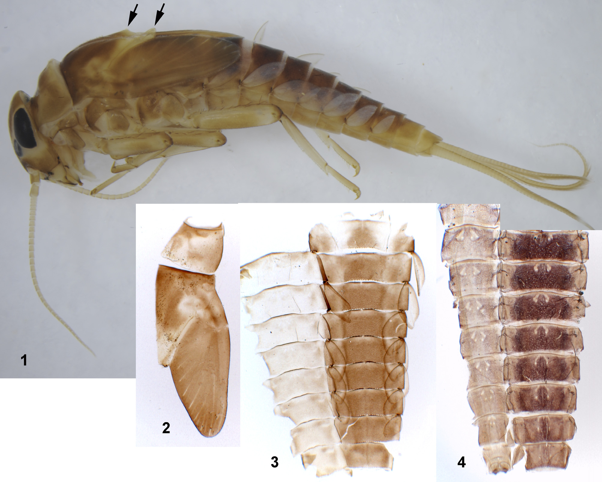
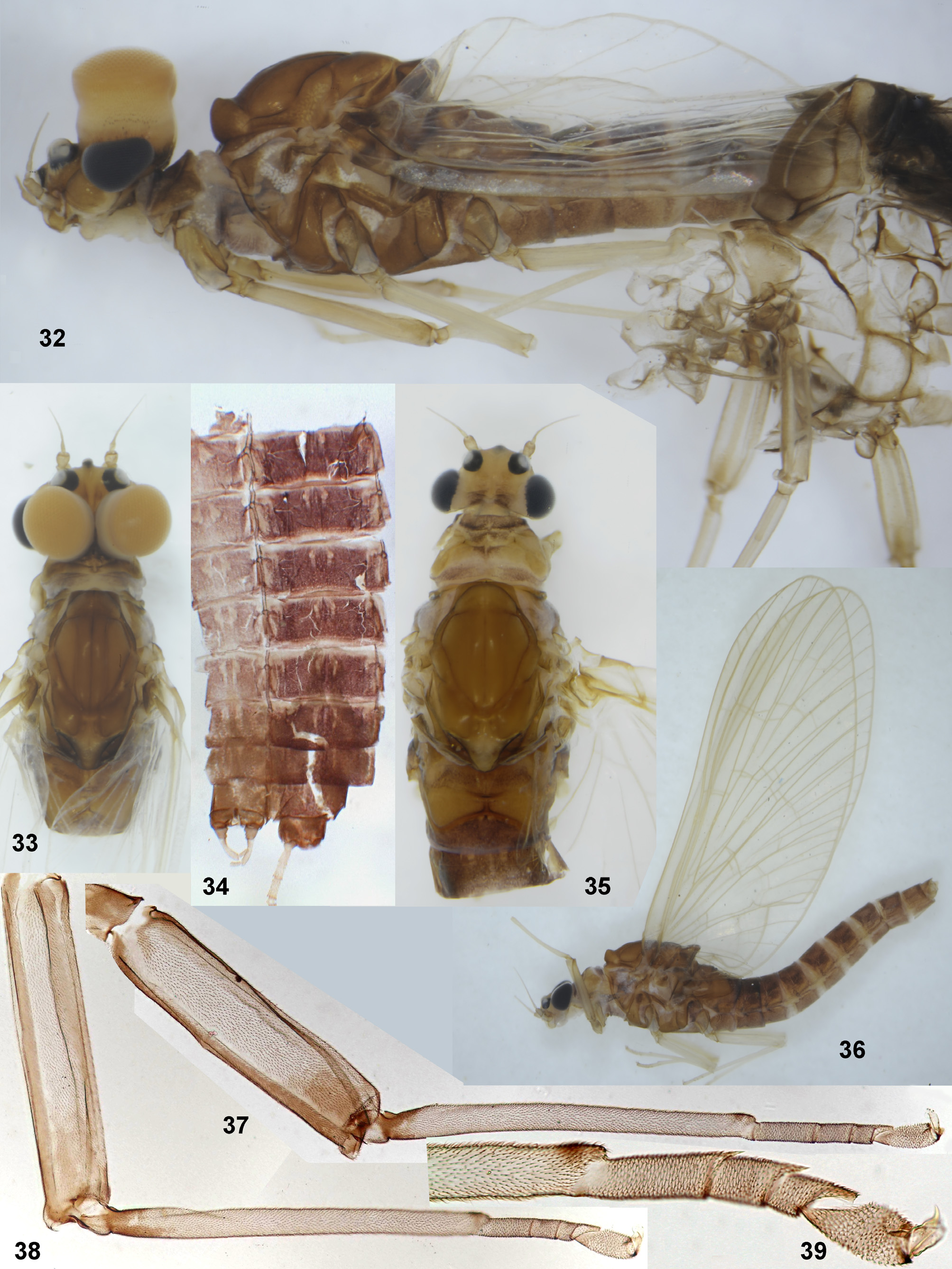
Descriptions. Larva. CUTICULAR COLORATION. Nearly unicolor brown ( Fig. 1 View FIGURES 1–4 ): head brown; pronotum and mesonotum brown with diffusive lighter areas ( Fig. 2 View FIGURES 1–4 ); thoracic pleura brown, with lighter membranes; legs unicolor brown; abdomen with terga unicolor brown, sterna unicolor light brownish (even sigilla on terga and sterna have the same color as background) ( Fig. 3 View FIGURES 1–4 ); cerci and paracercus unicolor brown.
HYPODERMAL COLORATION. In both sexes abdominal terga with intensive brown coloration as in winged stages ( Fig. 4 View FIGURES 1–4 ).
STRUCTURE. General appearance, including shape of head, thorax, legs, abdomen and caudalii ( Fig. 1 View FIGURES 1–4 ) similar to that of Baetis s.str.
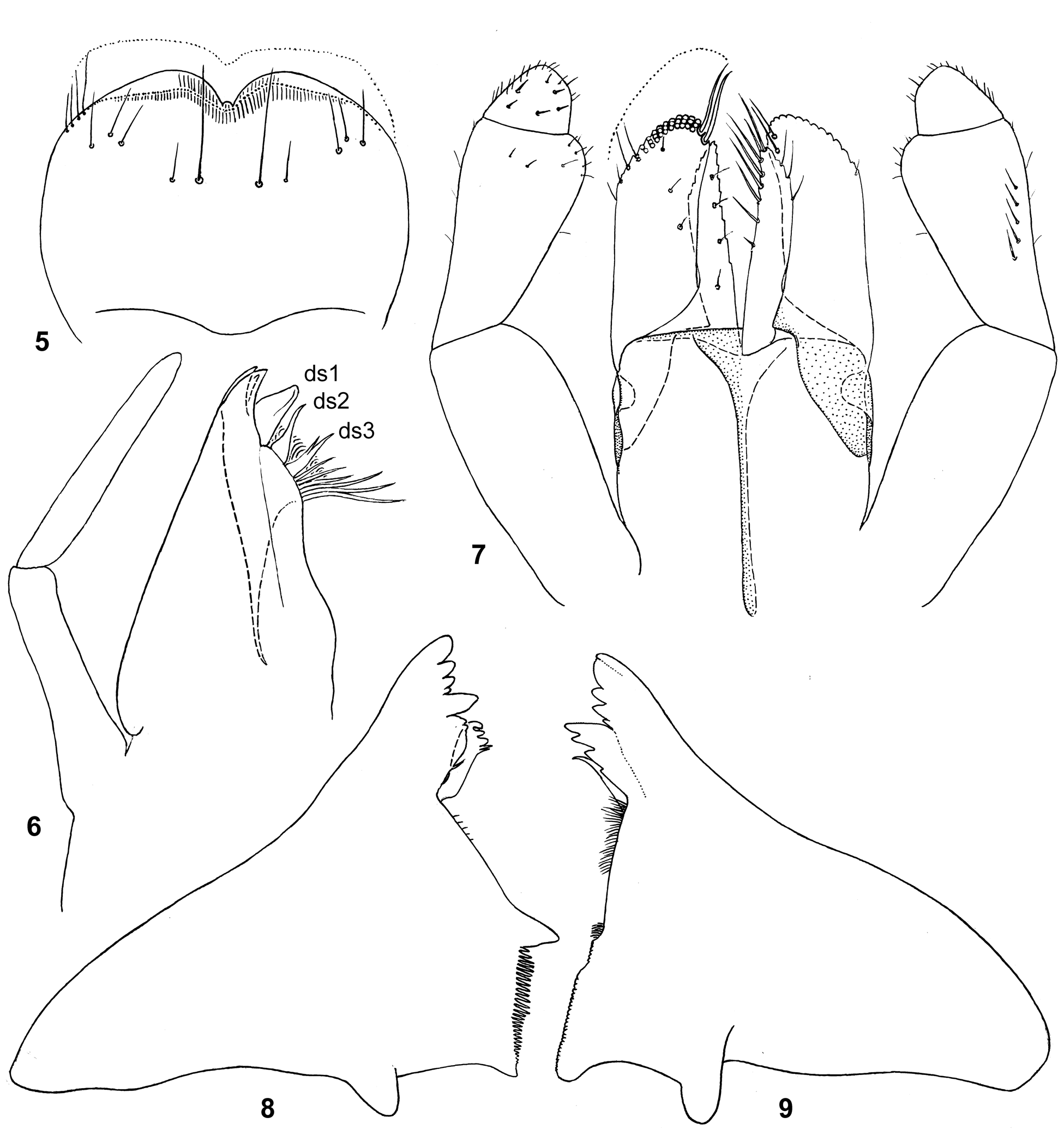
Frons not narrowed, frontal suture obtuse-angled ( Fig. 27 View FIGURES 10–27 ). Labrum roundish; dorsal surface with pair of submedian setae, pair of smaller setae by sides of them and 3 pairs of antero-lateral setae, not forming a row ( Fig. 5 View FIGURES 5–9 ). Mandibles elongated perpendicular to axis of rotation ( Figs 8–9 View FIGURES 5–9 ); on each mandible incisor fused with kinetodontium at most length; each incisor with 4 denticles, apical denticle longest; left prostheca with 3 blunt denticles and 2–3 pointed denticles; right prostheca pointed, adjacent to kinetodontium, with few slender processes (not visible on Fig. 9 View FIGURES 5–9 ); setae between prostheca and mola on right mandible dense, on left mandible few or absent. Maxilla ( Fig. 6 View FIGURES 5–9 ) of the « Baetis - type », i.e. 1 st dentiseta massive, canine-like and bent toward canines; maxillary palp 2-segmented. Labium ( Fig. 7 View FIGURES 5–9 ): paraglossae wider and slightly longer than glossae; latero-apical setae of paraglossa form 3 rows on its distal part; ventro-median setal row consists of 2 o 3 setae forming oblique row; dorso-median setal row reduced to 2 setae located close to apex. Labial palp with 2nd segment long, apically widened and projected medially, 3rd segment small ( Fig. 7 View FIGURES 5–9 ).
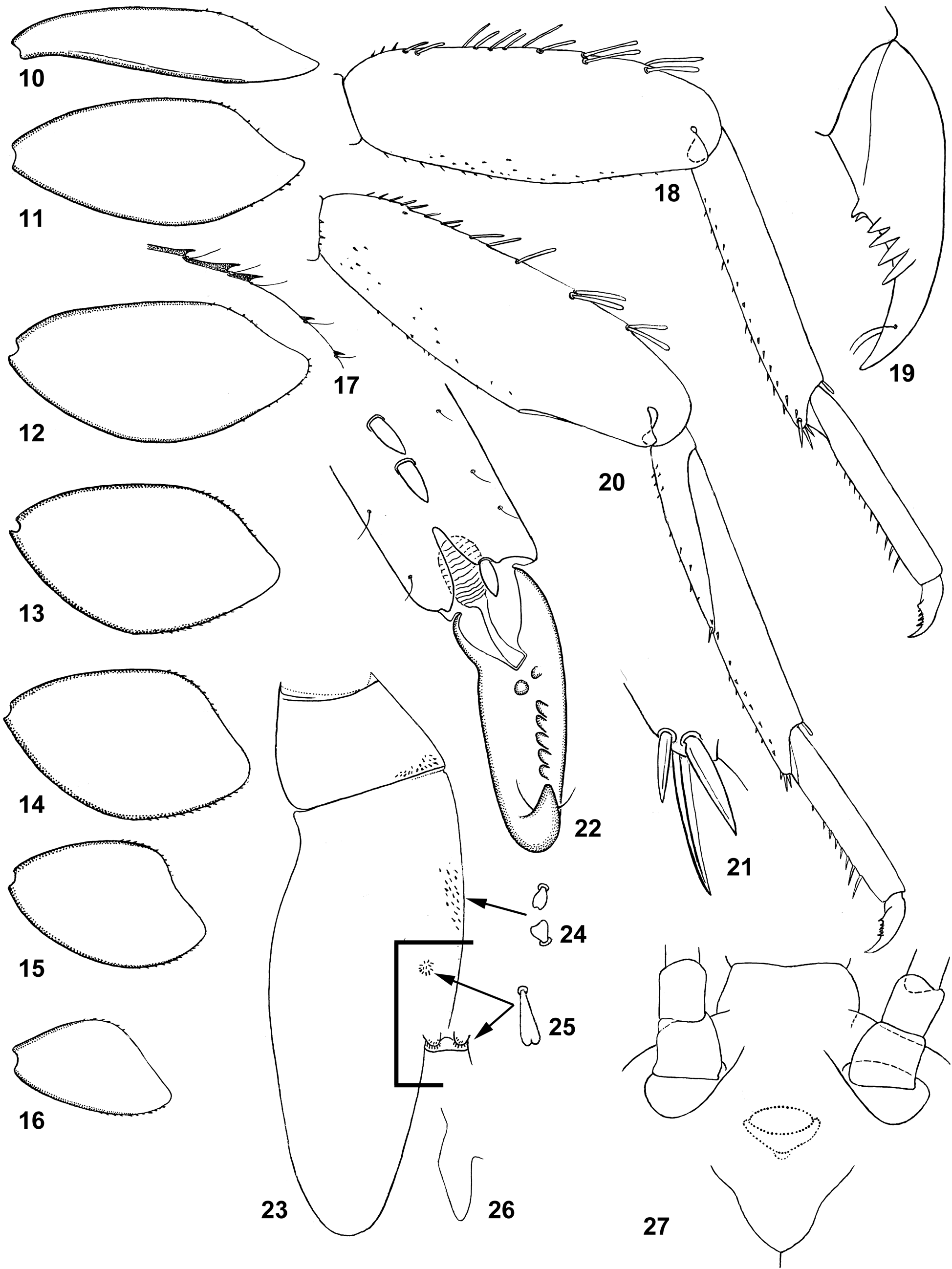
Pronotum with lateral margins rounded ( Fig. 23 View FIGURES 10–27 ). Mesonotum with 2 pairs of small protuberances: one pair near posterior margin between protoptera, another in front of them ( Figs 1 View FIGURES 1–4 , 23 View FIGURES 10–27 , 28 View FIGURES 28–31 ). Dorsal side of head, pronotum and mesonotum with minute triangular setae, each narrow at base and sharply widened apically ( Fig. 24 View FIGURES 10–27 , 28 View FIGURES 28–31 ); elongated setae of the same type form dense bunches on top of each protuberance of mesonotum ( Figs 25 View FIGURES 10–27 , 28 View FIGURES 28–31 ).
Leg setation as following ( Figs 18, 20–21 View FIGURES 10–27 ): Outer margin of femur in proximal half with one row of long stout blunt setae; in distal half with 4 such setae arranged it 2 pairs ( Figs 18, 20 View FIGURES 10–27 ). Patella-tibial suture absent on fore leg ( Fig. 18 View FIGURES 10–27 ), present on middle and hind legs ( Fig. 20 View FIGURES 10–27 ). Outer margin of tibia apically with one stout clavate seta. Inner margin of tibia with small sparse stout setae; apex of tibia with 3–5 larger unequal pointed stout setae ( Fig. 21 View FIGURES 10–27 ); apex of patella with 1–2 such setae (on fore leg, where patella-tibial suture is absent, these enlarged seta either present or not). Inner margin of tarsus with row of pointed stout setae progressively increased from most proximal to most distal one.
Claw asymmetric, with pair of proximal denticles, with 4–5 denticles of anterior row (posterior row of denticles absent), with symmetric pair of subapical setae ( Figs 19, 22 View FIGURES 10–27 ).
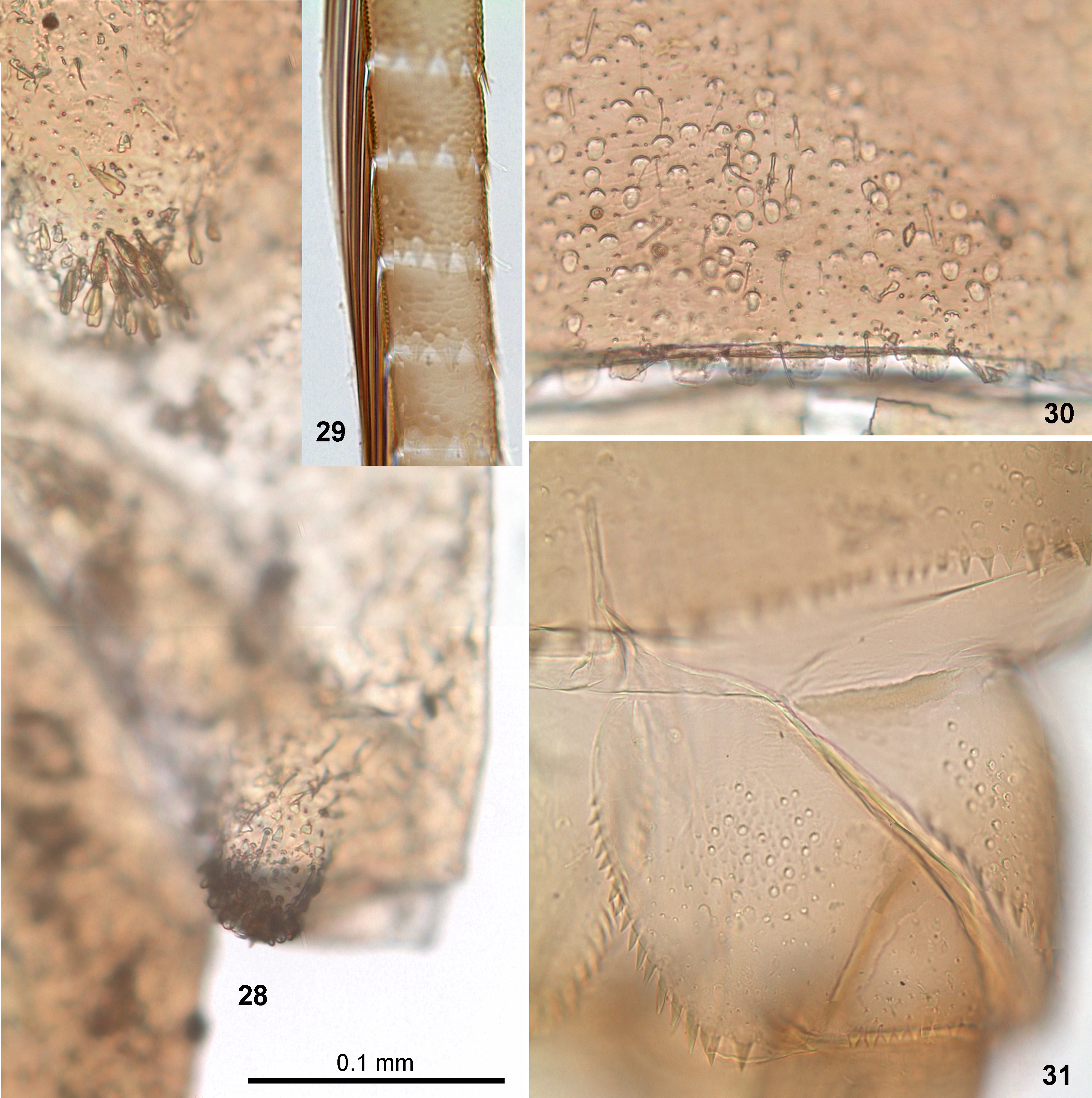
Metanotum and abdominal terga with short rounded scales in semicircular sockets; their sockets lack operculae ( Fig. 30 View FIGURES 28–31 ). Posterior margin of metanotum smooth, without denticles. Denticles on posterior margins of abdominal terga I–IX short, wide, blunt, nearly semicircular, membranous, colorless, form regular row ( Fig. 30 View FIGURES 28–31 ); on all terga, including tergum IX, this row not interrupted medially. On abdominal tergum X, projected median part without denticles on posterior margin; side parts with irregular pointed denticles. Sterna of posterior half of abdomen with scales in semicircular sockets, like that on terga. Posterior margins of abdominal sterna I–VII smooth; posterior margins of sterna VIII–IX and paraprocts with pointed sclerotized denticles ( Figs 31 View FIGURES 28–31 , 44 View FIGURES 44–47 ).
All tergalii I–VII obliquely truncate in such a manner that costal margin seems shorter than anal margin; both costal and anal margins sclerotized ( Figs 1, 3 View FIGURES 1–4 , 10–16 View FIGURES 10–27 ); in distal part sclerotized and non-sclerotized margins with few seta-bearing denticles ( Fig. 17 View FIGURES 10–27 ); tergalial tracheae invisible (very narrow, short and colorless); tergalius I narrow, tergalii III–VII progressively decrease in size.
Cerci and paracercus with well-developed primary swimming setae; segments of cerci with boundaries slightly inclined; denticles on their posterior margins triangular, slightly enlarged toward lateral side of cercus; secondary swimming setae absent ( Fig. 29 View FIGURES 28–31 ). Paracercus shorter than cerci ( Fig. 1 View FIGURES 1–4 ).
RESPIRATORY MOVEMENTS. Absent; tergalii unable to make rhythmical movements; being placed to stagnant water, larva cannot live for a long time.
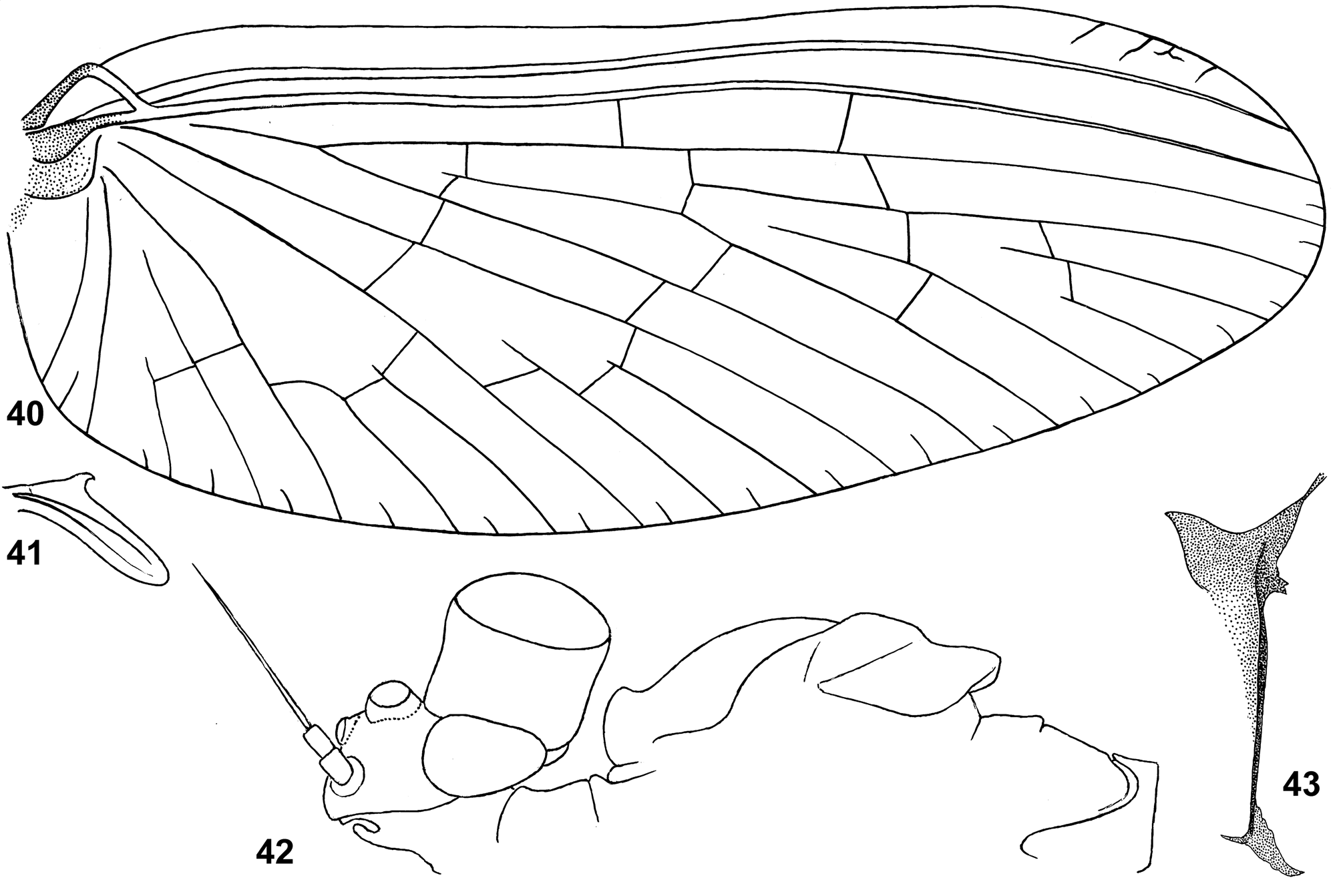
Subimago. CUTICULAR COLORATION. Pronotum brown; mesonotum brown with contrasting colorless medioparapsidal suture; thoracic pleura and sterna partly brown, partly colorless ( Fig. 32 View FIGURES 32–39 ). Shape of postsubalar sclerite as seen in Fig. 43 View FIGURES 40–43 . Wings brown due to brown rings around base of each microtrichion (as in Kluge 2012b: Fig. 22 View FIGURES 10–27 ). Legs with brown markings on femur and base of tibia ( Figs 37–39 View FIGURES 32–39 ). Abdomen nearly unicolor brown; each tergum with pair of slightly darker brown triangular markings; in male sternum IX and gonostyli lighter than other sterna and terga. Cerci colorless.
TEXTURE. On all legs of male and female all tarsomeres covered by pointed microlepides ( Fig. 39 View FIGURES 32–39 ).
Imago, male. Described by Kopelke (1980a).
COLORATION ( Figs 32–34 View FIGURES 32–39 ). Head brown. Turbinate eyes with facetted surface and proximal ¼ of stem orange, other part of stem yellow. Thorax with sclerites uniformly brown, membranes ocher. Wings colorless, veins light. Fore leg with femur brown, tibia and tarsus lighter brown. Middle and hind legs pale ocher, coxa and base of femur tinged with brown. All abdominal terga brown (both hypodermal and cuticular colorations), with medioanterior and medioposterior sigilla lighter; all abdominal sterna lighter brownish, also with medioanterior and medioposterior sigilla lighter ( Fig. 34 View FIGURES 32–39 ). Genitals brown ( Figs 48–52 View FIGURES 48–52 ). Cerci pale ocher, unicolor.
STRUCTURE. Turban eyes narrow and cylindrical ( Figs 32–33 View FIGURES 32–39 , 42 View FIGURES 40–43 ). Anteronotal protuberance pointed in lateral view ( Figs 32 View FIGURES 32–39 , 42 View FIGURES 40–43 ). Hind wing of the « Centroptilum - type »: narrow, with 2 longitudinal veins, with single hooked costal projection ( Fig. 41 View FIGURES 40–43 ). On middle and hind leg tarsus less than ½ of tibia length, with apical spine on 1 st and 2nd tarsomeres (initial 2nd and 3rd tarsomeres) (as in Fig. 39 View FIGURES 32–39 ).
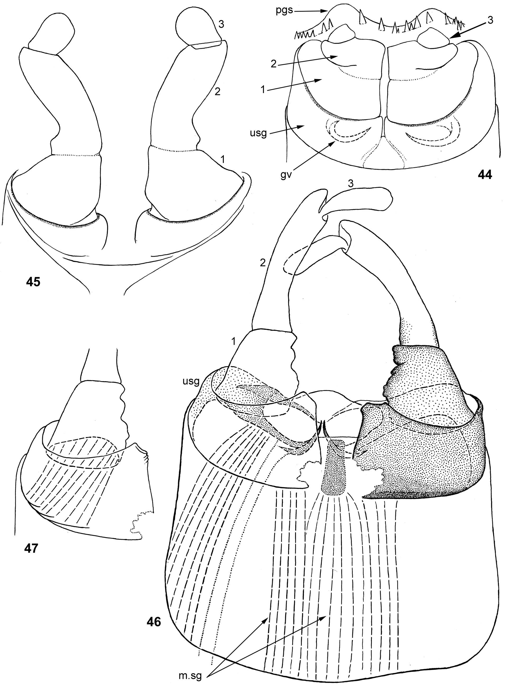
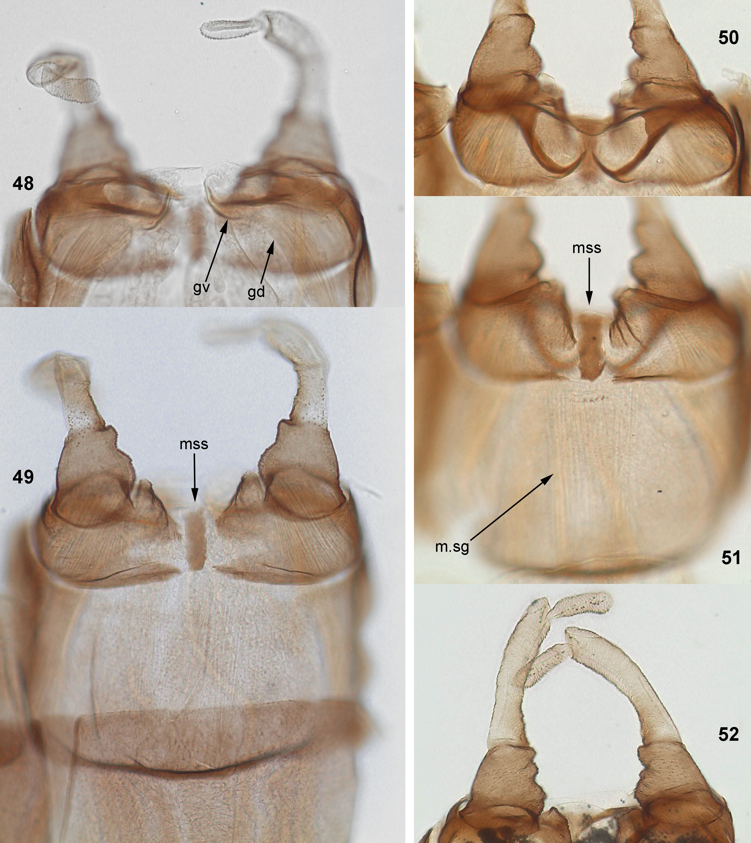
Male genitals ( Figs 44–52 View FIGURES 44–47 View FIGURES 48–52 ): Unistyligers with ventral surface sclerotized and pigmented except proximalmedian angle ( Figs 46 View FIGURES 44–47 , 49 View FIGURES 48–52 ); median styligeral sclerite present, elongate ( Figs 46 View FIGURES 44–47 , 49, 51 View FIGURES 48–52 ). Median styligeral muscle wide, attached to median styligeral sclerite and to unistyligers ( Figs 46 View FIGURES 44–47 , 51 View FIGURES 48–52 ). 1 st segment of gonostylus short and thick, with inner margin irregularly crumpled ( Figs 46 View FIGURES 44–47 , 49, 50, 52 View FIGURES 48–52 ). 3rd (distal) segment elongate ( Figs 46 View FIGURES 44–47 , 48, 52 View FIGURES 48–52 ). Gonovectes pointed, with apices sharply bent caudally and split at place of gonoduct attachment ( Figs 46 View FIGURES 44–47 , 48, 50 View FIGURES 48–52 ). Median part of penial bridge concave ( Figs 46 View FIGURES 44–47 , 50 View FIGURES 48–52 ).
Imago, female. Described by Kopelke (1980a). Coloration as in male ( Figs 35–36 View FIGURES 32–39 ). Eyes widely separated ( Fig. 35 View FIGURES 32–39 ). Fore tarsus with apical spine on 2nd and 3rd tarsomeres. Patella-tibial suture absent on fore leg ( Fig. 37 View FIGURES 32–39 ), present on middle and hind legs ( Fig. 38 View FIGURES 32–39 ).
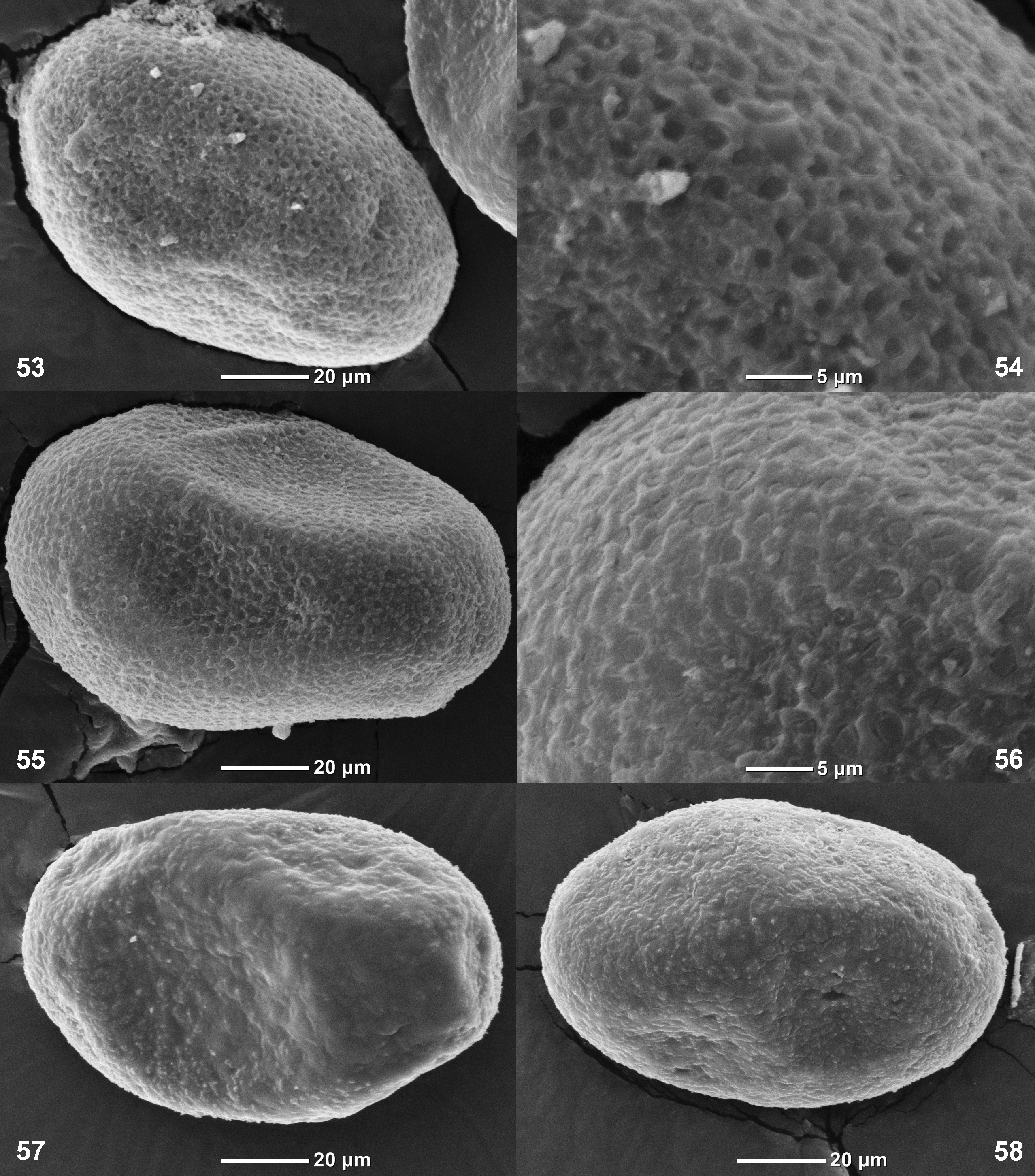
Eggs. Oval, 0.11 mm length. Chorion with poorly expressed net-like relief with small cells ( Figs 53–58 View FIGURES 53–58 ).
Dimensions. Fore wing length (and body length) 3.5–4.5 mm.
Distribution. Congo and Uganda.
Collecting place. Most part of larvae were collected in the river Munyaga 4 km upstream of the place where this river is served as the state boundary between Uganda and Democratic Republic of the Congo. Upstream of the collecting place, this small river runs in a dark forest of the Bwindi National Park; at this area its water is cold, mayflies are abundant, but their species diversity is limited, with domination of Tricorythus exophthalmus Kluge 2010 . Just downstream from the forest, this river runs by opened space in the village; here mayfly fauna has maximum species diversity and includes M. badium . Downstream from this place Munyaga continues to run by opened space among fields, its water becomes warmer, and mayfly fauna is poor.
No known copyright restrictions apply. See Agosti, D., Egloff, W., 2009. Taxonomic information exchange and copyright: the Plazi approach. BMC Research Notes 2009, 2:53 for further explanation.
|
Kingdom |
|
|
Phylum |
|
|
Class |
|
|
Order |
|
|
Family |
|
|
Genus |
Monocentroptilum badium (Kopelke 1980)
| Kluge, Nikita J. 2018 |
Centroptilum badium
| Kopelke 1980a : 113 |
Afroptilum (Afroptilum) badium:
| Gillies 1990a : 99 |