
Isognomon spathulatus ( Reeve, 1858 )
|
publication ID |
https://doi.org/10.11646/zootaxa.4107.2.2 |
|
publication LSID |
lsid:zoobank.org:pub:E36771E3-13D7-491C-8F57-73271113C8FD |
|
DOI |
https://doi.org/10.5281/zenodo.6061192 |
|
persistent identifier |
https://treatment.plazi.org/id/03D5613C-FFF2-FFEF-FF65-FAD545E88DE3 |
|
treatment provided by |
Plazi |
|
scientific name |
Isognomon spathulatus ( Reeve, 1858 ) |
| status |
|
Isognomon spathulatus ( Reeve, 1858)
Figures 1–16 View FIGURE 1 View FIGURE 2 View FIGURE 3 View FIGURE 4 View FIGURE 5 View FIGURE 6 View FIGURE 7 View FIGURE 8 View FIGURE 9 View FIGURE 10 View FIGURE 11 View FIGURE 12 View FIGURE 13 View FIGURE 14 View FIGURE 15 View FIGURE 16 , 18 View FIGURE 18 A, B
Perna spathulata Reeve, 1858 [November]: non-numbered page accompanying pl. 6, pl. 6, fig. 28.— Küster & Clessin, 1890 –1891 ( 18 August 1890; Lieferung 378): 39, pl. 14, fig. 2 [taxonomic revision of Isognomon ]; Paetel, 1890: 207 [shell collection catalog]; Fischer, 1891: 211 [127] [catalog of mollusks of southeast Asia]; Elera, 1896: 798 [University of Santo Tomas museum catalog and faunal checklist of the Philippines]; Hidalgo, 1905a: 25, 1905b: 369 [catalog of shelled mollusks of the Philippines and the Marianas]; Lynge, 1909: 52 [148] [as synonym of I. nucleus ( Lamarck, 1819) ; marine bivalves of Gulf of Thailand]; Fischer-Piette, 1976: 25, 39 [as synonym of I. isognomum ; taxonomic revision of Isognomon ].
Perna spatulata [ sic].— Martens, 1887: 203 [list of shelled mollusks from Mergui Archipelago].
Isognomon spathulata ( Reeve, 1858) — Cooke, 1886: 138 [as synonym of I. nucleus ]; Cotton, 1930: 231 [bivalves of southern Australia]; Fischer-Piette, 1976: 10 [as synonym of I. isognomum ; taxonomic revision of Isognomon ]; Lutaenko, 2000: 368 [bivalves of Vietnam in collection of Zoological Institute, St. Petersburg]; Tëmkin, 2010: 5 [molecular phylogeny of Pterioidea]; Xue et al., 2012: 350 [molecular phylogeny of Pteriomorphia Beurlen, 1944].
Isognomon spathulatum ( Reeve, 1858) — Rochebrune, 1881: 101 [malacofauna of southern Vietnam and Cambodia].
Pedalion spathulatum ( Reeve, 1858) — Faustino, 1928: 25 [review of mollusks of Philippines].
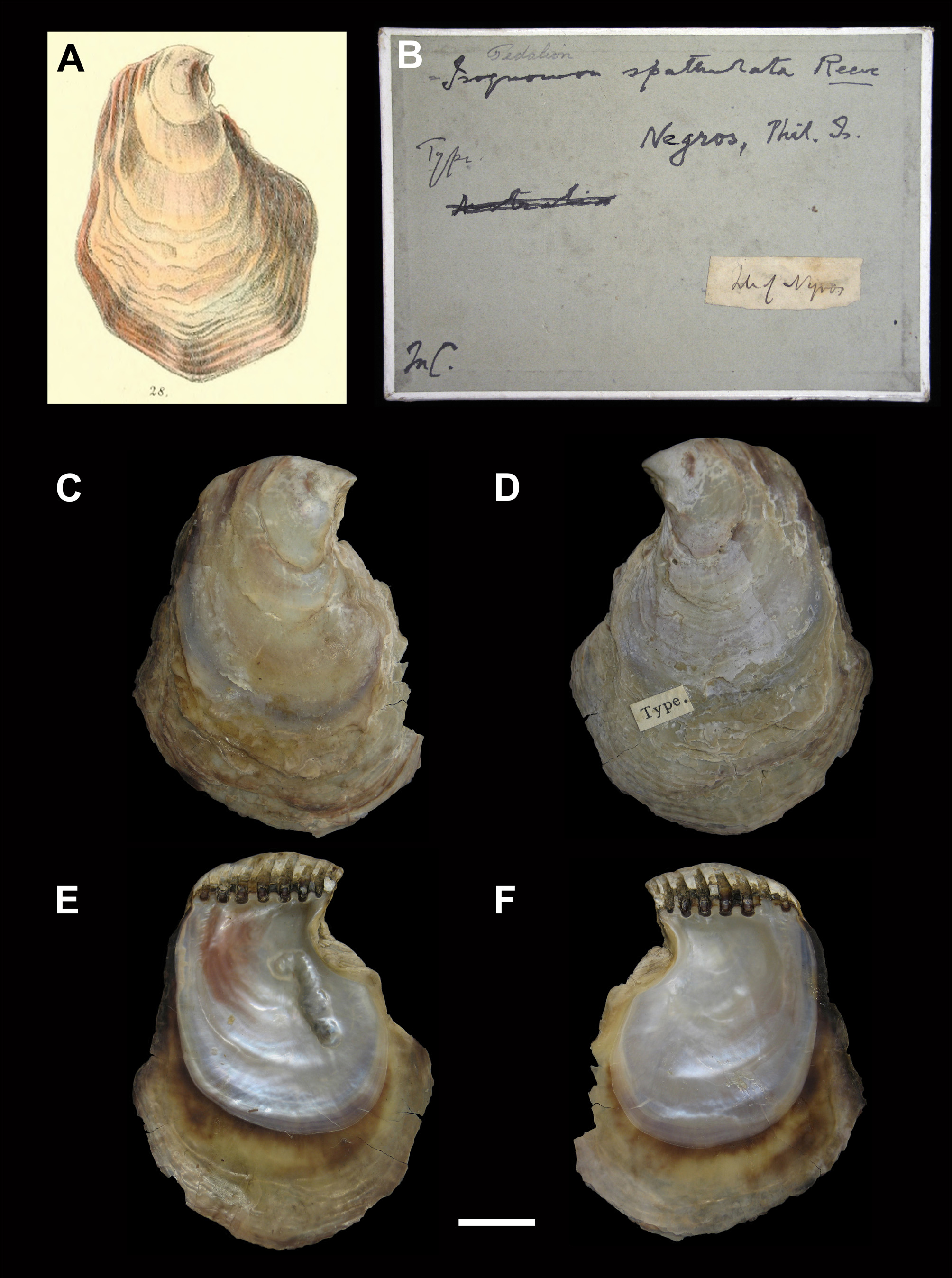
Type material examined. NHMUK 20130275/1 ( lectotype, designated herein; one pair of disarticulated valves; Fig. 1 View FIGURE 1 ); NHMUK 20130275/2–4 ( paralectotypes; three pairs of disarticulated valves and byssus).
Other material examined. Isognomon spathulatus: The Second International Marine Bivalve Workshop Study, Stn. KKB-01, southeastern part of Kungkrabaen Bay, eastern coast of Gulf of Thailand, Thamai District, Chantaburi Province, Thailand, 12º34.42´N, 101º 54. 25´E ( Bieler et al. 2008), byssally attached to prop roots of mangrove trees ( 10 specimens in 95% ethanol, AMNH 319254; 9 specimens 5% formalin fixed, 85% ethanol preserved, AMNH 319256; 10 specimens in 95% ethanol, AMNH 319257; 15 specimens in 95% ethanol, AMNH 319258; 2 dry pairs + 1 LV, AMNH 319367; 18 specimens in 95% ethanol, FMNH 311974; 50 pairs of disarticulated valves, ZMKU Bil 800001–80050); Bãi Dài beach, Vân Đồn Island, Quảng Ninh Province, Gulf of Tonkin, Vietnam ( 2 specimens in alcohol, ZISP 62048); Cát Bà Island, Hải Phòng province, Gulf of Tonkin, Vietnam (5 pairs in alcohol, ZISP 62049; 5 pairs in alcohol, ZISP 62050; 5 pairs in alcohol, ZISP 62051; 1 pair in alcohol, ZISP 62052; 1 pair in alcohol, ZISP 62053; 2 dry pairs, ZISP 62055; 1 pair in alcohol, ZISP 62059); Vĩnh Thực Island, Quảng Ninh Province, Gulf of Tonkin, Vietnam (1 pair in alcohol, ZISP 62054; 1 pair in alcohol, ZISP 62058); Cồn Lò Island, Gulf of Tonkin, Vietnam (1 dry pair, ZISP 62056); Hồng Gai City, Quảng Ninh Province, province, Gulf of Tonkin, Vietnam (2 pairs in alcohol, ZISP 62057).
Isognomon ephippium: The Second International Marine Bivalve Workshop Study, Stn. KKB-01, southeastern part of Kungkrabaen Bay, eastern coast of Gulf of Thailand, Thamai District, Chantaburi Province, Thailand, 12º34.42´N, 101º 54. 25´E, in clumps on mudflats fringing mangroves ( 33 specimens in 95% ethanol, AMNH 319250; 37 specimens in 95% ethanol, AMNH 319251; 11 specimens in 95% ethanol, AMNH 319252; 33 specimens in 95% ethanol, AMNH 319253; 3 dry pairs of disarticulated valves, AMNH 319366; 10 specimens in 95% ethanol, FMNH 311976; 50 pairs of disarticulated valves, ZMKU Bil 800051–800100); KKB-6, “midden material,” fishermen by-catch collected by trawl offshore of Kungkrabaen Bay area (6 RV + 12 LV dry valves, AMNH 319268).
Original diagnosis and description. “Species 28. (Mus. Cuming.) / PERNA SPATHULATA . Pern. testâ subpyriformi-ovatâ, / depressiusculâ, irregulari, versus marginem rudè tami- / natá; lutescente-albâ, purpureo marginatâ. / THE SPATHULATE PERNA . Shell somewhat pyriformly ovate, / rather depressed, irregular, rudely laminated / towards the margin; yellowish-white, edged with / purple. / Hab. Island of Negros, Philippines; Cuming. / Very rudely laminated in respect of sculpture, but pe- / culiar in form.” ( Reeve 1858: page opposite pl. 6, fig. 28.) [Note: “…/…” indicates line break as in the original text; diacritical marks in Latin words are copied from the original text.]
Emended diagnosis: Mangrove-associated isognomonid with equivalve, light brown-yellow to dark brown shell; sculpture of irregular commarginal lamellae (typically abraded); anterior auricles absent to greatly reduced, posterior auricles absent; anteroventral margins produced anteriorly; outline of nacreous layer characteristically comma-shaped; confluent scar of posterior adductor and retractor muscles with greatly elongated, narrow dorsal extremity; edentuous hinge bearing anterior terminal prodissoconch and 6–10 resilifers in adults; outer surface of mantle edge circumnavigated by dark brown stripe; inner surface of innermost mantle fold and adjacent proximal area dark brown-black; distal tips of ctenidia dark brown-black (in living specimens); gastric chamber with posteriorly facing ventral diverticulum; larger right and smaller left abdominal sense organs symmetrically placed with respect to anus in transverse plane.
Taxonomic and nomenclatural remarks. The type specimens of Isognomon spathulatus derive from the collection of Hugh Cuming ( 1791–1865), who travelled to the Philippines in 1836–1840 ( Dance 1980, 1986) and, possibly, obtained the shells sometime in 1837–1838, when he visited Negros Island. The type lot contained four pairs of valves, but the original description neither mentioned the actual number of specimens, nor included the terms “ holotype,” “the type,” or equivalent expressions that would fix the holotype by original designation (73.1.1; ICZN 1999: 79). Given the extent of variation in shell morphology and uncertainty regarding the identity of the type locality for all four specimens, a single specimen, corresponding to the illustration by G.B. Sowerby II in the original description ( Fig. 1 View FIGURE 1 ; Reeve 1858: pl. 6, fig. 28), is designated here as the lectotype of Perna spathulata .
The Latin adjective “ spathulatus ” derives from a diminutive form of the noun “spatha,” that comes from Ancient Greek spáthē ( σπάθη, f.), or "any broad blade, of wood or metal," that probably referred to the spoon-like shape of the shell of I. spathulatus . In the original combination with the generic epithet Perna , the adjective was properly applied in its feminine form, spathulata ; however, its use in combination with masculine genus names Pedalion and Isognomon resulted in species names that were uncoordinated in gender. The grammatical form “ spathulatum ,” that was occasionally applied to the masculine genus names, is an inflected form of the masculine adjective spathulatus . Therefore, Isognomon spathulatus is the proper combination.
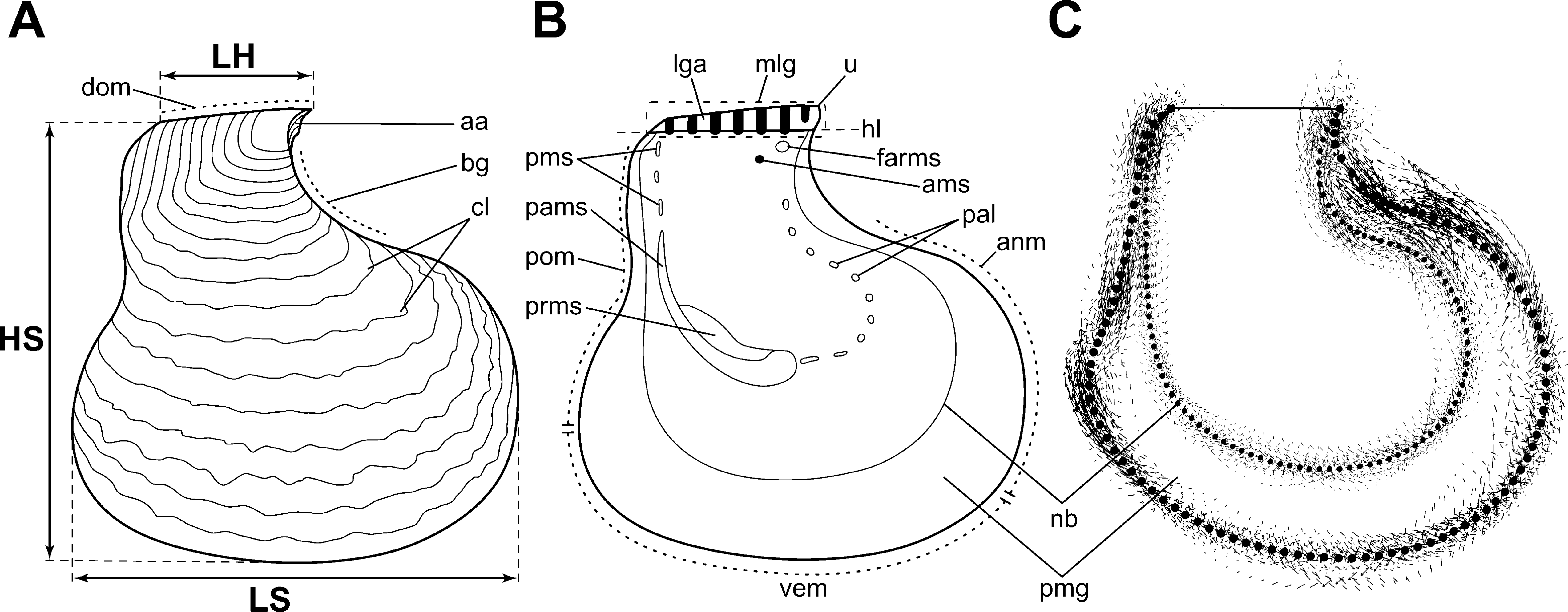
Description. Shell morphology. General features of the shell: Shell ( Figs 1 View FIGURE 1 C–F; 2; 3) strongly prosocline, inequilateral, weakly inequivalve, with left valve slightly more convex than nearly flat right valve. Region of maximum convexity approximately one fourth of valve height from hinge margin. Hinge line short ( Fig. 2 View FIGURE 2 B, hl), straight in adults (slightly arcuate in juveniles), with terminal umbones ( Fig. 2 View FIGURE 2 B, u) at anteriormost extremity of straight dorsal hinge margin ( Fig. 2 View FIGURE 2 A, dom). Shell outline variable, ventrally widening, with anteroventral margin curved anteriorly in most specimens ( Fig. 2 View FIGURE 2 C). Ventral prismatic margins ( Fig. 2 View FIGURE 2 B, C, pmg) wide, flexible in living specimens, allowing for tightly sealed shell closure by valve adpression. Anterior and ventral margins coincident ( Fig. 2 View FIGURE 2 B, anm, vem), broadly rounded; posterior margin ( Fig. 2 View FIGURE 2 B, pom) straight to weakly concave. Width of prismatic margin approximately equal between valves. Outline of inner nacreous layer ( Fig. 2 View FIGURE 2 B, C, nb) following outline of shell margins anteriorly and ventrally; posterior nacreous margin straight, producing characteristic comma-shaped outline of nacreous layer.
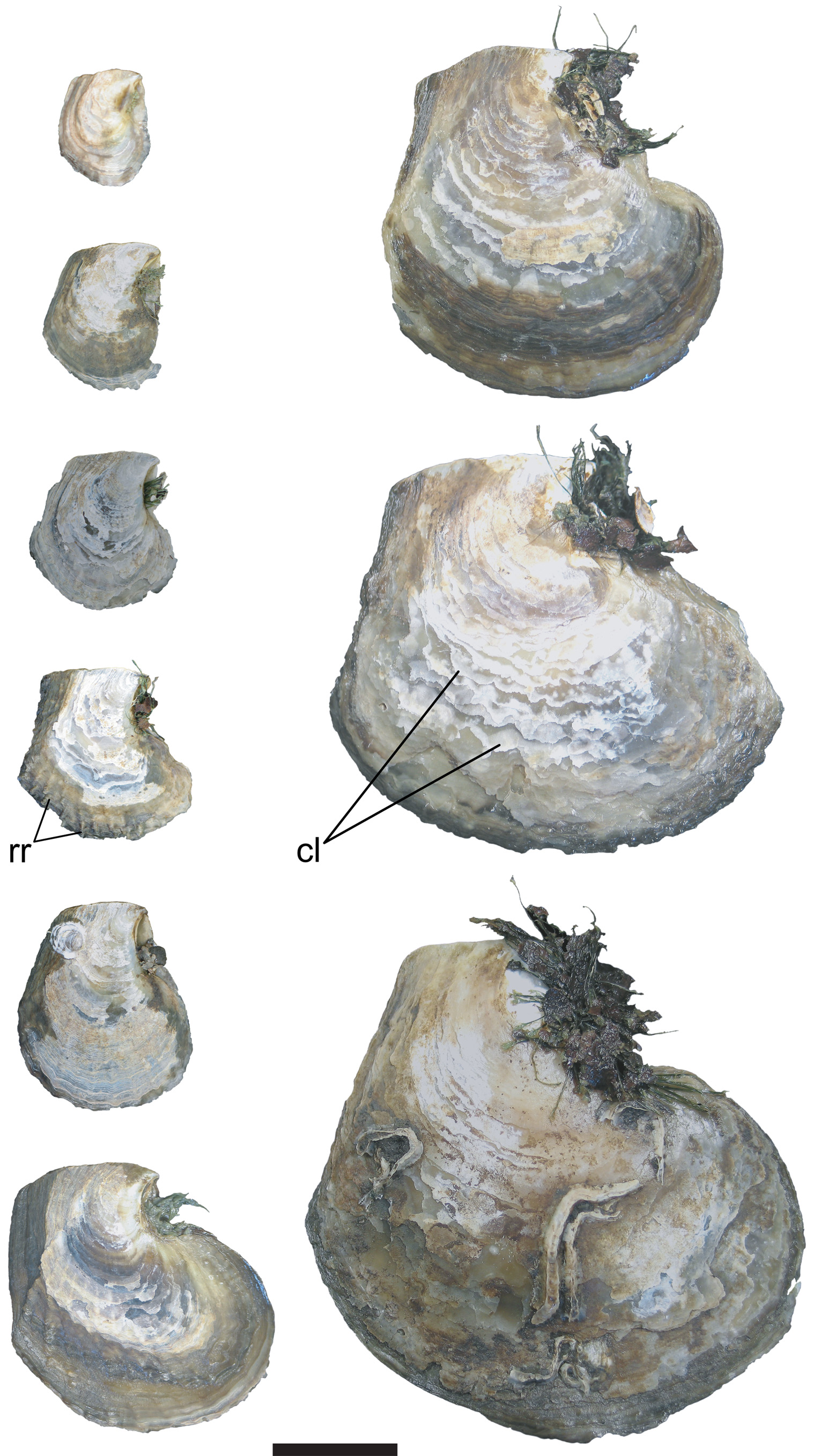
Height ( Fig. 2 View FIGURE 2 A, HS) exceeding length ( Fig. 2 View FIGURE 2 A, LS) in most specimens. Principal dissoconch growth gradients shifting from infracrescent growth in early growth stages to procrescent in later stages ( Fig. 3 View FIGURE 3 ). Externally, valves concordant in color, varying from light yellowish-brown or gray to dark brown and brown-red in adults, without specific pattern ( Figs 1 View FIGURE 1 C, D).
Juveniles (< 10 mm height) typically white. White coloration proximal to umbones often retained into adulthood and sharply demarcated from distal brown part of valves. Inner prismatic margin of same color as outer shell surface but generally lighter and often with irregularly shaped, dark brown or black blotches ( Figs 1 View FIGURE 1 E, F). Border of inner nacreous layer circumscribed by dark brown-back margin in most specimens. Nacre iridescent, silvery blue with light pink blotches proximally. Region of maximal shell thickness approximately at level of dorsalmost extremity of posterior adductor muscle. Occasional pearls small (diameter 0.4 mm; n = 1), white, with smooth surface of considerable luster but weak iridescence ( Fig. 4 View FIGURE 4 A).
Shell mean dimensions (from single population, ZMKU Bil 800001–800050, n = 50): height ( Fig. 2 View FIGURE 2 A, HS) 42.86–69.68 mm, mean 58.95 ± 5.81 mm SD; length (= width; Fig. 2 View FIGURE 2 A, LS) 41.89–68.53mm, mean 55.98 ± 7.71mm SD; hinge length ( Fig. 2 View FIGURE 2 A, LH) 15.67–27.35 mm, mean 22.05 ± 2.52 mm SD.
Auricles, byssal notch, and gapes: Anterior auricles ( Fig. 2 View FIGURE 2 A, aa) absent or inconspicuous in adults. In juveniles, anterior auricles ( Fig. 5 View FIGURE 5 A, aa) small, narrow (higher than long), subtriangular, symmetrical between valves, extending ventrally from umbones at acute angle with hinge line, with densely lamellose surface. Anterior auricles demarcated from concave anterodorsal shell margin by groove, more distinct in right valve. Posterior auricles absent.
Commissural line straight or slightly wavy along the ventral margin. Byssal notch ( Fig. 5 View FIGURE 5 A, bn) present only in right valves of juveniles (< 25 mm in height). Byssal notch broad, weakly demarcated, with ventral border coincident with anterior shell margin. Byssal gape ( Fig. 2 View FIGURE 2 A, bg) facing anterodorsally, symmetrical between valves, formed along concave shell margin between beak and convex anterior shell margin. Posterior gape absent.
Sculpture: Irregular lamellae ( Figs 2 View FIGURE 2 A; 3, cl) projecting from broadly spaced, irregular, wavy commarginal lirae. Lamellae fragile, largely or entirely abraded in most specimens, resulting in nearly smooth exterior shell surface or low-relief irregular commarginal sculpture. Occasional specimens with low-relief radial ribs (costae; Fig. 3 View FIGURE 3 , rr). External sculpture concordant between valves.
Muscle scars: Posterior adductor muscle (PAM) and posterior pedobyssal retractor muscle (PRM) attachment sites forming a single, large, elongated, confluent muscle scar ( Fig. 2 View FIGURE 2 B, pams, prms) in posteroventral region of nacreous surface at approximately 40–45º relative to hinge axis, with two subequal lobes along anterior margin and elongated narrow dorsal extremity. PAM and PRM scars symmetrical between valves. PAM scar crescent-shaped, widest in ventralmost extremity and gradually tapering dorsally. PRM scars suboval, laterally compressed, often with concave anterior margin, adjacent to concave anterior border of PAM scars. PRM scars extending from ventralmost border to approximately two thirds of PAM scar height. Two small, subequal, rounded scars formed by fused extremities of anterior pedobyssal retractor (FARM; Fig. 2 View FIGURE 2 B, farms) and pedo-byssal accessory ( Fig. 2 View FIGURE 2 B, ams) muscles, with former situated slightly anterodorsal to latter in subumbonal cavity of each valve. Pallial line ( Fig. 2 View FIGURE 2 B, pal) discontinuous, consisting of multiple attachments of sinuses formed by merged radial pallial retractor muscles in both valves. Pallial line extending from anteroventral extremity of PAM scar to area proximal to FARM scar, roughly following outline of anterior shell margin. Relatively indistinct small pallial muscle scars ( Fig. 2 View FIGURE 2 B, pms), occasionally fused, forming straight, narrow, secondary pallial line, stretching from dorsal extremity of PAM scar toward posterior extremity of hinge line.
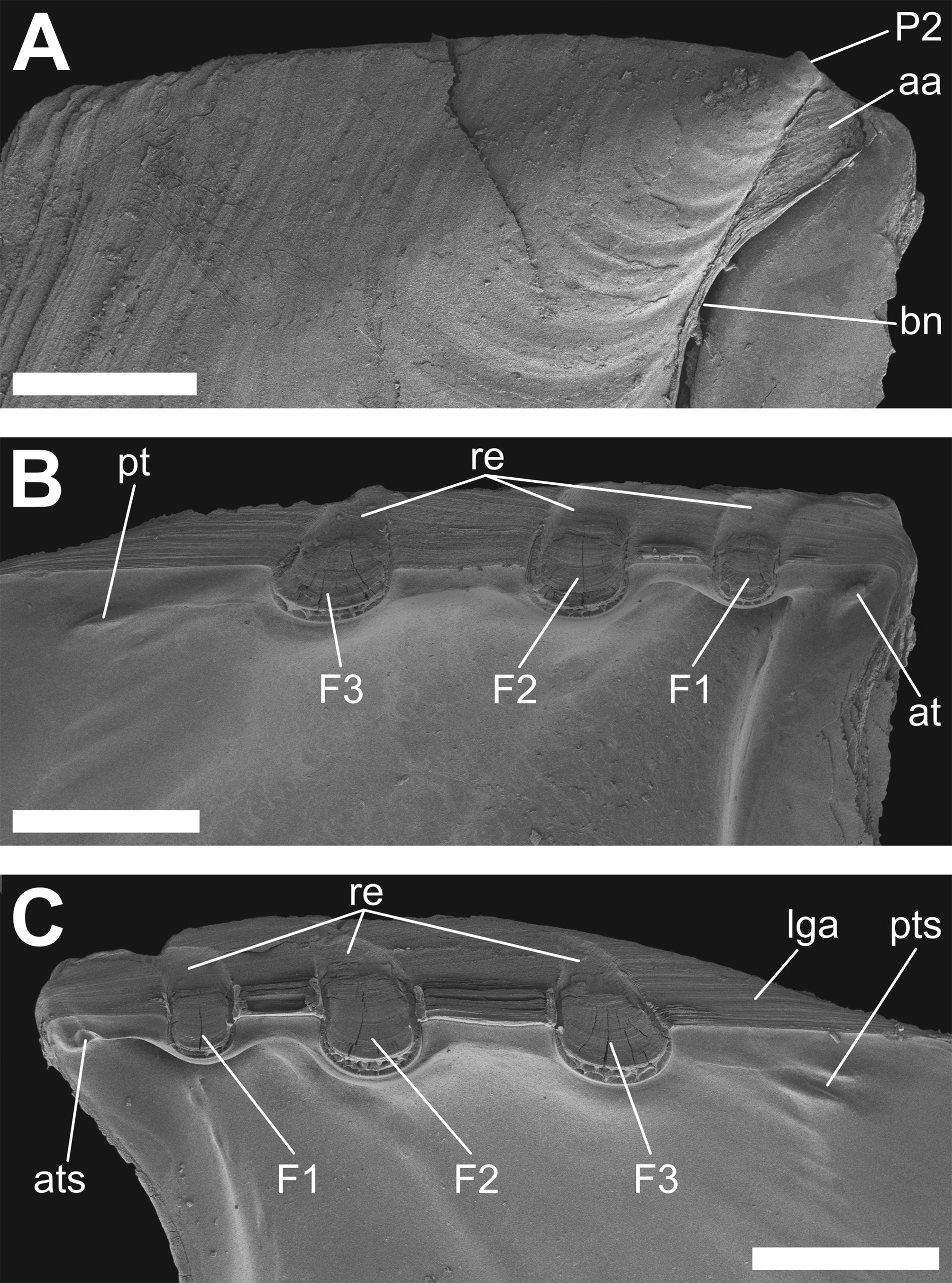
Hinge and ligament: Hinge in juveniles ( Figs 5 View FIGURE 5 , 6 View FIGURE 6 ) with posteriorly oblique, single, anterior subumbonal tooth ( Figs 5 View FIGURE 5 B; 6B, at) and posteriorly oblique, ridge-like, posterior submarginal tooth ( Fig. 5 View FIGURE 5 B, pt) in left valve and corresponding sockets in right valve ( Fig. 5 View FIGURE 5 C, ats, pts). Hinge teeth in adults less distinct and frequently absent entirely.
Ligamental (cardinal) area ( Figs 2 View FIGURE 2 B, 5C, lga) symmetrical, occupying dorsal part of valves along entire length of hinge axis, extending to dorsal shell margin, and typically gradually decreasing posteriorly. Ligamental areas of opposing valves separated by broad, V-shaped cleft (resulting from interumbonal growth), allowing opening of valves about hinge axis. Dorsal margin ( Fig. 2 View FIGURE 2 A, dom) typically severely damaged in adult specimens.
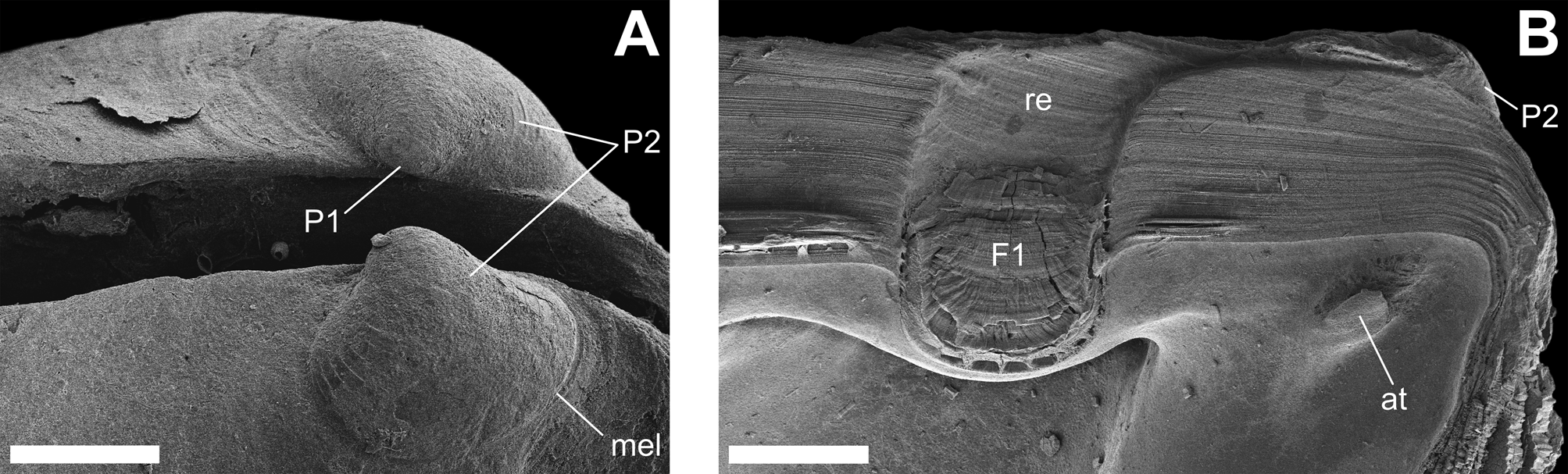
Ligament ( Fig. 2 View FIGURE 2 B, mlg) multivincular, opisthodetic, and dorsal submarginal. Greater part of ligamental area dorsal to hinge axis occupied by older, non-functional part of ligament; active part of ligament restricted to area at and slightly ventral to hinge axis. Fibrous components of ligament (resilia; Figs 5 View FIGURE 5 B, C; 6B, F1–3) deposited in resilifers ( Figs 5 View FIGURE 5 B, C; 6B, re); lamellar component of ligament occupying flat ligamental area between resilifers. Fibrous and lamellar layers continuous across valves, with former produced more ventrally than latter. Resilifers perpendicular to hinge axis, slightly curving anteriorly in their dorsal part, typically uniform in width, and deposited at relatively equal intervals in same individual. Resilifers monoserial, rounded in cross section, breached along ventral surface in large and crenulated in small individuals ( Figs 5 View FIGURE 5 B, C, 6B). One or two anteriormost resilifers in adult shells generally narrower and frequently not reaching hinge axis in adults. Number of resilifers in adult individuals (from single population, ZMKU Bil 800001–800050, n = 50): 6–10 (mean 7.62 ± 1.03 mm SD).
Ontogenetically first (anteriormost) resilifer (F1) originating below center of larval hinge (provinculum), projecting first posteriorly nearly parallel to hinge line and gradually widening, then abruptly turning ventrally (perpendicular to hinge axis), and from that point no longer expanding in width ( Fig. 6 View FIGURE 6 B). All subsequent resilifers developing posterior to F1 along hinge axis along similar growth trajectory. Ligament growth pattern formula ( Malchus 2004): [1c a -p, 2p-pv, 3p-pv, n p-pv].
Larval shell: Prodissoconch ( Figs 5 View FIGURE 5 A; 6, P2) terminal, tilted ventrally relative to hinge axis, with homogeneous surface texture. Prodissoconch 1 (P1) D-shaped, equilateral, devoid of commarginal sculpture; length 58 Μm (n = 1) ( Fig. 6 View FIGURE 6 A, P1). P1 distinguished from prodissoconch 2 (P2) by first commarginal lira. P2 very slightly opisthogyrate, subtriangular in outline, with posterior margin slightly broader and rounder than anterior margin; average length 266.5 ± 0.5 Μm SD (n = 2). Exterior surface of P2 with conspicuous, regularly widely spaced commarginal lirae. P2 sharply demarcated from early dissoconch by metamorphic line ( Fig. 6 View FIGURE 6 A, mel).
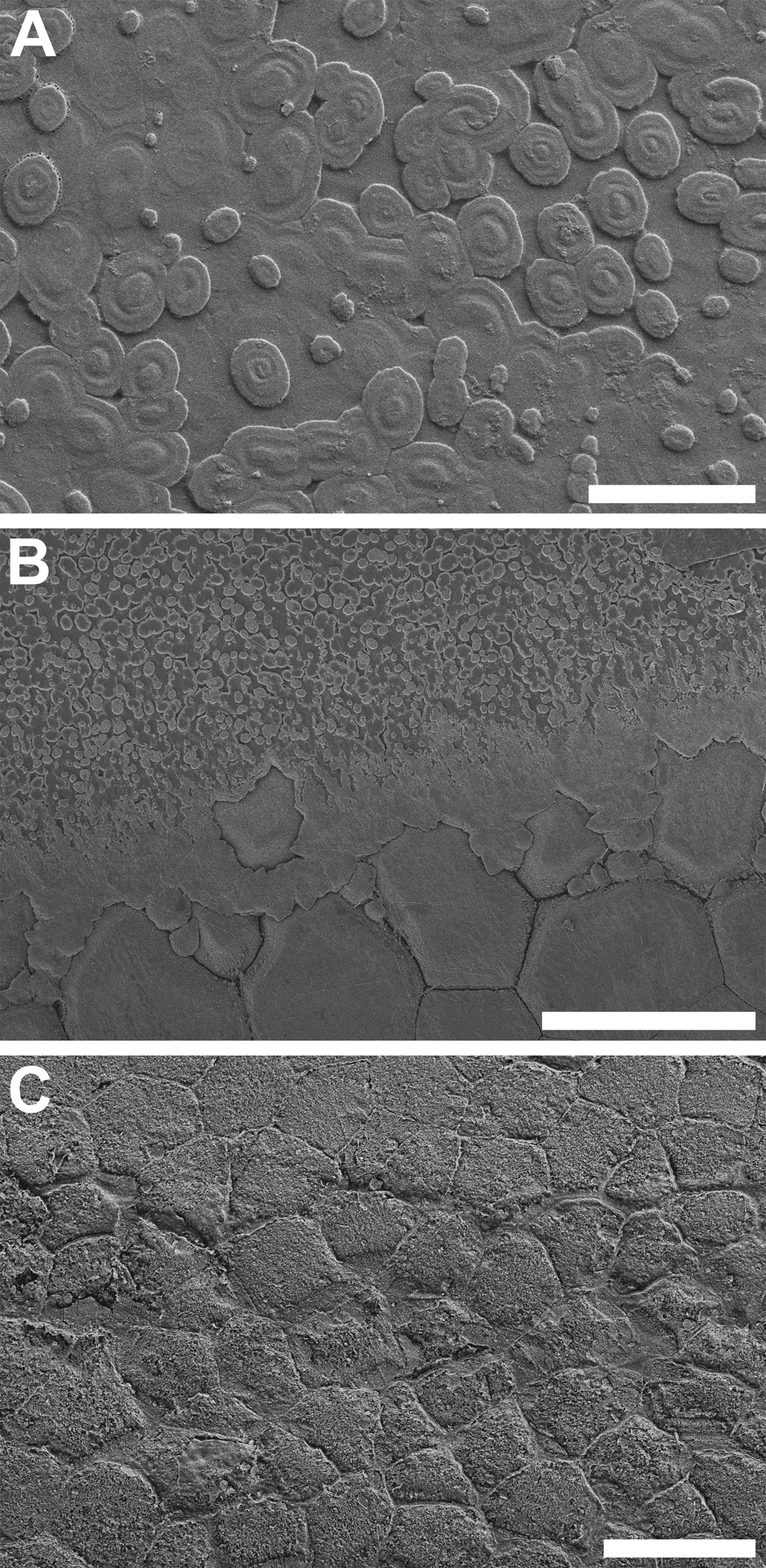
Shell microstructure ( Fig. 7 View FIGURE 7 ): Microstructure prismatonacreous, with outer shell layer composed of simple, vertical, polygonal (predominantly pentagonal and hexagonal, frequently irregular) prisms ( Figs 7 View FIGURE 7 B, C) and inner sheet nacreous layer. Individual nacre tablets ( Figs 7 View FIGURE 7 A, B) regularly oval in outline and varying in size depending on growth stage; growing predominantly by nucleation with subsequent coalescence and, occasionally, by dextral or sinistral spiral growth. Prismatic layer in umbonal region often abraded, revealing underlying nacreous layer.
Anatomy. Visceral mass ( Fig. 8 View FIGURE 8 B, vm) relatively small compared to size of shell, situated ventral to hinge, produced posteroventrally toward anterior surface of posterior adductor muscle and connected to it by loose connective tissue. Visceral mass occupied by digestive glands, enveloping centrally placed gastric chamber and gonadal tissue.
Mantle: Mantle lobes occupying most of area between valves extending from hinge line ( Fig. 8 View FIGURE 8 A, hl) around circumference of shell nacreous border ( Figs 8 View FIGURE 8 A, C, nb), with four distinct regions: mantle isthmus, central, pallial, and marginal zones.
Margins of mantle lobes fused dorsally forming translucent, colorless mantle isthmus ( Fig. 8 View FIGURE 8 B, mi) with irregularly crenulated outline. Mantle isthmus crenulations corresponding to ventrally protruding extremities of resilia. Area of mantle lobe fusion continuous with mantle isthmus, extending ventrally at approximately right angle relative to hinge axis for approximately 3–4 mm anteriorly and 10–15 mm posteriorly. Remaining mantle margins free. Anterior part of mantle isthmus and anterodorsal margins of mantle lobes forming pallial hood concealing lips, mouth, dorsal part of labial palps, and foot. Inner surfaces of mantle lobes connected by short supramyal septum ( Figs 8 View FIGURE 8 B, C, ss), stretching from dorsalmost surface of posterior adductor muscle to posterior surface of visceral mass dorsal to intestine. Area between inner surfaces of mantle lobes circumscribed by supramyal septum and dorsal surface of visceral mass, and filled with loose connective tissue.
Central zone of mantle lobes ( Figs 8 View FIGURE 8 A; 9C, mlh) homogeneous, colorless, semitranslucent, with inner surfaces fused to visceral mass, pericardium, and kidneys. Mantle-visceral mass fusion extending to proximal edges of outer labial palps anteriorly and ventral border of pericardial wall posteriorly. Mantle lobes perforated by pallial retractors, posterior adductor, pedobyssal retractor and accessory muscles. In larger individuals, mantle central zone ventral to visceral mass thicker, denser, and non-translucent, creamy pale yellow to light brown. Large eosinophilic secretory cells with distinct granules ( Fig. 9 View FIGURE 9 B, ec) lining epithelium of outer surface of central zone and extending into connective tissue of pallial zone.
Contractile zone of mantle lobes ( Figs 8 View FIGURE 8 A; 9C, mlc) colorless, translucent, free from fusion; widest at posteroventral part and narrowing towards mantle isthmus. Outer surface of contractile zone translucent-white, demarcated distally by light brown rim running proximal to mantle margin. Inner surfaces of contractile zone of mantle lobes light brown except for white, wide cilial trail, stretching medially from posterior mantle edge to area proximal and posterior to ventral edges of palps.
Marginal zone of mantle (mantle edge; Figs 8 View FIGURE 8 A; 9C, mm) surrounded by single layer of columnar epithelium ( Fig. 9 View FIGURE 9 A, ce) and subdivided into four folds with periostracal groove ( Figs 9 View FIGURE 9 A, D, pgr) separating pairs of inner (IF) and outer folds (OF). Periostracum ( Figs 9 View FIGURE 9 A, D, p) secreted from cleft between OF-1 and IF-1 ( Figs 9 View FIGURE 9 A, D). Bundles of pallial muscles ( Figs 9 View FIGURE 9 A, B, pm) penetrating into IF-2 and OF-2. Connective tissue containing numerous muscle fibers ( Fig. 9 View FIGURE 9 A, mfb).
Innermost fold (IF-2; Figs 9 View FIGURE 9 A, D, if-2) largest in height and width, longitudinally grooved, and with muscle bundles underlying epithelia of inner and outer surfaces. Margin of IF-2 with equally spaced small, digiform, conical, non-branched tentacles ( Figs 9 View FIGURE 9 A, D, t) of approximately equal size. Bases of IF-2 tentacles sunken below margin for approximately one fourth of fold height along its outer surface. Tentacles and both surfaces of IF-2 dark brown-black. Subtriangular infoldings of IF-2 of opposing mantle lobes forming pallial fold ( Fig. 8 View FIGURE 8 A, pf) at posteroventral part of mantle margin at points of contact with distal tips of ctenidia. Exterior surface of IF-2 and adjacent area of mantle margin dark brown-black, resulting in conspicuous dark color rim on periphery of mantle.
Middle mantle fold (IF-1; Figs 9 View FIGURE 9 A, D, if-1) slightly shorter and narrower than IF-2; with alternating nonbranched, digiform, conical tentacles ( Figs 9 View FIGURE 9 A, D, t) and shorter, non-branched, wide, flat, lobate tentacles, with latter situated on outer and former on inner sides of fold margin. IF-1 conical tentacles approximately 2–3 times taller than IF-2 tentacles. IF-1 colorless except for faint, irregular, small dark-colored blotches on inner surface. Pallial muscles extending along outer surface to edge of IF-2. IF-1 outer surface with longitudinal folds and grooves. Subepithelial pigmented (eosinophylic) and secreting cells concentrated in proximal part of the IF-1 outer surface bordering periostracal groove. Both inner folds covered by ciliated epithelium.
Outer fold proximal to periostracal groove (OF-1; Figs 9 View FIGURE 9 A, D, of-1) shorter and narrower than other mantle folds; covered by smooth, thin epithelium. Connective tissue with large secretory and eosinophilic cells, and diffuse muscle fibers. Marginal dark pigmented spots (possibly pallial ocelli) present on OF-1 margin, but not localized histologically. No distinct nerves innervating pigmented spots were detected. Inner and outer surfaces of OF-1 not pigmented.
Outermost fold (OF-2; Figs 9 View FIGURE 9 A, D, of-2) slightly exceeding OF- 1 in height and approximately of same height as IF-1. Subepithelial secretory and pigmented eosinophilic cells situated along outer surface of OF-2. Loose muscle fibers dispersed throughout connective tissue. Surfaces of OF-2 not pigmented; outer surface circumnavigated by distinct dark brown, narrow stripe. Both outer folds covered by non-ciliated epithelium, lacking tentacles, and not invested by branches of pallial muscles.
Inner and outer pairs of mantle folds diminishing in size and gradually fusing towards mantle isthmus, producing two folds in each lobe. More proximal to mantle isthmus, inner folds of opposing mantle margins fuse, forming medial ridge, bounded on either side by outer mantle folds of opposing mantle margins; subsequently, outer mantle folds fusing with medial ridge, forming mantle isthmus.
Cilial trail stretching medially on inner surfaces of mantle lobes from pallial fold to area proximal to ventral edges of labial palps and providing cilial connection between inner surfaces of mantle lobes and dorsal edges of outer demibranchs.
Musculature: Adults monomyarian with posterior adductor muscle ( Fig. 8 View FIGURE 8 C, pam). Posterior adductor muscle large, elongated, narrow, and crescent-shaped in lateral view; placed posteroventrally within nacreous layer. Narrow dorsal extremity of posterior adductor parallel to posterior shell margin; wider ventral extremity curving anteriorly. Posterior adductor heterogeneous, comprised of subequal semitransclucent anterior ( Fig. 8 View FIGURE 8 B, amq) and opaque posterior ( Fig. 8 View FIGURE 8 B, amc) lobes, corresponding to “quick” and “catch” muscle fibers, respectively.
Two symmetrical posterior pedobyssal retractor muscles ( Figs 8 View FIGURE 8 B, C, prm) extending and widening posteroventrally from lateral surfaces of byssal root to concave anteroventral surface of posterior adductor muscle.
Left and right pairs of symmetrical, fused anterior pedobyssal retractor muscles ( Fig. 8 View FIGURE 8 B, farm) extending from dorsal side of byssal root to attachment points in subumbonal cavities in each valve, with anterior branches running parallel to and on inner side of posterior branches. Symmetrical accessory pedobyssal muscles branching off anterior pedobyssal retractors at approximately right angles in area proximal to esophagus and forming attachments slightly posteroventral to anterior pedobyssal retractor attachment sites.
Web-like network of radial pallial retractor muscles ( Fig. 8 View FIGURE 8 A, rprm) penetrating contractile and marginal zones. Pallial retractors fusing proximally forming sinuses attached to inner nacreous shell layer (pallial line). Fibers of pallial muscles underlying inner and outer surfaces of mantle tissue, penetrating into IF-2 and OF-2 mantle folds. Paired longitudinal ctenidial retractor muscles passing through ctenidial axes ( Fig. 11 View FIGURE 11 , crm).
Foot and byssus: Foot ( Fig. 8 View FIGURE 8 C, f) short, muscular, tongue-shaped, somewhat flattened dorsally, slightly tapering distally, non-pigmented, excepting occasional, irregular, orange-brown spots on dorsal surface. Foot emerging from anterior surface of visceral mass with distal tip pointing dorsally. Byssal groove with regular transverse ridges on inner surface, extending medially on ventral surface of foot from wide byssal opening to distal tip; conspicuous distal pit absent.
Byssus ( Fig. 8 View FIGURE 8 B, by) consisting of non-mineralized, individual byssal threads ( Fig. 8 View FIGURE 8 C, byt), spreading fanwise in plane of shell commissure. Byssal threads dark bronze-green, laterally compressed, flat (ribbon-like), approximately uniform in width along entire length, except for slightly wider distal extremity proximal to adhesive disks. Byssal threads fused in byssal stem and root. Byssal threads terminating distally with translucent, yellowish or brown-green, flat adhesive disks ( Fig. 8 View FIGURE 8 C, ad). Adhesive disks wide, roughly oval or leaf-shaped in outline, formed by expansion of byssal threads in direction of their long axis, with edges typically damaged around periphery. Dorsal surface of adhesive disks smooth, with faint radial striations and single conspicuous, frontal buttress ridge, continuous with margin of byssal thread, and not extending to disk’s periphery. Adhesive disks of individual byssal threads often fused, producing common, irregularly shaped attachment surfaces. Byssal thread rootlets plumate, with proximally tapering narrow axis (not extending beyond posteriormost pair of side branches). Byssal stem dark bronze, subcylindrical, slightly laterally compressed, composed of tightly consolidated byssal threads, and slightly extending from byssal opening. Posterior part of byssal stem continuous with short byssal root, but clearly demarcated from it by sharp change in color. Byssal root cream-yellow, translucent (opaque when dried), embedded in midventral part of visceral mass; composed by tightly packed, dorsoventral stack of byssal thread rootlets; bifurcated distally with side branches of rootlets divided into subequal left and right bundles. Side branches of rootlets fused, forming vertical lamellae of byssal root. Byssal root lamellae penetrating byssal gland and interdigitating with bundles of pedobyssal retractor muscles.
Labial palps: Paired symmetrical labial palps ( Figs 8 View FIGURE 8 B, C, lp) projecting dorsoventrally from their connection to lips to level of base of foot on left and right anterolateral surfaces of visceral mass. Each palp consisting of inner and outer elongated triangular folds, attached to visceral mass along longer side, wider at base and narrowing dorsally. Outer and inner folds continuous with upper and lower lips respectively; inner surfaces with 35–40 ciliated, parallel, transverse ridges ( Fig. 10 View FIGURE 10 , ri); exterior surfaces smooth. Aboral side of pulp ridges with secondary parallel ledges ( Fig. 10 View FIGURE 10 , le). Outer surfaces of outer folds light brown in living specimens. Anterior filament of inner demibranch not inserted into or fused with distal oral groove (labial palp-ctenidia association of Category III; Stasek 1963).
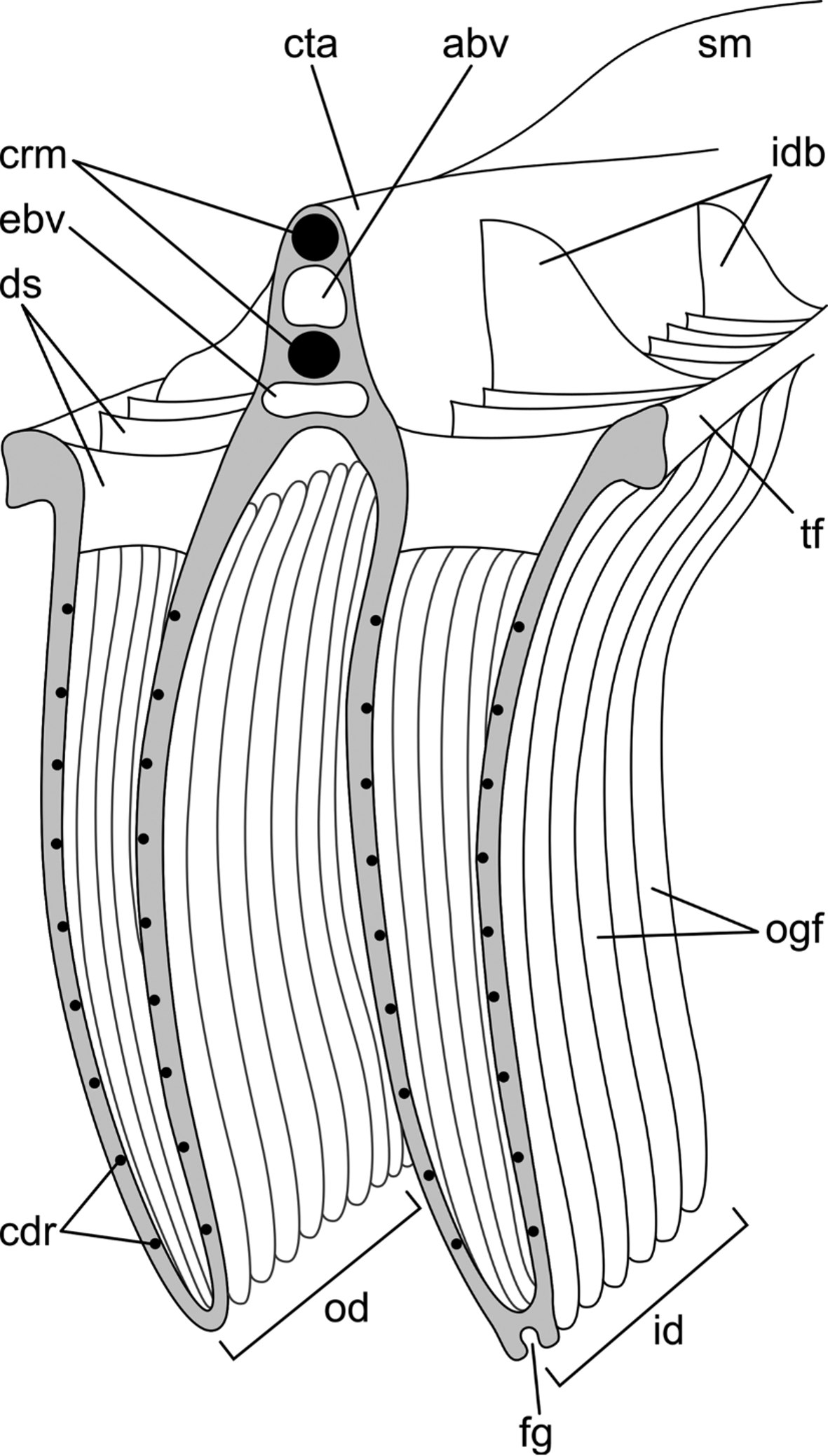
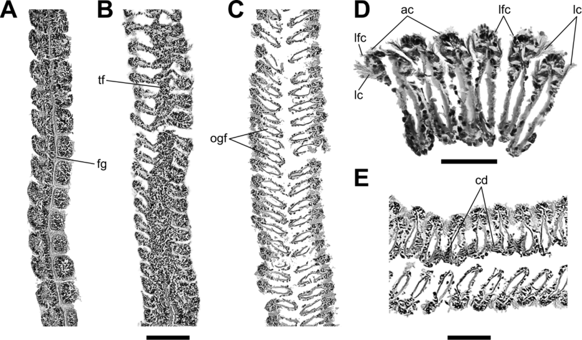
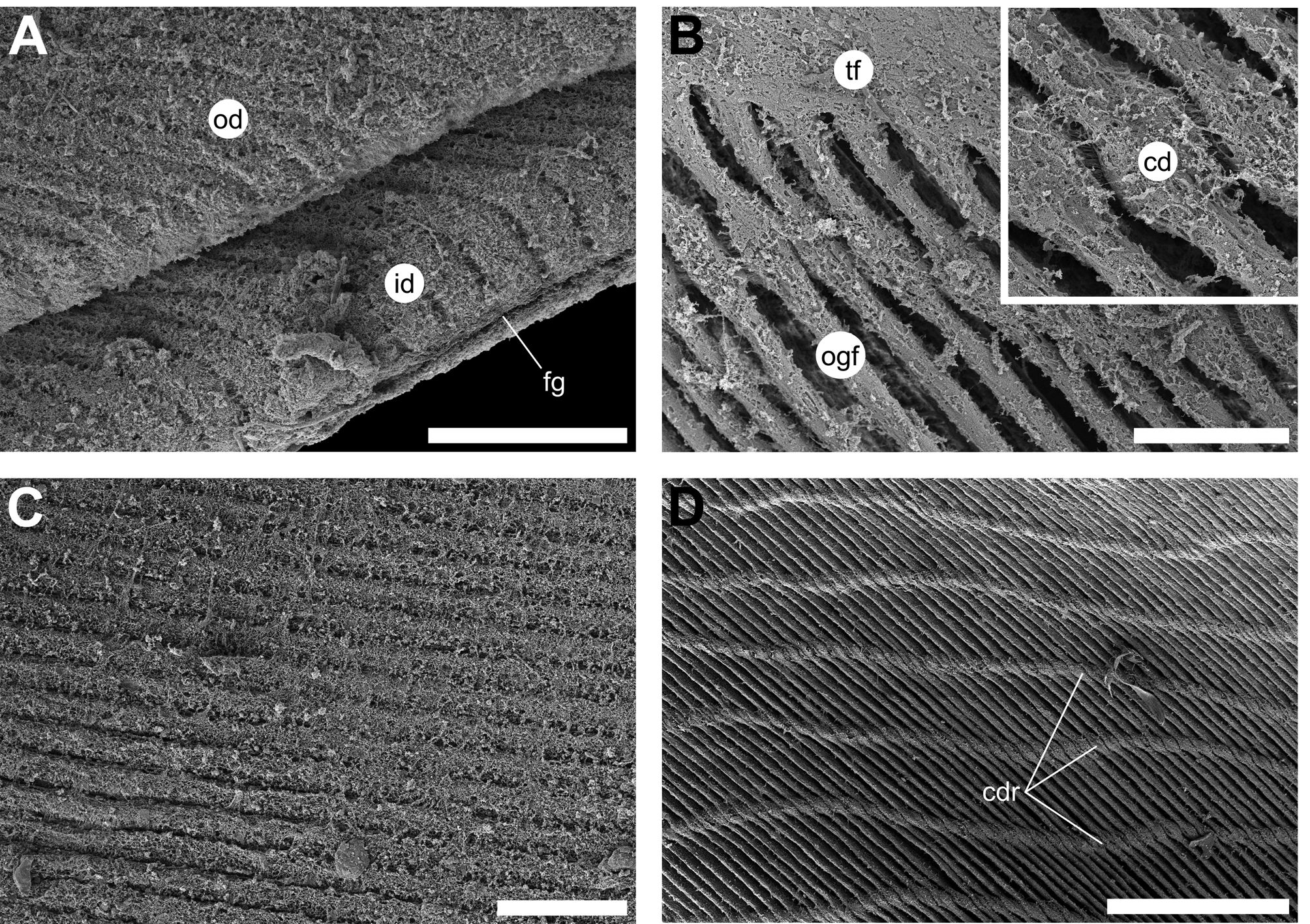
Ctenidia: Paired ctenidia ( Figs 8 View FIGURE 8 B, C, ct) large, translucent-white, non-plicate, broadly sickle-shaped, encircling visceral mass and posterior adductor muscle ventrally, roughly following outline of nacreous border. Distal tips of ctenidia frequently dark brown-black in living specimens. Inner demibranchs ( Figs 11 View FIGURE 11 ; 12; 13A, id) with marginal food grooves ( Figs 11 View FIGURE 11 ; 12A; 13A, fg), very slightly taller (more dorsoventrally extended) than outer demibranchs ( Figs 11 View FIGURE 11 ; 13A, od). Dorsal edges of descending lamellae within demibranch fused forming membranous ctenidial axis. Ctenidial axis ( Fig. 11 View FIGURE 11 , cta) fused to visceral mass and connected to adductor muscle by suspensory membrane ( Figs 8 View FIGURE 8 B, C; 11, sm). Suspensory membrane translucent to faint brown in preserved specimens, stretching from area proximal to labial palps to anteroventral side of posterior adductor muscle.
Filamental organization homorhabdic ( Figs 11 View FIGURE 11 ; 12C; 13B, ogf) and eleutherorhabdic ( Figs 11–13 View FIGURE 11 View FIGURE 12 View FIGURE 13 ), with adjacent filaments connected by ciliated disks ( Figs 12 View FIGURE 12 E; 13B, cd), produced from lateral thickening of filaments and distributed in rows (> 10 in adults; Figs 11 View FIGURE 11 ; 13D, cdr). Serial tissue fusion of filaments restricted to regions proximal to ctenidial axes ( Fig. 13 View FIGURE 13 B, tf), dorsal edges of ascending lamellae ( Fig. 11 View FIGURE 11 , tf), and ventral margins of demibranchs at point of junction of ascending and descending lamellae ( Fig. 12 View FIGURE 12 B, tf). Distalmost extremities of filaments unfused. Dorsal edges of ascending lamellae of inner demibranchs united medially via ciliated junctions from posteriormost extremities to area ventral to foot; dorsal edges of ascending lamellae of outer demibranchs attached laterally to mantle by ciliated junctions along transversely ridged cilial trail on inner surfaces of contractile zone of mantle lobes. Abfrontal surfaces of dorsal margins of ascending and descending lamellae within demibranchs united by interlamellar dorsal septa ( Fig. 11 View FIGURE 11 , ds), occurring at intervals of 12–15 filaments. At varied intervals (1–4 septa), dorsal septa expanding into narrow interdemibranchial buttresses ( Fig. 11 View FIGURE 11 , idb), stretching dorsoventrally to varying extent along lateral surfaces of gill axes, frequently reaching their dorsalmost extremities. Filaments possessing frontal (apical; Fig. 12 View FIGURE 12 D, ac), laterofrontal ( Fig. 12 View FIGURE 12 D, lfc), and lateral ( Fig. 12 View FIGURE 12 D, lc) cilia; abfrontal cilia absent.
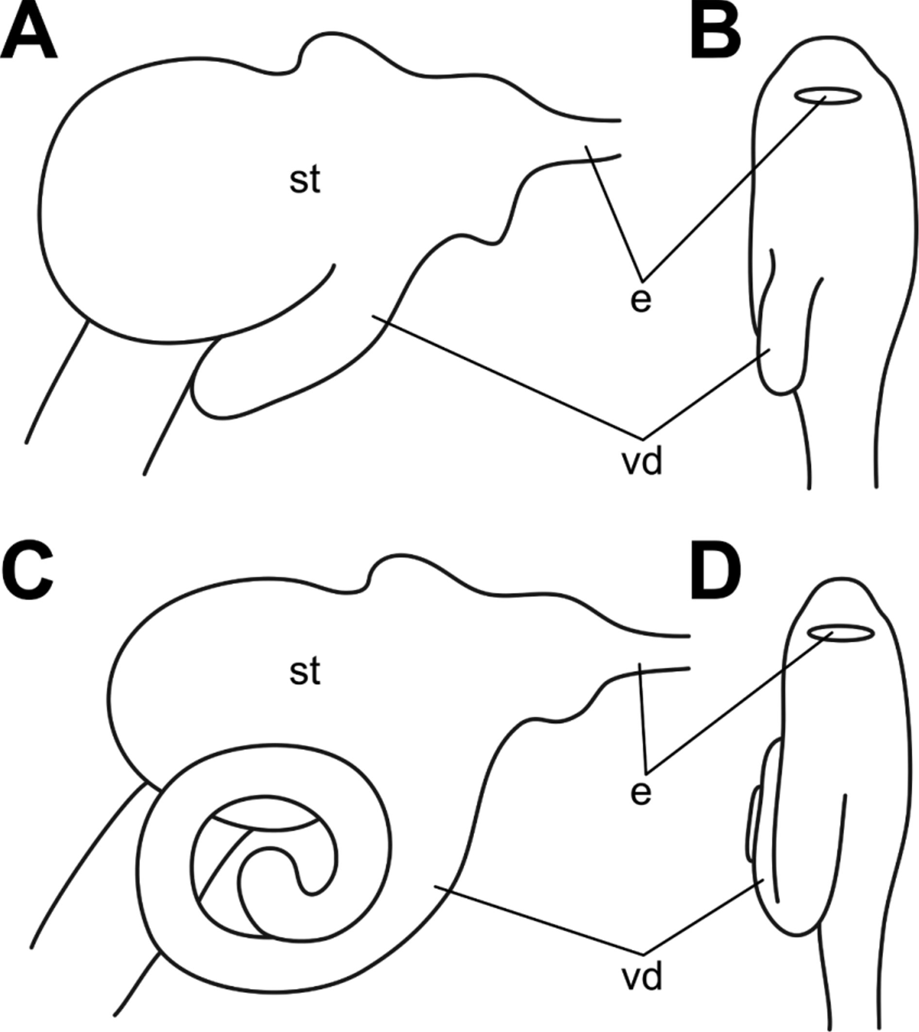
Alimentary system: Mouth ( Fig. 8 View FIGURE 8 C, mo) dorsoventrally compressed, concealed by anteriorly extending lips with ventral lip slightly more extensive and curling dorsally towards smaller dorsal lip. Inner lip surfaces smooth and non-pigmented; outer lip surfaces light brown in living specimens. Mouth opening into dorsoventrally flattened esophagus ( Figs 8 View FIGURE 8 C; 14; 18A, e) with parallel longitudinal grooves and ridges. Esophagus entering gastric chamber (“stomach”; Figs 8 View FIGURE 8 C; 14; 18A, B, st) anterodorsally though slit-like opening demarcated by esophageal lip.
Gastric chamber ( Fig. 14 View FIGURE 14 ) globular, length slightly exceeding height, enveloped by digestive gland. Antechamber ( Fig. 14 View FIGURE 14 B, ant) small, compressed, placed anteriorly relative to capacious main chamber ( Fig. 14 View FIGURE 14 , mst), markedly demarcated from main stomach chamber by fleshy fold on left wall ( Fig. 14 View FIGURE 14 A, ff). Base of fleshy fold fusing with floor of main chamber, devoid of folds and finger-like processes. Posterior wall of main stomach chamber with short, irregular, transverse folds and grooves ( Fig. 14 View FIGURE 14 A, trf). Conspicuous ventral diverticulum ( Figs 14 View FIGURE 14 B; 18A, B, vd) extending from midventral surface of main chamber posteriorly, nearly reaching intestine.
Wide anterior and posterior transverse dorsal folds ( Fig. 14 View FIGURE 14 B, df) on surface of anterior region of main chamber demarcating shallow dorsal hood ( Fig. 14 View FIGURE 14 A, dh). Left pouch ( Fig. 14 View FIGURE 14 A, lpo) small, shallow, situated on anterolateral surface of main chamber proximal to fleshy fold, concealed by gastric shield. Region of left pouch ventral to gastric shield diagonally grooved. Anterior dorsal fold investing left pouch, gradually narrowing ventrally, separating dorsal hood from dorsal groove ( Fig. 14 View FIGURE 14 A, dgr).
Crystalline style projecting from opening of co-joined style sac and midgut ( Fig. 14 View FIGURE 14 A, stys/in) into stomach lumen, located medially in posteroventral side of main chamber. Food-sorting caecum ( Fig. 14 View FIGURE 14 A, fsc) narrow, extending dorsoventrally from stomach floor to level of esophageal opening, with posterior wall formed by fleshy fold.
Cuticle ( Fig. 13 View FIGURE 13 A) occupying most of left surface of main chamber, slightly extending anteriorly onto ventral surface of dorsal hood. Gastric shield ( Fig. 14 View FIGURE 14 A, gs) occupying surface of conspicuous, semispherical, bulbous extension on anterolateral surface of main chamber proximal to fleshy fold; distinct teeth of gastric shield absent. Cuticular ridge ( Fig. 14 View FIGURE 14 A, cr) extending from left wall, separated by cleft from opposing gastric shield.
Minor typhlosole ( Fig. 14 View FIGURE 14 B, tym) merging with right wall upon entry into lumen of main chamber. Major typhlosole ( Fig. 14 View FIGURE 14 , ty) forming broad, low ridge, passing along right ventral side of main chamber. In its course, major typhlosole extending into ventral diverticulum along its posterior wall to apex, emerging along anterior wall of diverticulum onto right ventral side of main chamber, skirting around base of fleshy fold, passing proximal duct of digestive diverticula in right anterior part of gastric chamber, crossing floor of main chamber, extending dorsally into food sorting caecum, and terminating at its apex proximal to esophagus.
Typhlosolar guard ridge ( Fig. 14 View FIGURE 14 , tgr) originating from inconspicuous thickening in area between typhlosoles, where they emerge from intestinal opening, circumnavigating major typhlosole along its entire length, and terminating inside ventral diverticulum on left side of typhlosole.
Intestinal groove ( Fig. 14 View FIGURE 14 B, ig) splitting into two tracks on ventral floor in posteroventral surface on main chamber: left track following course of major typhlosole, forming narrow groove between major typhlosole and typhlosolar guard ridge ridge; right track diverging to right stomach wall, diving into ventral diverticulum, then ascending in S-shaped curve along right wall of antechamber, passing ventral to base of anterior dorsal fold, crossing roof of gastric chamber to left side, and descending along left wall into funnel-like stirring hollow ( Fig. 14 View FIGURE 14 A, sh). Two conspicuous, parallel, adjacent, transverse ventral folds ( Fig. 14 View FIGURE 14 A, vf) extending left from area proximal to emergence on major typhlosole on posteroventral floor of stomach with anteriormost ridge, extending slightly dorsally onto left wall of main chamber.
Patch of very fine diagonal striated area ( Fig. 14 View FIGURE 14 B, sta) situated in slightly recessed area in anterodorsal region of right wall of main chamber between dorsal folds.
Exact number of ducts of digestive diverticula ( Fig. 14 View FIGURE 14 , ddd) could not be determined. Anterior duct field in antichamber consisting of individual ducts opening into food-sorting caecum and into small embayment on right ventral surface of antechamber. Posterior duct fields consisting of embayment with two ducts connected by several fine parallel grooves and situated on left midventral side of main chamber between posteroventral extremity of fleshy fold and anterior ventral ridge. Single ducts located inside stirring hollow and distalmost extremity of ventral diverticulum.
Style sac and midgut merged, forming single tube ( Fig. 14 View FIGURE 14 A, stys/in). Intestine ( Fig. 8 View FIGURE 8 C, in) descending from gastric chamber straight to posteroventral extremity of visceral mass, making short loop anteriorly, and ascending parallel to descending arm on left side, crossing over descending arm to right, and proceeding along curve to posterodorsal side of visceral mass; upon exiting visceral mass, intestine perforating ventricle and supramyal septum, passing medially and terminating on posteroventral surface of posterior adductor muscle. Intestinal sheath light brown. Anus ( Figs 8 View FIGURE 8 C; 15, an) with short, colorless, semitranslucent, subtriangular anal funnel ( Figs 8 View FIGURE 8 C; 15, af). In some specimens anal funnel bifurcated, possibly due to damage by predators ( Fig. 15 View FIGURE 15 inset).
Vascular and excretory systems: Heart ( Fig. 8 View FIGURE 8 C, h) enclosed within pericardium ( Fig. 15 View FIGURE 15 , pc), adjacent to posterodorsal surface of visceral mass; consisting of white, large, dorsal ventricle ( Figs 8 View FIGURE 8 B; 15, v) and two dark gray, symmetrical, ventral auricles ( Figs 8 View FIGURE 8 B; 15, au). Chambers of ventricle and auricles traversed by numerous crisscrossing muscle fibers. Ventricle with smooth surface, penetrated by intestine in dorsal extremity. Walls of auricles papillose, fused lateroventrally and connecting to ventral pericardium via membranous extensions.
Paired branchial afferent ( Fig. 11 View FIGURE 11 , abv) and efferent ( Fig. 11 View FIGURE 11 , ebv) blood vessels passing longitudinally through ctenidial axes. Anterior aorta running mid-dorsally through mantle isthmus from ventricle to anteriormost extremity of pallial hood, and subdividing into left and right circumpallial arteries ( Fig. 9 View FIGURE 9 A, cpa). Posterior aorta following course of intestine posteriorly and branching into interior of adductor muscle above anus.
Paired nephridia (kidneys; Figs 8 View FIGURE 8 C; 15, k) laterally compressed, inconspicuous, symmetrical, transparent sacs with small brown speckles, connected to ventral extremities of auricles via reno-pericardial canals, expanding anteroventrally into suspensory membranes and fused to posterolateral surfaces of visceral mass. Ureter ( Figs 8 View FIGURE 8 A, C, ur) elongated, passing along gentle curve anterior to ventral lobe of posterior adductor muscle and opening into excurrent chamber via renal pore.
Central nervous system and sense organs: Central nervous system conforming to general bivalve bauplan. Paired cerebropleural ganglia located on inner surfaces of fused anterior pedobyssal retractor muscles, where pedobyssal accessory muscles branching off and joined by circumesophageal intercerebral commissure, arching over esophagus slightly posterior to upper lip. Paired cerebropedal, cerebrovisceral, and anterior pallial nerves branching off cerebropleural ganglia. Anterior pallial nerves projecting into mantle in area dorsal to dorsal lip, passing between fused anterior pedobyssal retractor muscles. Adjacent pedal ganglia located in visceral mass proximal to foot. Pedal ganglia connected to cerebropleural ganglia by cerebropedal nerves and giving rise to pedal nerves penetrating foot. Visceral ganglia joined by intervisceral transverse commissure, located on ventral surface of posterior adductor muscle. Circumpallial nerve ( Fig. 9 View FIGURE 9 A, cpn) passing through mantle margin slightly proximal to circumpallial artery. Paired abdominal sense organs ( Fig. 15 View FIGURE 15 , aso) forming narrow ridges, partly colored by small, light gray speckles in preserved specimens, symmetrically placed proximal to anus on posteroventral surface of posterior adductor and stretching along gentle arc ventrally and toward lateral edges of adductor muscle. Right abdominal sense organ slightly larger than left. Small dark pigmented spots, possibly pallial ocelli capable of photoreception, distributed along mantle OF-1 margin. Paired symmetrical ctenidial ocelli (small dark pigmented spots) localized proximal to bases of anteriormost filaments of inner demibranchs.
Distribution. Geographic range —Coastal regions of Gulf of Thailand and South China Sea, Southeast Asia. Type locality —Negros Island, Philippines ( Reeve 1858). Other localities— Malabon, Morong, Bataan Province, Luzon Island; Manapla, Negros Island; Saguisi, Mindanao Island; Philippines ( Hidalgo 1905a, b); Côn Sơn Island, southeastern Vietnam (“Poulo-Condor”; Rochebrune 1881); Hồng Gai City, Vân Đồn Island, Vĩnh Thực Island, Quảng Ninh Province; Cát Bà Island, Hải Phòng province; Cồn Lò Island, Gulf of Tonkin, Vietnam; Kadan (“King”) and Thayawthadangyi (“Elphinstone”) Islands, Mergui Archipelago, Burma ( Martens 1887); Kungkrabaen Bay, Thamai District, Chantaburi Province, Gulf of Thailand ( Printrakoon & Tëmkin 2008; this study); Salak Kok Bay, Chang Island, Trat Province, Gulf of Thailand (Printrakoon & Valentich-Scott unpubl. data).
Habitat and ecology ( Fig. 16 View FIGURE 16 ). Individuals pleurothetic on right valve, byssally attached (singly or in small clusters) to prop roots of mangrove trees or wedged into crevices formed by upside-down forks of prop roots ( Printrakoon & Tëmkin, 2008: 82).
Comparative remarks. Among living species of Isognomonidae , I. spathulatus most closely resembles I. ephippium ( Fig. 17 View FIGURE 17 ), a common Indo-Pacific species, with which the former is typically synonymized ( e.g., Huber 2015).
Previous morphometric analysis of linear shell measurements of the shell revealed statistically significant differences between I. ephippium and I. spathulatus (referred to as “mudflat” and “mangrove” Isognomon , respectively): the individuals of the former are significantly larger (by about 1.5 times), but nearly twice as thin as the latter ( Printrakoon & Tëmkin 2008), have more rounded outlines, and frequently possess a distinct radial color pattern ( Fig. 17 View FIGURE 17 A, rs). There are also pronounced differences in color: in I. spathulatus , the nacre is silvery blue and the inner prismatic margin is brownish; in I. ephippium , nacre has a purple tint and the margin has a color gradient from black distally to ochre brown proximally ( Figs 17 View FIGURE 17 B, D, pmg). Occasional pearls in I. ephippium are small (diameter 0.4 and 0.9 mm; n = 2), weakly iridescent, creamy white with yellowish tint ( Figs 4 View FIGURE 4 B, C).
Anterior auricles are absent or greatly reduced in adults of both species ( Figs 2 View FIGURE 2 A; 17A, C, aa). In I. ephippium , posterior auricles flattened and vary in extent from being weakly sinuated ( Figs 17 View FIGURE 17 C, pas; Jackson 1890: pl. 26, fig. 16) to entirely absent ( Figs 17 View FIGURE 17 A, pa); in I. spathulatus , they are entirely absent.
In I. spathulatus , shell sculpture consists of irregular lamellae projecting from the lirae; in I. ephippium , lamellae develop into extremely fragile, short, flat, broad, overlapping calcitic scales with rounded tips, typically abraded in adults ( Figs 17 View FIGURE 17 A, C, ccs). In some specimens, the overlapping scales form low-relief radial costae along distal shell margin.
In both species the confluent attachments of the posterior adductor muscle and the posterior pedo-byssal retractor muscles form a trilobed muscle scar. In I. ephippium , the middle lobe of the scar is larger than the ventral lobe and, occasionally, is coincident with it ( Fig. 17 View FIGURE 17 D, pams, prms); the dorsal extremity is much shorter, wider, and with a rounded tip. The PAM/PRM scar in I. ephippium is placed more centrally within the nacreous layer, so that the posterior pallial line ( Fig. 17 View FIGURE 17 D, pms) is placed more anteriorly, forming an arc that stretches dorsally towards the middle of the hinge line, then turns posteriorly, and proceeds to its posterior end.
The number of the resilifers, counted in adult individuals of the same population, was on average larger in I. ephippium than in I. spathulatus : 9–16 (mean 11.66 ± 1.92 mm SD, n = 50; ZMKU Bil 800051–800100) in the former compared to 6–10 (mean 7.62 ± 1.03 mm SD, n = 50; ZMKU Bil 800001–800050) in the latter. Whereas in I. spathulatus resilifers are typically uniform in width and deposited at relatively equal intervals along hinge axis in same individual, in I. ephippium they gradually diminish in height and become more widely spaced toward the posterior end of the hinge axis ( Fig. 17 View FIGURE 17 D, mlg).
The morphology of the prodissoconch is very similar between the two species, but there is a notable size difference: in I. ephippium , the lengths of P1 and P2 are 67 Μm (n = 1) and 303.5 ± 2.12 Μm SD (n = 2), respectively, exceeding corresponding dimensions for I. spathulatus : 58 Μm (n = 1) for P1 and 266.5 ± 0.5 Μm SD (n = 2) for P2.
A number of differences between the two species were also observed in various aspects of soft anatomy. Individuals of I. spathulatus have largely non-pigmented (white) foot and gills, in individuals of I. ephippium the dorsal surface of the foot is largely brown, whereas the lateral surfaces of the gills are gray, typically with white ventral margins. In I. spathulatus , the tentacles and the exterior surface of the IF-2 fold, as well as the adjacent area of the mantle margin, are dark brown-black, resulting in conspicuous dark color rim on the periphery of the inner surface of the mantle. In I. ephippium , the exterior surface of the IF-2 fold has a yellow-light brown narrow rim. The edges of the exterior surfaces of the mantle margins are light yellow-brown in I. ephippium and dark brownblack in I. spathulatus .
The morphology of the byssus is very similar between I. ephippium and I. spathulatus , although the fan of byssal threads appears to be wider and more laterally compressed in the former. Prior quantitative comparison of the byssus between the two species showed statistically significant differences in meristic characteristics ( Printrakoon & Tëmkin 2008: 78, 81, 82, fig. 5).
The left and right abdominal sense organs are approximately equidistant from the anus in I. spathulatus , but in I. ephippium , the position of the organs as asymmetric, with the left ASO placed noticeably more ventrally than the right one relative to the anus.
The most striking feature of internal anatomy that distinguishes I. spathulatus from I. ephippium is the morphology of the ventral diverticulum of the gastric chamber. In I. spathulatus , it comprises a short, posteriorly directed blind appendix, extending from the ventral surface of the main gastric chamber ( Figs 14 View FIGURE 14 B; 18A, B, vd). In I. ephippium , the diverticulum is considerably longer and peculiarly spirally coiled in the sagittal plane on the right side of the gastric chamber ( Figs 18 View FIGURE 18 C, D, vd).


No known copyright restrictions apply. See Agosti, D., Egloff, W., 2009. Taxonomic information exchange and copyright: the Plazi approach. BMC Research Notes 2009, 2:53 for further explanation.
|
Kingdom |
|
|
Phylum |
|
|
Class |
|
|
Order |
|
|
Family |
|
|
Genus |
Isognomon spathulatus ( Reeve, 1858 )
| Tëmkin, Ilya & Printrakoon, Cheewarat 2016 |
Pedalion spathulatum (
| Faustino 1928: 25 |
Perna spatulata
| Martens 1887: 203 |
Isognomon spathulata (
| Xue 2012: 350 |
| Temkin 2010: 5 |
| Lutaenko 2000: 368 |
| Fischer-Piette 1976: 10 |
| Cotton 1930: 231 |
| Cooke 1886: 138 |
Isognomon spathulatum (
| Rochebrune 1881: 101 |