
Parvodinium umbonatum (F. Stein) Carty (2008: 106)
|
publication ID |
https://doi.org/10.11646/phytotaxa.509.2.1 |
|
persistent identifier |
https://treatment.plazi.org/id/03D587FF-D218-9D37-3AD4-D9A6EA64FEFA |
|
treatment provided by |
Marcus |
|
scientific name |
Parvodinium umbonatum (F. Stein) Carty (2008: 106) |
| status |
|
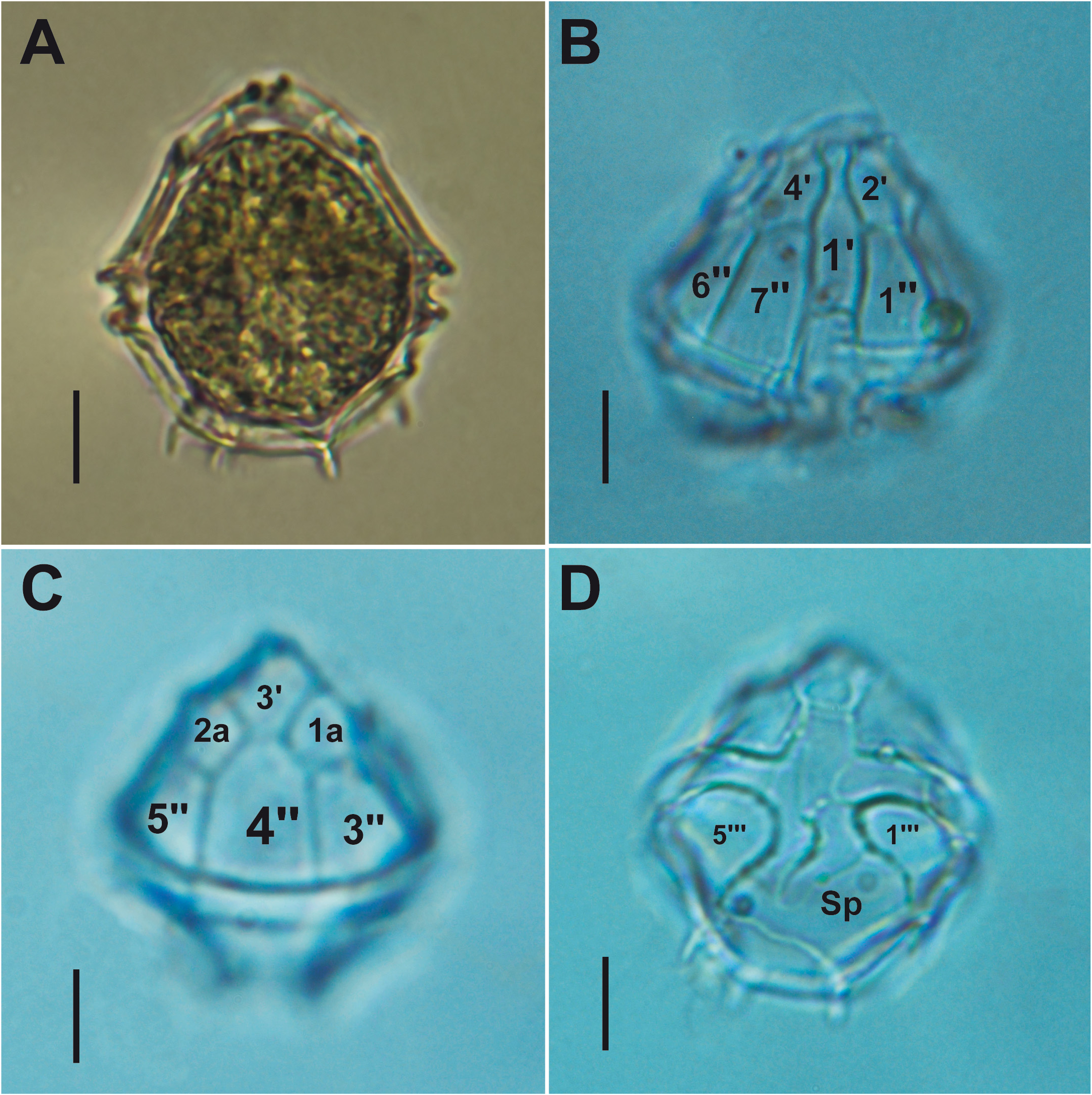
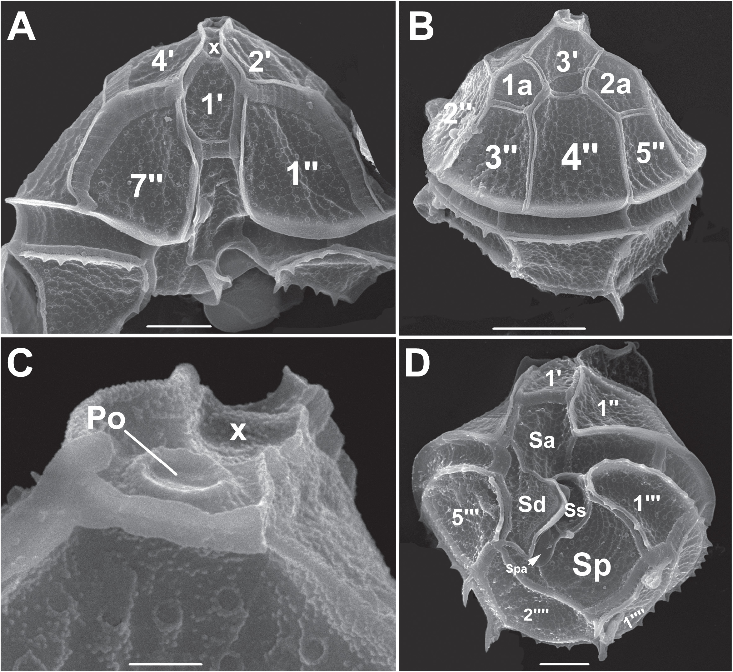
Parvodinium umbonatum (F. Stein) Carty (2008: 106) . Figures 7–8 View FIGURE 7 View FIGURE 8
Basionym: Peridinium umbonatum F. Stein (1883: 12)
Synonyms:
Peridinium minimum in A. J. Schilling (1891: 293)
Peridinium umbonatum var. inaequale in Lemmermann (1910: 669)
Properidinium inaequale in Meunier (1919: 62)
Peridinium caudatum var. guildfordense in Playfair (1920: 800
Glenodinium guildfordense in Er. Lindemann (1931: 700)
Peridinium caudatum var. planktonicum in Playfair (1920: 801)
Peridinium geminum in Playfair (1920: 803)
Glenodinium kamptneri in J. Schiller (1955: 51)
Dimensions: L: 36–47 µm, Td: 28–35 µm.
Cell shape: Sub-oval, epitheca shaped like a slightly polygonal dome ( Fig. 7B View FIGURE 7 ) and slightly taller than the hypotheca and on the latter four spines are observed with LM ( Fig. 7A View FIGURE 7 ), which in reality represent multiple spines located on the edges of the antapical and post-cingular plates ( Fig. 8D View FIGURE 8 ). Descending cingulum. The sulcus extends to the antapex, widening strongly backwards ( Fig. 7D View FIGURE 7 ).
Thecal characteristics:Tabulation, Po, X, 4ʹ, 2a, 7ʹʹ, 5C, 5S, 5ʹʹʹ, 2ʹʹʹʹ. Configuration of the 3ʹ and 4ʹ plates, conjuctum type ( Figs. 7C View FIGURE 7 and 8B View FIGURE 8 ). The edges of the 2ʹʹ, 3ʹʹ and 4ʹʹ plates that surround the apical pore channel resembling an apical semi-neck ( Fig. 8C View FIGURE 8 ).
Cingulum and sulcus: Narrow equatorial cingulum, formed by 5 plates. The cingular lists slightly prominent with crenulated margins, distal ribs not always present. Sulcus formed by 5 plates, the large, broad plate Sa penetrating deeply in the epitheca ( Fig. 8A and 8D View FIGURE 8 ). The Sd plate has a crescent moon-shaped fin partially covering the Ss and Spa plates, is not in contact with the 2ʹʹʹʹ plate. The Sp plate, very wide at its distal end, extends to the antapex ( Fig. 8D View FIGURE 8 ).
Ornamentation: The plates present a smooth network of rows of tiny nodules and pores between these lines surrounded by similar nodules. The edges of the post-cingular plates are fimbriated and has spines at the corners of the antapical plates ( Fig. 8 View FIGURE 8 ).
Locality: La Ayantuna lake in Pacific island.
Comments: This species varies widely in terms of its shape ( Borics et al. 2005; Carty 2008; Lefèvre 1932; Moestrup & Calado 2018). Among the morphological variations described by Lefèvre (1932), Moestrup & Calado (2018) recognize several varieties. Our specimens found in La Ayantuna has multiple spines in the epithecal sutures, around the cingulum and along the sulcal edges, like Parv. umbonatum var. spiniferum (M. Lefèvre) Moestrup comb. nov ( Moestrup & Calado 2018).
World distribution: Wide distribution in USA ( Carty 2014). It is also reported for Europe, China, and New Zealand ( Moestrup & Calado 2018), for South America in Brazil ( Ramos et al. 2016; Cardoso et al. 2010) and Colombia ( Canosa & Pinilla 2007; Cardozo et al. 2005; Montoya 2011; Pinilla et al. 2007).
No known copyright restrictions apply. See Agosti, D., Egloff, W., 2009. Taxonomic information exchange and copyright: the Plazi approach. BMC Research Notes 2009, 2:53 for further explanation.
|
Kingdom |
|
|
Phylum |
|
|
Class |
|
|
Order |
|
|
Family |
|
|
Genus |
Parvodinium umbonatum (F. Stein) Carty (2008: 106)
| Bustamante-Gil, Carolina, Boltovskoy, Andrés, Rengefors, Karin, Tavera, Rosaluz, Amat, Eduardo & Ramírez-Restrepo, Jhon J. 2021 |
Parvodinium umbonatum (F. Stein)
| Carty, S. 2008: ) |