
Antarctoperlinae Enderlein, 1909
|
publication ID |
https://doi.org/10.11646/zootaxa.4671.1.2 |
|
publication LSID |
lsid:zoobank.org:pub:292E9F1A-FF84-433F-9297-ECDA12A00903 |
|
persistent identifier |
https://treatment.plazi.org/id/03D7197F-197D-FFD4-51F6-FB45FB6CF828 |
|
treatment provided by |
Plazi |
|
scientific name |
Antarctoperlinae Enderlein |
| status |
|
Antarctoperlinae Enderlein View in CoL
Araucanioperla brincki (Froehlich)
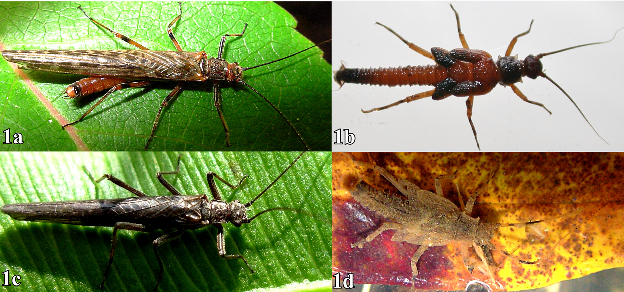
Last instar larva (Figs: 1b, 2a–g, 3a–e, 4a, b)
Measurements (mm, n=8): body length 9.5 –10.3; antenna 5.4–6.5; cercus 1.1–1.3; head width 1.6–1.8; pronotum width 1.7–1.8; pronotum length 1.1–1.3; mesonotum length 1.2–1.3; metanotum length 1.1–1.2; mid femur length 1.2–1.3; mid tibiae length 1.5–1.6; hind femur length 1.7–1.8; hind tibiae length 2.0–2.1.
Colour ( Fig. 1b View FIGURE 1 ): Brown, head, antennae, pronotum, wing pads, abdomen segments VIII to X, tibia and tarsus, darker. Remainder of abdomen, thorax, and femur reddish–brown. Larvae frequently covered with a thin film of detritus.
Head: Globose, covered by spatulate setae, slightly depressed in the frons. Antennae reaching the apex of the hind wings pads. Labrum ( Fig. 2b View FIGURE 2 ) transverse, twice as wide as long, of rounded angles, ventrally with an anteromedial lobe without setae, each side with an acute setae patch, which decrease in size from distal to proximal, with a group of 4 to 6 thick and acute proximal setae. Mandibles as in figure 3a with four incisors, right mandible with a group of setae at the base of incisors. Hypopharynx globose ( Fig. 3b View FIGURE 3 ). Maxillary palp with 5 segments ( Figs. 3 View FIGURE 3 c–e), distal segment lanceolate, as long as the previous 3 segments combined. Galea with apex dilated with a group of short setae, outer margin with a row of setae short and curves. Lacinia with three apical teeth and a marginal row of 14 acute setae, the four distally laminar in shape, others progressively more cylindrical ( Fig. 3d View FIGURE 3 ). Labium ( Figs. 3 View FIGURE 3 f–c) with glossae and paraglossae short and of similar length, distal segment oval.
Thorax: Pronotum trapezoidal, sides slightly convergent anteriorly, with the four angles projected in an acute triangular process ( Fig. 2d View FIGURE 2 ). Meso and metanotum with straight posterior margin ( Fig. 2a View FIGURE 2 ). Fore wing pads with parallel sides, slightly expanded to the sides. Hind wing pads slightly divergent. Notal surface and all thorax margins with short cylindrical setae. Legs with tibiae and femora covered with short and robust setae. Tibiae with dark band proximally. Tarsi with short setae, first segment twice as long as wide, second short and third twice the sum of the previous two. Curved claws ( Fig. 2c View FIGURE 2 ).
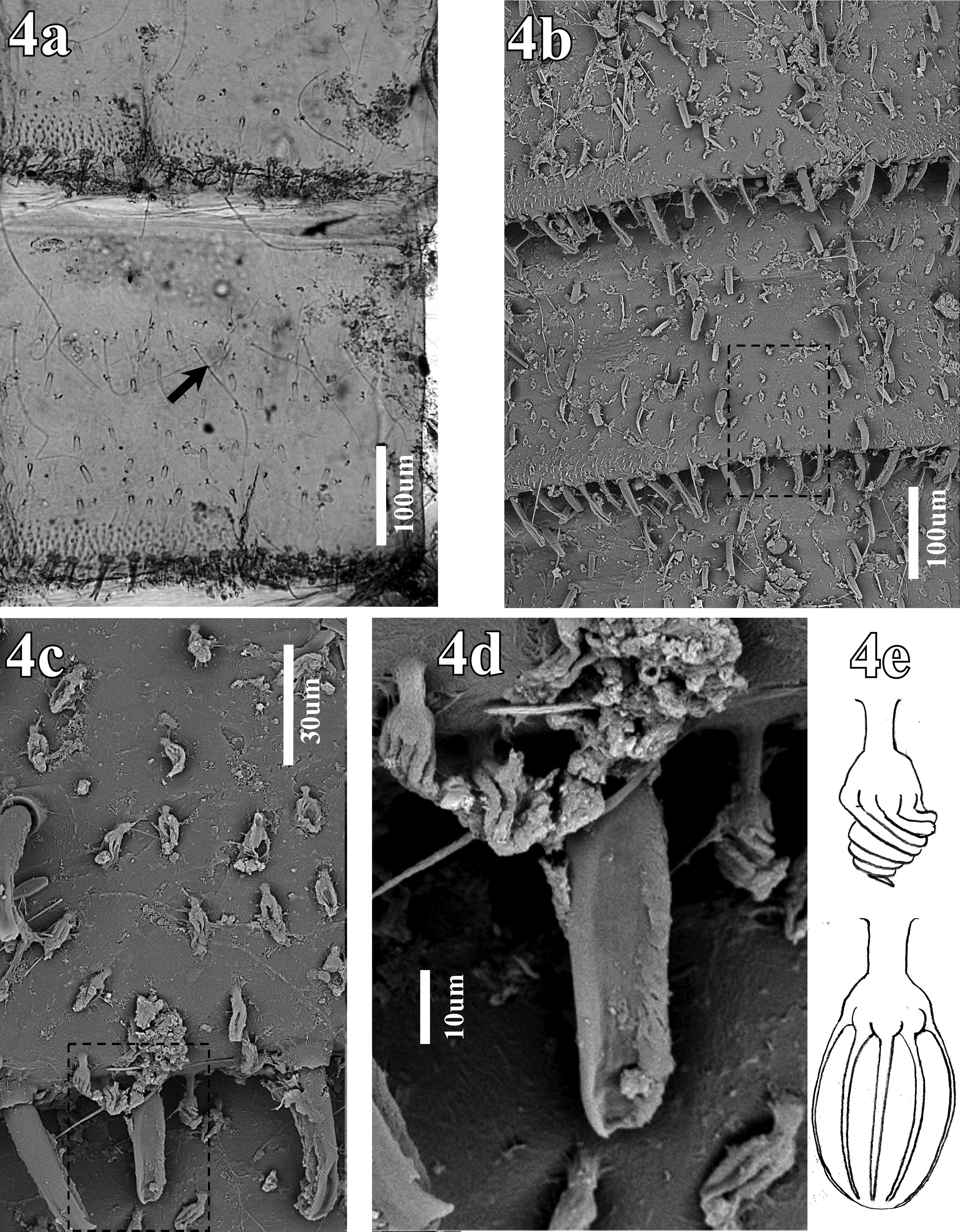
Abdomen: Segments cylindrical, medially constricted ( Figs. 2a, f View FIGURE 2 ). Tergites with cylindrical and truncated setae on the posterior margin ( Fig. 2e View FIGURE 2 ), in several larvae, these setae collapsed, appearing spatulated. Surface with similar setae, some lanceolate, and abundant chloride cells ( Figs. 4c, d View FIGURE 4 ) with six cuticular rods ( Fig. 4d, e View FIGURE 4 ). Tergite X as long as the three previous segments, with parallel sides, rounded posterior margin, with similar setae to other segments but more abundant. Triangular subanal lobe with acute apical spines ( Fig. 2g View FIGURE 2 ). Gill, large, rosette in shape.
Remarks: The larva morphology is similar to that of A. bullocki . The distinctive brown–reddish color in A. brincki is evident only in the mature last instar larva. Earlier instars are uniformly light brown as with A. bullocki ( Fig. 1d View FIGURE 1 ). The antennae and legs are shorter in A. brincki than in A. bullocki , the wing pads are slightly expanded in A. brincki unlike the flanged wing pads of A. bullocki . Both species share similar specializations of the abdomen, but in A. bullocki each segment has a row of filamentous setae before the medial constrictions ( Fig. 4a View FIGURE 4 ). These setae are absent in A. brincki ( Fig. 4b, c View FIGURE 4 ).
Araucanioperla bullocki ( Navás 1933)
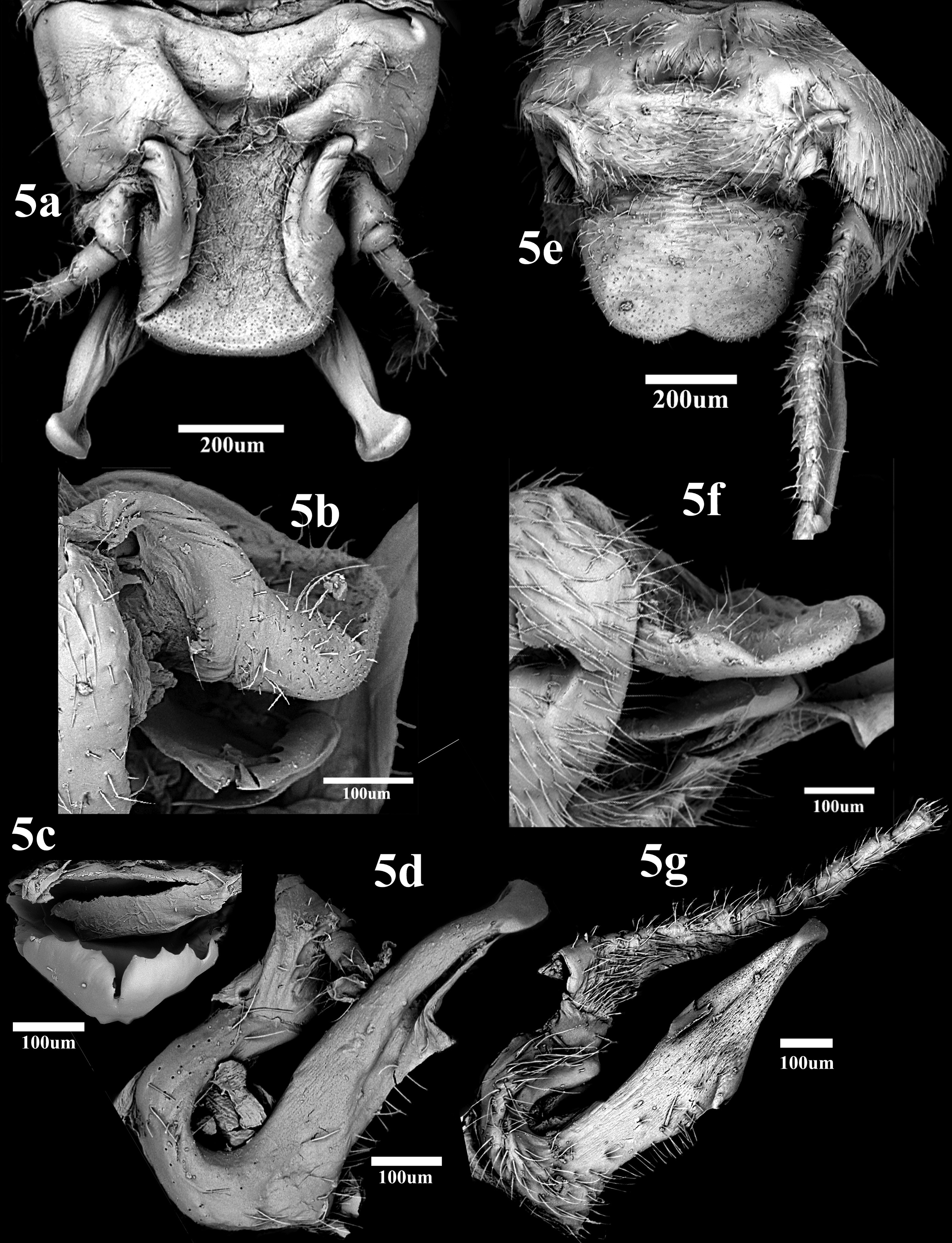
Male genitalia ( Fig. 5 View FIGURE 5 a–d)
Abdomen: Tergum X in dorsal view with lateral sclerites large and sclerotized. Anterior sclerite trapezoidal and small. Central sclerite expanded as a robust rectangular lamina, dorsal surface slightly sclerotized, apex truncated without medial emargination ( Figs. 5a, b View FIGURE 5 ). Posterior sclerite absent. Epiproct narrow, shovel shaped, dorsal margin bent, with 4–5 sharp teeth on each side ( Fig. 5c View FIGURE 5 ), with a thin medial ventral hook. Paraprocts ( Fig. 5d View FIGURE 5 ) robust, wide at the base, narrowing towards apex, with a triangular ventromedial membrane, apex curved with a large sclerotized mass, in dorsal the mass is transverse and rounded.
Remarks: The male genitalia of both species are distinctive. Unlike A. bullocki , A. brincki has a thin median sclerite with a rounded and emarginated apex ( Fig. 5e View FIGURE 5 ), the epiproct is wide and small dorsal teeth ( Fig. 5f View FIGURE 5 ), and paraprocts are not curved or sharp in lateral view, but with a smaller oval apical structure ( Fig. 5g View FIGURE 5 ).


No known copyright restrictions apply. See Agosti, D., Egloff, W., 2009. Taxonomic information exchange and copyright: the Plazi approach. BMC Research Notes 2009, 2:53 for further explanation.
|
Kingdom |
|
|
Phylum |
|
|
Class |
|
|
Order |
|
|
Family |