
Chaetonotus (Hystricochaetonotus) iratus, Križanová & Vďačný, 2022
|
publication ID |
https://doi.org/ 10.5852/ejt.2022.840.1941 |
|
publication LSID |
lsid:zoobank.org:pub:CE89365D-A3C5-483D-9C80-E5CAECCA740F |
|
DOI |
https://doi.org/10.5281/zenodo.7195234 |
|
persistent identifier |
https://treatment.plazi.org/id/1A749F30-3FDF-4A02-8923-1DAE313BAB8E |
|
taxon LSID |
lsid:zoobank.org:act:1A749F30-3FDF-4A02-8923-1DAE313BAB8E |
|
treatment provided by |
Felipe |
|
scientific name |
Chaetonotus (Hystricochaetonotus) iratus |
| status |
sp. nov. |
Chaetonotus (Hystricochaetonotus) iratus View in CoL sp. nov.
urn:lsid:zoobank.org:act:1A749F30-3FDF-4A02-8923-1DAE313BAB8E
Figs 31‒32 View Fig View Fig ; Supp. file 1: Table S7 View Table 7
Morphological diagnosis
Body elongated and about 124 µm long. Head slightly wider than neck, separated from trunk by an inconspicuous neck constriction. Cephalion, epipleurae, and hypopleurae clearly demarcated. Trunk widest at ca U61, gradually tapers towards furca base (U82). Mouth ventral, one central cuticular tooth. Hypostomium bears two parallel, horizontally arranged lamellae accompanied by tear-shaped protuberances. Pharynx without dilatations. Intestine straight, with a marked anterior section. Scales spined, three-lobed, slightly overlapping, distributed in about 12 columns, 22 scales per column. Scales and spines increase gradually in size in a posterior direction. Dorsal surface covered by scales from posterior end of cephalion (ca U4) to furca branches (ca U83). Furca branches slightly shorter than adhesive tubes, lateral margins more or less straight, furcal indentation V-shaped, adhesive tubes comparatively short.
Molecular diagnosis
18S rRNA gene: 1716 C. ITS2: 38 C, 61 T, 67 C, 68 A, 85 T, 86 G, 91 C, 103 C, 113 T, 114 A, 124 A, 129 G, 155 C. 28S rRNA gene: 465 C, 656 T, 674 G. Cytochrome c oxidase subunit I (codon ordinal numbers are followed by the corresponding span of nucleotide positions in parentheses): 123 (367‒369) GTG, 159 (475‒477) CGG, 186 (556‒558) CTA, 216 (646‒648) ATC.
Reference molecules are shown in Supp. file 1: Figs S5 View Fig , S 11A, S View Fig 17 View Fig . All diagnostic molecular autapomorphies are marked by arrows. Reference alignments with corresponding nucleotide positions are in Supp. file 1: Alignments 1‒4.
The p -distance from species described in the present study is 0.05‒4.33% in 18S, 14.44‒37.76% in ITS2, 0.25‒9.79% in 28S, and 5.62‒12.74% in COI. There are 1‒16 CBCs (except for Ch. (H). arcanus sp. nov., Ch. (H). luxus sp. nov., and Ch. (H). slavicus sp. nov., where there are no CBCs) in the 18S rRNA molecule, 1‒3 CBCs in the ITS2 molecule, and 1‒18 CBCs in the first two domains of the 28S rRNA molecule (except for Ch. (H). luxus sp. nov. and Ch. (H). slavicus sp. nov., where there are no CBCs).
Etymology
The Latin adjective ‘ iratu · us, - a, - um ’ [m, f, n] (‘angry, irate’) refers to the spiny appearance of the new species.
Material examined
Holotype SLOVAKIA • adult (photomicrographs, hologenophore); Shallow section of the River Váh , Stankovany, Veľká Fatra Mts; 49°08′26.3″ N, 19°10′14.6″ E; CU-FNS- 21-09-20 /HO. GoogleMaps
Photomicrographs of the holotype are available at the Department of Zoology, Comenius University in Bratislava at https://fns.uniba.sk/en/gastrotricha/. The holotype is shown in Fig. 32 View Fig .
Type material
A DNA sample of the holotype specimen STV 65 has been deposited in the Natural History Museum, Vajanského nábrežie 2, 810 06 Bratislava, Slovakia (ID Collection Code 01427609).
Type locality
Shallow section of the River Váh near the village of Stankovany, Veľká Fatra Mts, Slovakia, 49°08′26.3″ N, 19°10′14.6″ E.
Gene sequences
The nuclear 18S and ITS 1-5.8S-ITS2-28S rDNA sequences as well as the mitochondrial COI sequence of the holotype specimen STV 65 have been deposited in GenBank under the following accession numbers: OM 421720 View Materials , OM 421696 View Materials , and OM 424075 View Materials , respectively.
Description
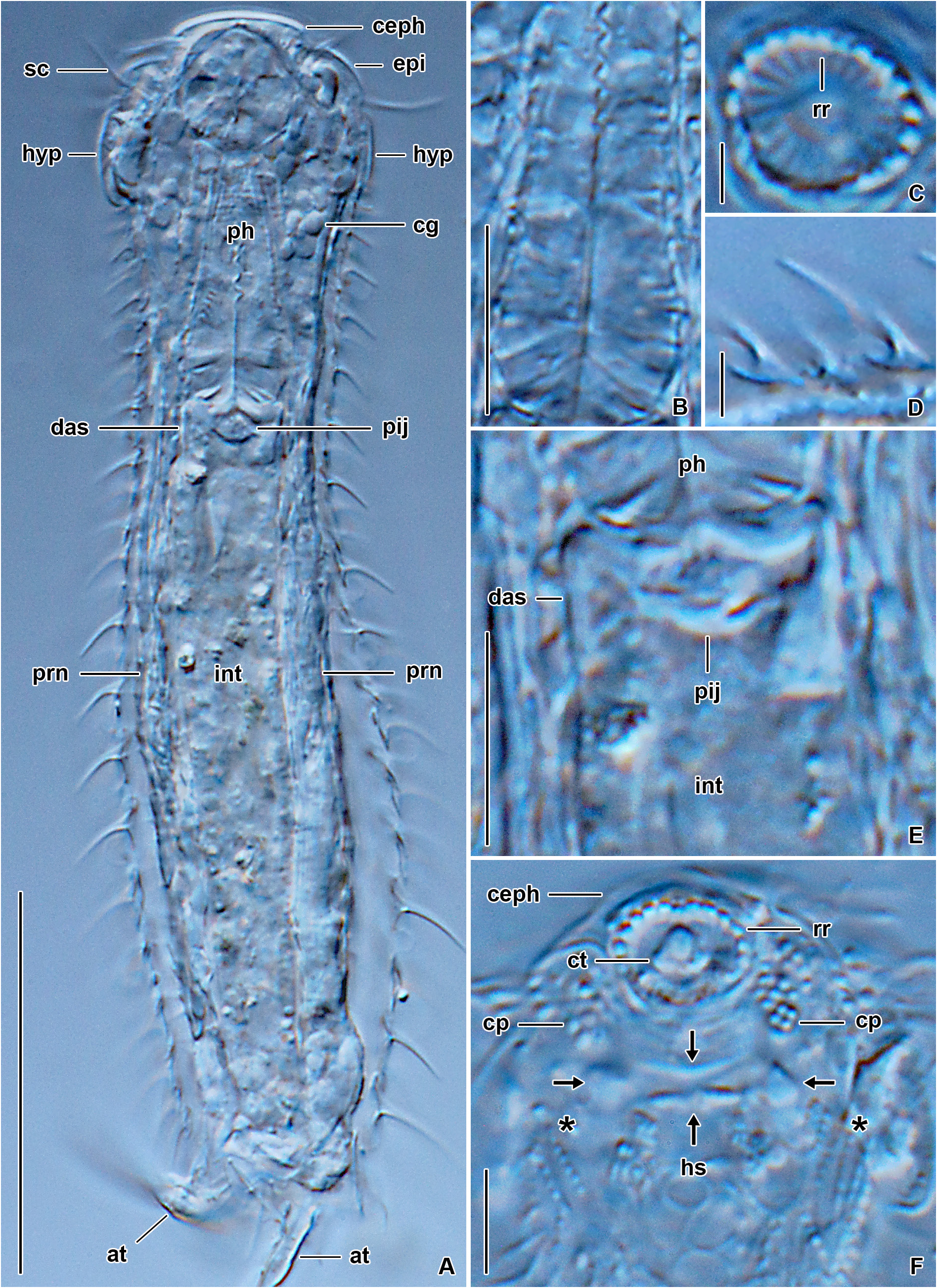
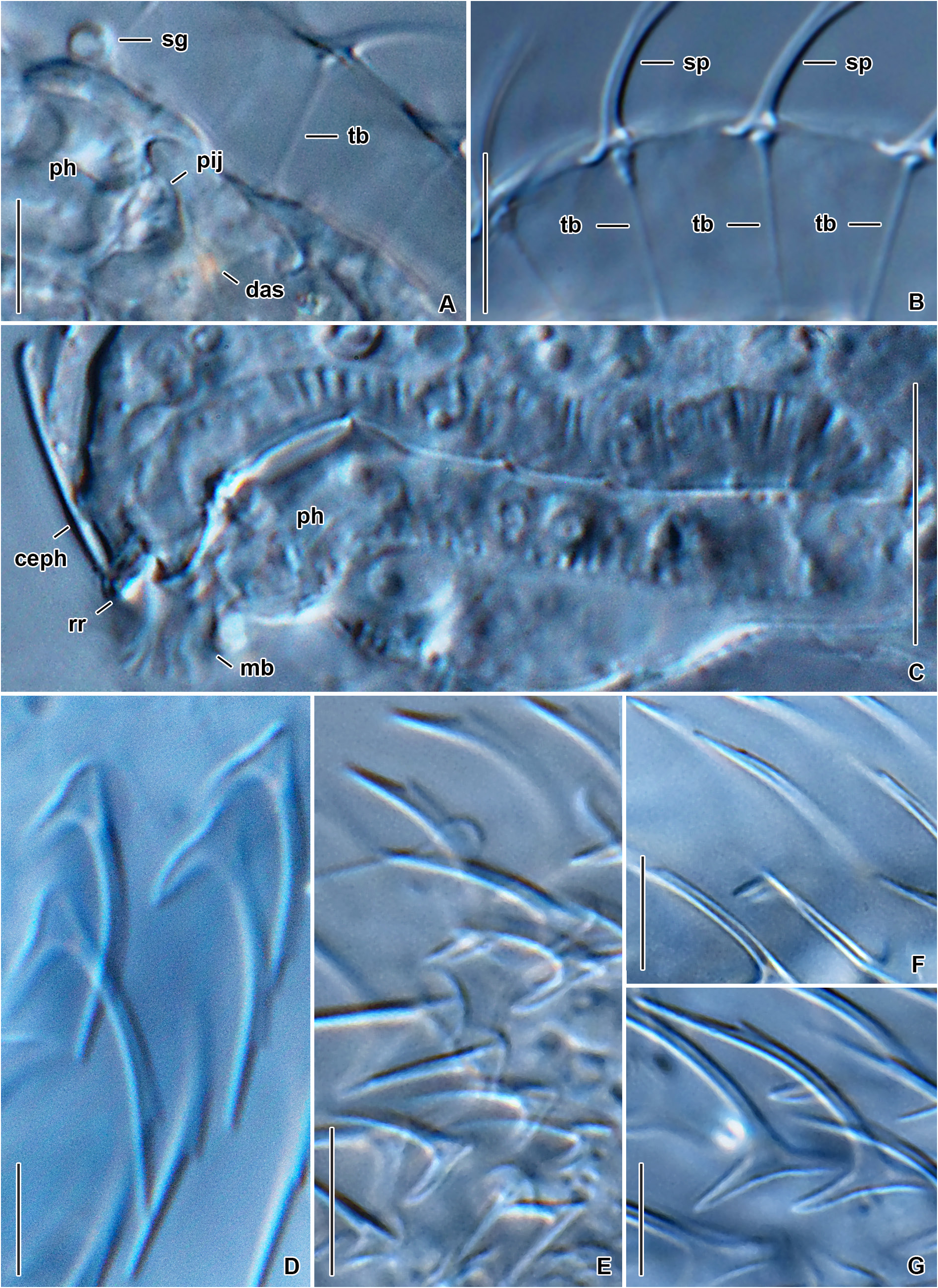
HABITUS. Chaetonotus (Hystricochaetonotus) iratus sp. nov. is about 124 µm long and has a slender elongated body, with a head region slightly broader than the neck and trunk ( Figs 31A, J View Fig , 32A View Fig ). Body width is about 21.5 µm at U10, 20.0 µm at U50, and 22.5 µm at U60. The head is relatively wide (20.2 µm at U6), with a plate-like cephalion. Epi- and hypopleurae are clearly demarcated in the head outline ( Figs 31A, J View Fig , 32A View Fig ). The neck (ca U13–U34) is only inconspicuously marked and smoothly continues to the trunk. In comparison with the head, the trunk is comparatively slender, gradually dilatating from about U35 to U61, where it reaches the maximum width. Then, the trunk gradually narrows towards U82, where more or less straight margins of the furca branches start to form. Dorsal sensory bristles were not observed. The furcal indentation is deeply V-shaped and approximately 19.4 µm long. Welldeveloped adhesive tubes are straight and approximately 10.7 µm long ( Fig. 31A, J View Fig ).
HEAD. The cephalion (U1) is clearly demarcated in the body outline, distinctly flattened, and surrounds the mouth ventrally like a bib. The epipleurae are formed approximately at U3–U7 while the hypopleurae at U8–U14. The latter structures are well recognizable in the head outline ( Figs 31A, J View Fig , 32A View Fig ). Two pairs of cephalic ciliary tufts (6.9–18.8 µm) emerge laterally between the cephalion and the epipleurae edge (ca U3) as well as between the epi- and the hypopleurae edge (U7). The mouth ring is oval, approximately 6.9 µm in the largest diameter, and located subapically at U2–U5. There are strong but short, rod-like reinforcements lining the walls of the mouth ring and inner delicate structures directed towards the center of the mouth ring ( Figs 31I–J View Fig , 32C, F View Fig ). One cuticular tooth is clearly visible in the center of the mouth ring ( Figs 31I View Fig , 32F View Fig ). The hypostomium (ca U5–U9) is composed of two more or less parallel, horizontally arranged lamellae whose lateral sides are accompanied by tear-shaped protuberances. Moreover, the lateral sides of the hypostomium are lined from U3 to U8 by relatively wide patches of irregularly arranged basal bodies ( Figs 31I View Fig , 32F View Fig ).
INTERNAL MORPHOLOGY. The pharynx extends from ca U5 to U31, is 28 µm long and 6.2–8.8 µm wide, sinuous, and without dilatations ( Figs 31J View Fig , 32A–B View Fig ). The intestine runs from U22 to U87 and has a separate, well-differentiated anterior section (U32–U35). Transversal bands connected to the base of dorsal scales are well recognizable. The adhesive gland is placed right behind the terminal part of the intestine (ca U85–U87), forming a short dichotomy at the subtle furca base.
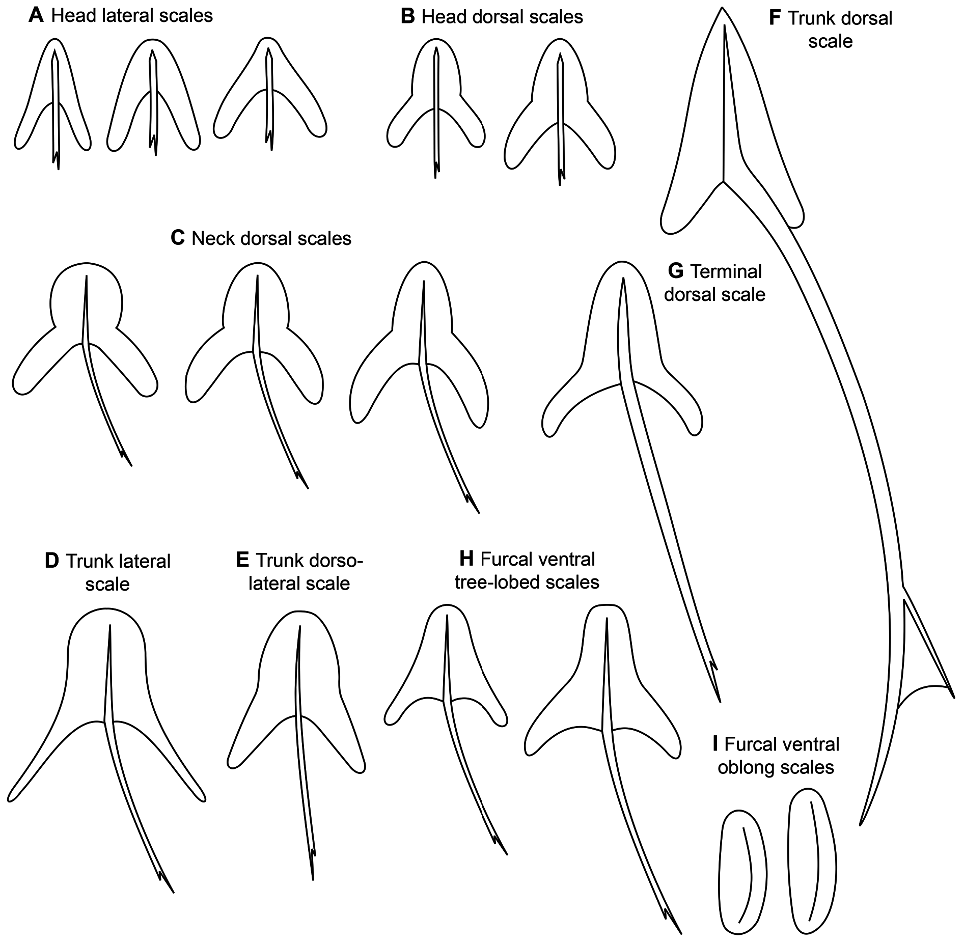
SCALES AND SPINES. Almost the entire body is covered by slightly overlapping, mostly three-lobed scales that adhere to the basal cuticle layer along either all or most of their perimeter. Scales are very densely packed, forming a minimum of 12 longitudinal rows on the dorsal side, with 22 scales in the central row. They have a rounded anterior lobe and elongated posterior lobes narrowly rounded distally. The transition between the anterior and posterior lobes is marked except for the ventro- and dorsolateral scales in which the transition is indistinct ( Fig. 31D‒G View Fig ). The size of scales increases from 2.3×2.1 µm to 7.4×3.1 µm in a posterior direction. Dorsal furca branches scales are 4.3 ×3.4 µm in size, threelobed and spined but their anterior lobe is more elongated, their posterior lobes are slightly shorter and narrower, the transition between the anterior and posterior lobes is continuous and indistinct ( Fig. 31H View Fig ). Spines do not differentiate into various types, only their length slightly increases from 2.3 µm to 6.5 µm in a posterior direction ( Figs 31A‒H, J View Fig , 32A, D View Fig , Supp. file 1: Table S7 View Table 7 ). Spines are slightly narrower posteriorly but do not become hair-like terminally.A lateral denticle is not developed ( Figs 31B–C View Fig , 32A, D View Fig ).
VENTRAL CILIARY BANDS AND VENTRAL INTERCILIARY FIELD. Ventral ciliary bands commence almost right behind the hypostomium (U10) and terminate at ca U87. Their anterior region is broadened as typical of most species described herein. The ciliary bands are accompanied by two ventrolateral rows of threelobed scales that start at U13. They are 3.6–5.3× 2.5–3.1 µm in size and have a similar morphology as the dorsal and dorsolateral scales but the transition between the anterior and posterior lobes is continuous and hence indistinct ( Fig. 31G View Fig ). Unfortunately, no further features of the ventral side were observed.
| OM |
Otago Museum |
No known copyright restrictions apply. See Agosti, D., Egloff, W., 2009. Taxonomic information exchange and copyright: the Plazi approach. BMC Research Notes 2009, 2:53 for further explanation.
|
Kingdom |
|
|
Phylum |
|
|
Order |
|
|
Family |
|
|
Genus |
|
|
SubGenus |
Hystricochaetonotus |