
Epistylis riograndensis, Utz, Laura R. P., Farias, Ana Carolina Silva Rodrigues, Freitas, Eduarda Correa & Araújo, Gabriella Oliveira De, 2014
|
publication ID |
https://doi.org/ 10.11646/zootaxa.3869.5.5 |
|
publication LSID |
lsid:zoobank.org:pub:F446648A-E5BE-43E8-90AD-781BC570F590 |
|
DOI |
https://doi.org/10.5281/zenodo.5696069 |
|
persistent identifier |
https://treatment.plazi.org/id/03D74F12-FB59-D62F-FF61-FEA1035FF1CD |
|
treatment provided by |
Plazi |
|
scientific name |
Epistylis riograndensis |
| status |
sp. nov. |
Epistylis riograndensis n. sp.
Diagnosis. Freshwater Epistylis bearing green algae in its cytoplasm. The species exists as dichotomously branched, umbellate colonies of elongate zooids measuring on average 162 µm in length and 45 µm in width, with a C-shaped macronucleus that lies transversally in the adoral half of the cell. All infundibular polykineties consisted of 3 rows of kinetosomes; PK2 terminated at the adstomal curvature of PK1, and PK3 terminated midway between the adstomal ends of PK1 and PK2.
Type locality. Artificial lake in the Botanical Garden located in Porto Alegre municipality (30.03’08’’ S; 51.10’40’’ W), Rio Grande do Sul, Brazil.
Etymology. The specific epithet refers to the state of Rio Grande do Sul.
Deposition of slide. One slide with protargol-stained specimens was deposited in the Protist Collection of the Museum of Science and Technology of the Pontifícia Universidade Catolica do Rio Grande do Sul, Brazil under the number MCTP02.
Morphology of live specimens. Colonies of E. riograndensis were alternating irregular branched with secondary and tertiary branches terminating at the same level to create an overall flabellate shape ( Figure 1 View FIGURES 1 – 9 and 10 View FIGURE 10 – 11 A). Neither the basal stalk nor the branches were contractile. The basal stalk had a smooth surface without ridges or protuberances ( Figure 1 View FIGURES 1 – 9 ), and was approximately the same diameter as the primary branches ( Table 1 View TABLE 1 ) that were in general longer than the basal stalk. Colonies had up to 182 zooids, but usually consisted of 20–30 zooids of similar dimensions. Zooids were elongate and ranged from 115 to 221.5 µm in length, and from 35.7 to 50 µm in width ( Table 1 View TABLE 1 ; Figure 2 View FIGURES 1 – 9 ). When contracted, the zooid was transversely folded in the region near the scopula ( Figure 10 View FIGURE 10 – 11 B) The peristomial lip was wider than the body and the epistomial disk was moderately elevated above it ( Figure 3 View FIGURES 1 – 9 ). A single “C-shaped” macronucleus was observed lying transversely in the adoral half of the cell ( Figure 4 View FIGURES 1 – 9 ).
Morphology of stained specimens. Infraciliary characteristics and the macronucleus of E. riograndensis were easily revealed by protargol staining. Reticulate somatic myonemes extended from the epistomial disk to the scopula of the zooid ( Figure 5 View FIGURES 1 – 9 ). The trochal band was located 10 to 15 µm, on average adoral of the scopula ( Figure 6 View FIGURES 1 – 9 ). The oral infraciliature was typical of peritrich ciliates and presented an inner polykinety and an outer haplokinety that made 1 ½ circuits around the peristome before entering the infundibulum. Three infundibular polykinetids (PK1, PK2, and PK3) all consisting of three rows of kinetosomes, were observed ( Figure 7 View FIGURES 1 – 9 ): rows of PK1 were equal in length and ended adstomally at the margin of the cytostome ( Figure 8 View FIGURES 1 – 9 ). Rows of PK2 also were equal in length, terminating at the adstomal curvature of PK1 on one end and merging with the rows of PK1 abstomally ( Figure 8 View FIGURES 1 – 9 and 11 View FIGURE 10 – 11 ). PK3 consisted of three long rows of kinetosomes of equal length that terminated approximately midway between the adstomal ends of PK1 and PK2 ( Figure 9 View FIGURES 1 – 9 and 11 View FIGURE 10 – 11 ).
Molecular analyses. The Epistylis riograndensis 18S sequence allowed an assessment of its phylogenetic position within Peritrichia. All phylogenetic trees generated with different methods were congruent ( Figure 12 View FIGURE 12 ), showing that E. riograndensis clustered with other Epistylis species within the order Vorticellida.
Character Mean (µm) SD (µm) Mode CV (%) Range Total length of the body from epistomial disk to aboral end 52.8 6.2 50 11.7 45 – 62.5 Width of the body at midpoint between oral and aboral ends 42 6.9 45 16.0 32.5 – 50 Distance between Trochal Band and Scopula 12.2 1.9 12.5 16.1 10 – 15 Length of Basal Stalk 85.0 8.6 87.5 10.1 75 – 100 Width of Basal Stalk 10.5 3.2 12.5 30.8 5 – 12.5 Length of the Macronucleus 21.4 4.1 25 19.5 12.5 – 25 Width of Macronucleus at Midpoint 10.8 3.0 12.5 28.4 7.5 – 15
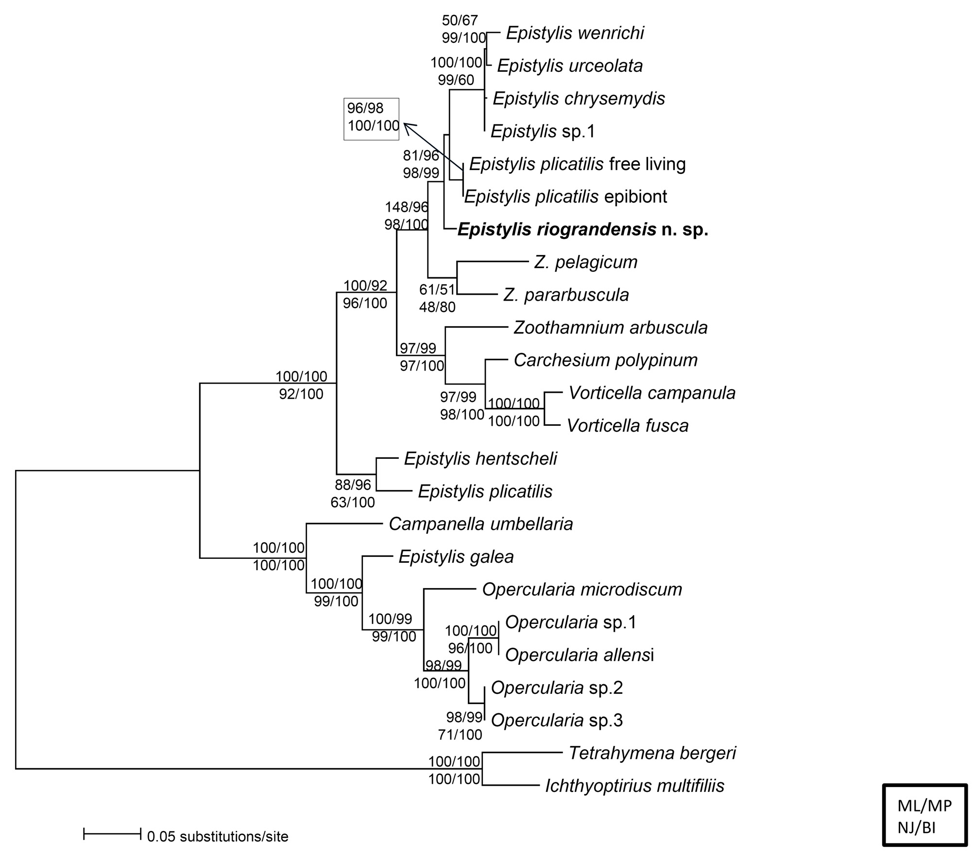
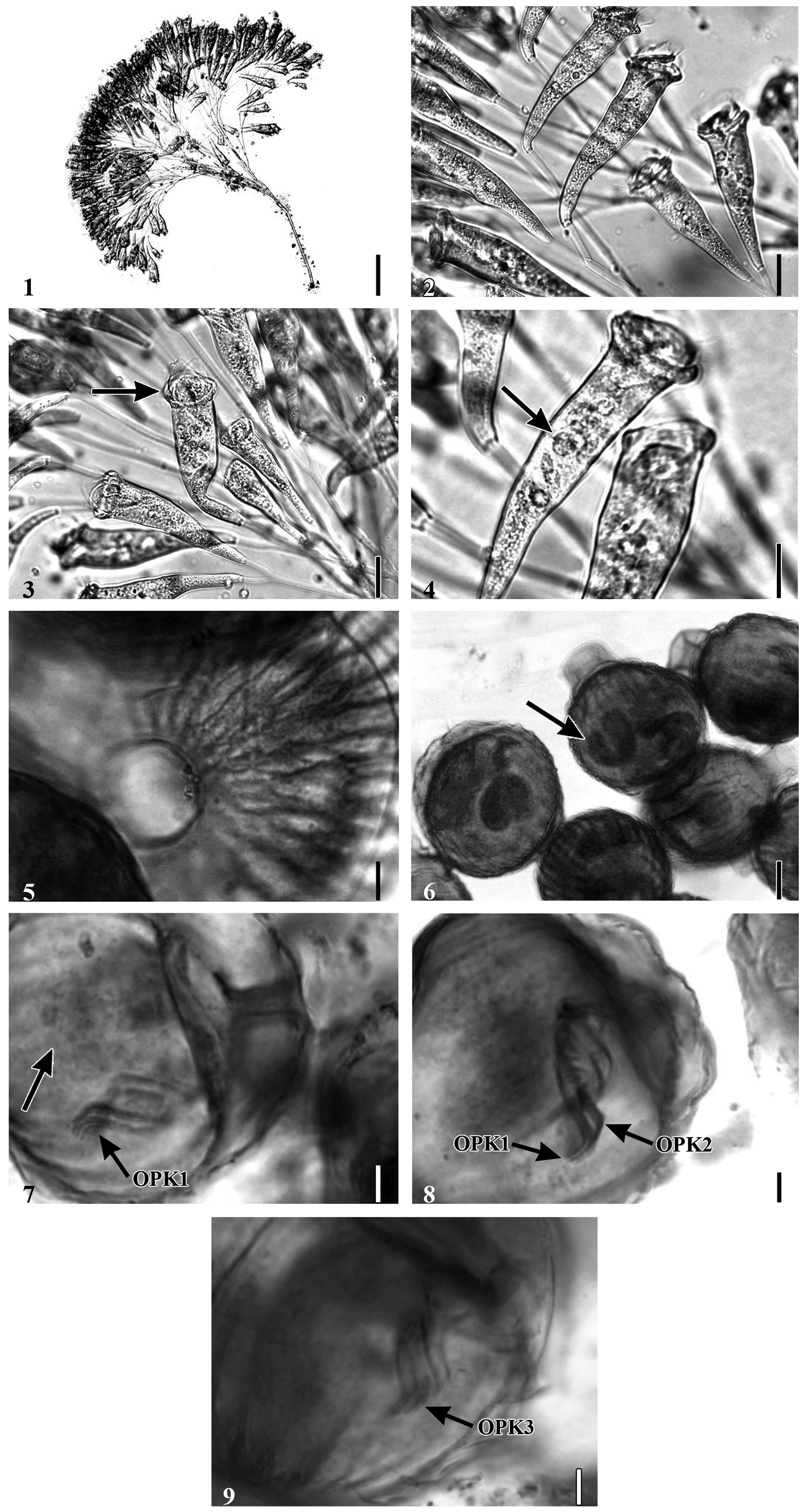
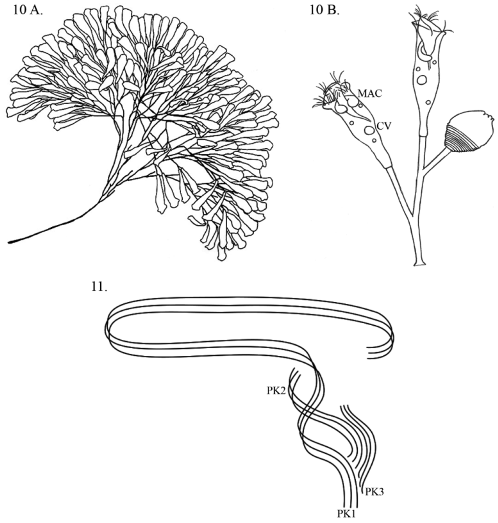
TABLE 1. Measurements of live colonies of Epistylis riograndensis attached to coverslips collected from an artificial lake in the Botanical Garden, Southern Brazil. A total number of 25 zooids were measured for each character.
| Character | Mean (µm) | SD (µm) | Mode | CV (%) Range |
|---|---|---|---|---|
| Length of the Body from epistomial disk to aboral end | 161.7 | 23.5 | 155 | 14.5 115 – 212.5 |
| Length of the body from peristomial lip to aboral end | 133.8 | 20.3 | 122.5 | 15.2 90 – 180 |
| Width of the Body below the peristomial lip | 44.9 | 3.7 | 47.5 | 8.2 37.5 – 50 |
| Width of the body at midpoint between oral and aboral ends | 47 | 4.0 | 50 | 8.5 37.5 – 52.5 |
| Width of peristomial Lip | 52.5 | 4.6 | 50 | 8.8 40 – 62.5 |
| Width of epistomial disk | 37.1 | 3.0 | 37.5 | 8.1 32.5 – 45 |
| Thickness of peristomial Lip | 10.7 | 1.5 | 10 | 14.3 7.5–12.5 |
| Width of scopula | 11.9 | 1.3 | 12.5 | 10.9 10–15 |
| Length of basal stalk | 323.5 | 142.7 | 410 | 44.1 82.5–590 |
| Width of basal stalk | 17.5 | 1.8 | 17.5 | 10.3 15 – 22.5 |
No known copyright restrictions apply. See Agosti, D., Egloff, W., 2009. Taxonomic information exchange and copyright: the Plazi approach. BMC Research Notes 2009, 2:53 for further explanation.