
Trizocheles spinidigitus, Komai, Tomoyuki & Chan, Tin-Yam, 2016
|
publication ID |
https://doi.org/10.11646/zootaxa.4088.3.1 |
|
publication LSID |
lsid:zoobank.org:pub:D055AB86-A0A2-4E30-8671-4B0990C478FB |
|
DOI |
https://doi.org/10.5281/zenodo.6076087 |
|
persistent identifier |
https://treatment.plazi.org/id/03D78781-E245-FF88-D4A4-FD5609A59AC9 |
|
treatment provided by |
Plazi |
|
scientific name |
Trizocheles spinidigitus |
| status |
sp. nov. |
Trizocheles spinidigitus View in CoL n. sp.
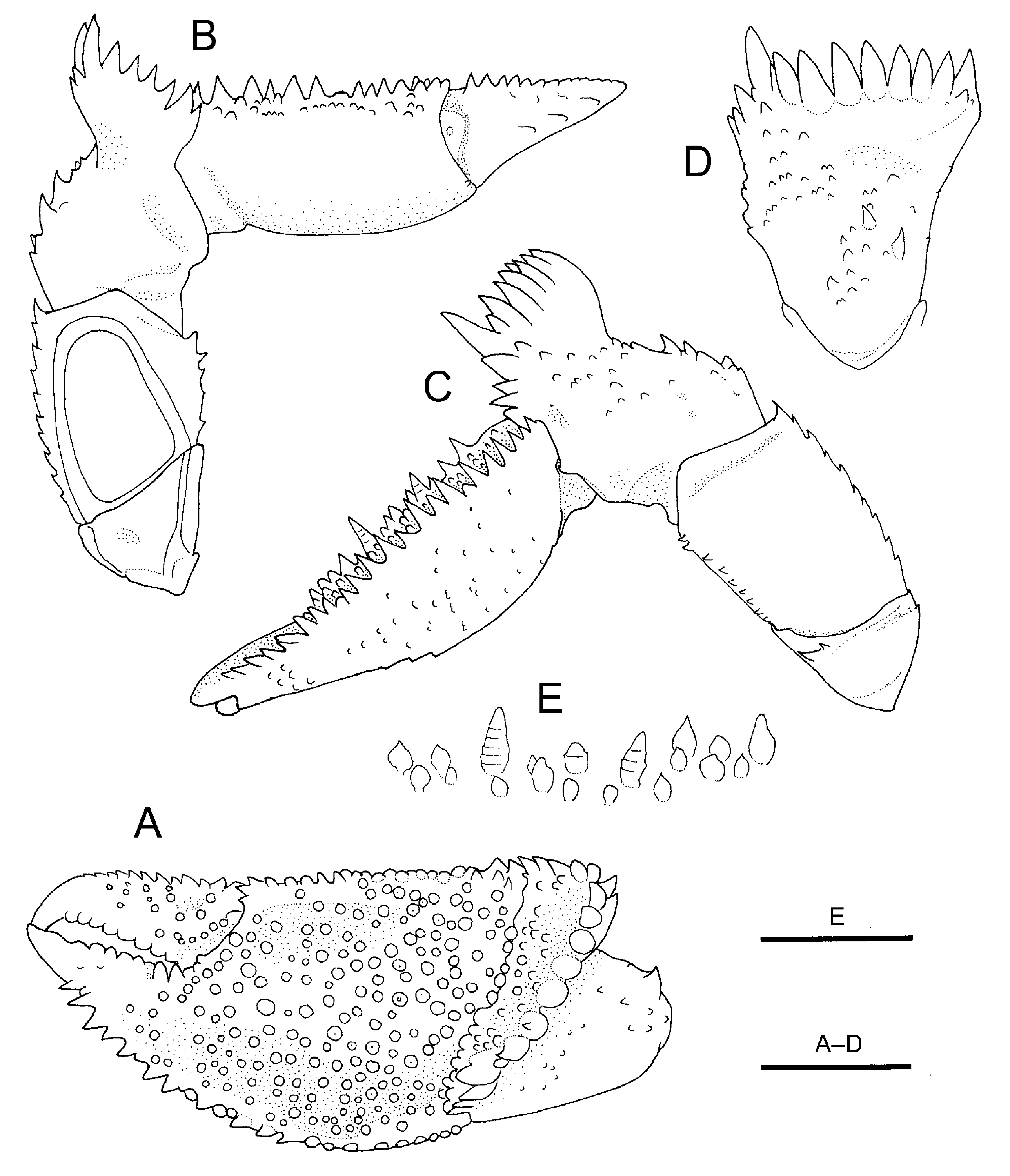
( Figs. 6–8 View FIGURE 6 View FIGURE 7 View FIGURE 8 )
Material examined. Holotype: BIOPAPUA, stn DW 3745, S of Bougainville, 05°33’S, 154°00’E, 369–377 m, 12 October 2010, male (sl 5.2 mm), MNHN-IU-2014-12696
Paratypes: same data as holotype, 2 males (sl 2.3, 4.0 mm), 3 females (sl 2.9–4.2 mm), 3 ovigerous females (sl 3.5–3.8 mm), MNHN-IU-2014-12697
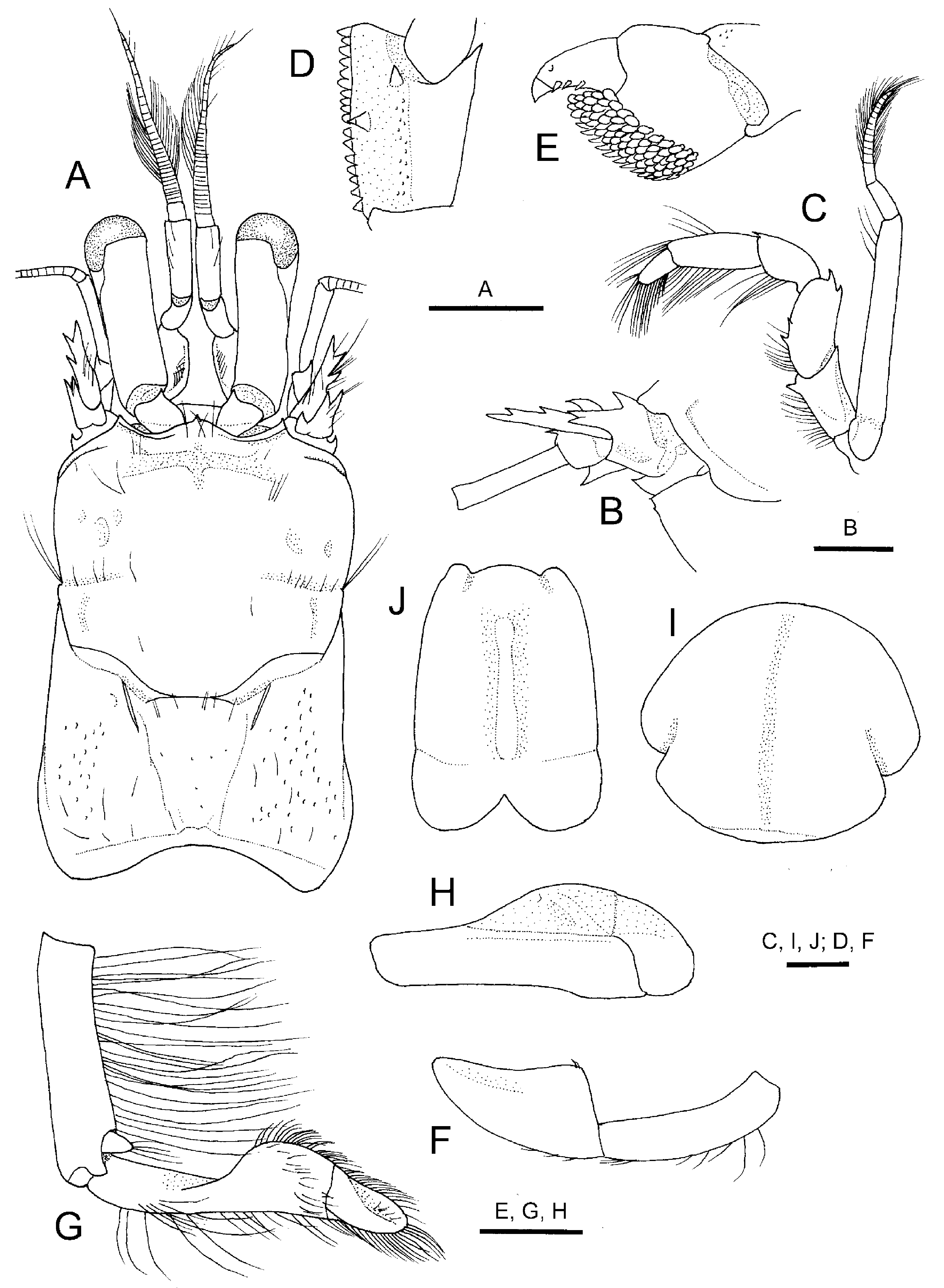
Description. Shield ( Fig. 6 View FIGURE 6 A) slightly wider than long and distinctly longer than posterior carapace; dorsal surface with moderately long, deep transverse groove subrostrally; lateral margins each with small notch posterior to midlength; cervical groove clearly delineated laterally. Rostrum broadly triangular, with small marginal spine medially, extending as far as or slightly overreaching lateral projections. Lateral projections well developed, each with marginal, prominent spine. Posterior median plate trapezoidal, narrowed posteriorly, clearly delimited, moderately well calcified; sulci cardiobranchialis apparent ( Fig. 6 View FIGURE 6 A). Branchial region with scattered shallow pits. Branchiostegite calcified in part adjacent to margins, central part largely membranous; dorsal margin unarmed; anterior margin triangular, terminating in minute spine.
Ocular peduncle ( Fig. 6 View FIGURE 6 A) about 0.7 length of shield, not constricted at midlength, with uncalcified longitudinal suture extending from base of cornea to base of peduncle on lateral surface; cornea slightly inflated, width about 0.4 of peduncular length. Ocular acicle drawn out into acute spine, separated by basal width of one acicle. Interocular lobe clearly visible in dorsal view.
Antennular peduncle ( Fig. 6 View FIGURE 6 A) moderately short, overreaching distal corneal margin by half-length of ultimate segment. Ultimate segment subequal in length to penultimate segment. Basal segment with 1 small spine on anterodorsal margin of statocyst lobe and 1 spinule on ventrodistal margin.
Antennal peduncle ( Fig. 6 View FIGURE 6 A, B) falling slightly short of or just reaching lateral corneal base. Fifth segment cylindrical, unarmed. Fourth segment with small spine on dorsodistal margin. Third segment with prominent spine at ventrodistal margin. Second segment with dorsolateral distal angle produced, terminating in bifid spine; lateral margin unarmed or armed with 1 small spine; dorsomesial distal angle with prominent spine; dorsal surface deeply depressed proximally. First segment with tiny spine at dorsolateral distal angle; ventrodistal margin with 1 spinule lateral to excretory pore. Antennal acicle nearly reaching midlength of ocular peduncle, terminating in simple spine, mesial margin with 1 spine proximodorsally, lateral margin with 2 spines in distal half. Antennal flagellum not reaching tip of cheliped, with 3–5 short to long setae on distal margin of each article.
Third maxilliped ( Fig. 6 View FIGURE 6 C) moderately slender. Basis with 1 or 2 minute denticles on mesial margin. Ischium with well-developed crista dentata and 1 small accessory tooth on mesial face ( Fig. 3 View FIGURE 3 C, D); dorsodistal angle and ventrodistal angle each with small spine. Merus with 2 small spines on dorsal margin distally and 2 small spines on ventral margin. Carpus with small dorsodistal spine, ventrodistal margin unarmed. Propodus and dactylus unarmed, dactylus about half-length of propodus. Exopod overreaching distal margin of merus by 0.3 length; flagellum welldeveloped.
Chelipeds ( Fig. 7 View FIGURE 7 A–C) subequal and symmetrical, with propodal-carpal rotation approximately 45°; narrow proximal hiatus between dactylus and fixed finger. Dactylus slightly shorter than palm, surfaces with tufts of short to long stiff setae; dorsal surface with row of moderately large, corneous-tipped spines decreasing in size distally on midline, dorsomesial margin delimited with row of small to moderately large, corneous-tipped spines; mesial surface with row of small corneous-tipped spines adjacent to dorsal margin ( Fig. 7 View FIGURE 7 D); ventral surface unarmed; cutting edge with row of rounded calcareous teeth, terminating in large corneous claw. Fixed finger with cutting edge bearing row of moderately strong, rounded or subacutely pointed calcareous teeth, terminating in large corneous claw. Palm distinctly longer than carpus; dorsomesial margin with row of 7–9 large, often unequal, corneous-tipped spines; dorsal surface nearly flat, with numerous, scattered short to long stiff setae, and 4 or 5 irregular longitudinal rows of small to moderately strong spines, spines in lateralmost row extending onto fixed finger; lateral surface nearly perpendicular, with some scattered tiny tubercles or protuberances proximally and sparse tufts of short to long, stiff setae; mesial surface with some small spines or tubercles dorsally; ventral surface with few low protuberances distally and tufts of long setae extending onto fixed finger. Carpus subtrapezoidal; vast majority of spines corneous-tipped; dorsomesial margin with 3 prominent spines increasing in size distally (distal 2 spines particularly strong); dorsal surface with 2 irregular longitudinal rows of large, corneous-tipped spines mesial to midline and tufts of short to long setae, dorsolateral margin delimited by row of small corneous-tipped spines; lateral surface with numerous, scattered stridulating tubercles (vast majority of tubercles bearing corneous tip), ventrodistal angle with 1 or 2 tiny tubercles ( Fig. 7 View FIGURE 7 E); mesial surface with 1 moderately large spine near base of second spine on dorsomesial margin, oblique row of small tubercles adjacent to dorsomesial margin and few tufts of stiff setae; ventral surface slightly concave, without conspicuous armature or setae. Merus trigonal in cross section, with shallow transverse groove along distal margin; dorsal margin with row of small spines decreasing in size proximally; dorsodistal margin with small spine directed forward; lateral surface unarmed, with sparse short setae distally and ventrally, ventrolateral margin slightly delimited, with 1 tiny spine subdistally; mesial surface also smooth, with few short setae proximally, ventromesial margin bluntly carinate, with row of small spines; ventral surface nearly flat, with row of short to moderately long setae laterally. Ischium with row of small spines or spiniform tubercles on ventromesial margin.
Second pereopod ( Fig. 8 View FIGURE 8 A, B) overreaching chelipeds by half-length of dactylus, strongly compressed laterally, similar from right to left; spines corneous-tipped. Dactylus subequal in length to propodus, nearly straight, terminating in strong, curved corneous claw; dorsal margin armed with single or double row of 6–10 moderately large spines and tufts of short to moderately long stiff setae (these spines decreasing in size distally); lateral surface unarmed, with rows of tufts of moderately long setae adjacent to dorsal and ventral margins; mesial surface with 2–4 small but prominent spines on midline proximally and some tufts of moderately long setae; ventral margin with 4–7 widely spaced small corneous spines and 1 stiff seta at base of corneous claw (this seta slightly shorter than claw). Propodus about 2.7–3.1 times longer than wide; dorsal surface with 2 rows of large spines (7 or 8 spines in outer row, 5–9 spines in inner row) and tufts of long stiff setae; lateral surfaces unarmed, with row of tufts of short to long stiff setae; mesial surface with stridulating apparatus composed of dorsal row of prominent tubercles and ventral row of prominent, short to moderately long, transverse rods; ventral surface with low protuberance subdistally and few tufts of long stiff setae, ventrodistal margin with 1 corneous spinule (sometimes submarginal). Carpus with row of 6–8 long spines and tufts of long stiff setae on dorsal margin; lateral surface shallowly sulcate medially, with longitudinal rows of tufts of setae adjacent to dorsal margin and on midline; mesial surface with series of 5 transverse stridulating rods (increasing in length distally), none subdivided into short elements. Merus with row of tufts of moderately long setae on dorsal margin; lateral surface with sparse very short setae, smooth; mesial surface flat, almost glabrous; ventral margin with row of tiny, widely spaced spines and tufts of long stiff setae. Ischium unarmed, dorsal margin with short feathered setae, ventral margin with tufts or individual stiff setae.
Third pereopod ( Fig. 8 View FIGURE 8 B, D) generally similar to second pereopod in shape and setation, but armature weaker. Dactylus 1.1 times as long as propodus; dorsal margin with single row of 7 spines; ventral margin with 5 widely spaced corneous spines and 1 stiff seta at base of corneous claw. Propodus with single row of 5–7 spines + 1–4 proximal mesial spines on dorsal surface; mesial surface with scattered short, sharp tubercles or short vertical ridges, representing vestige of stridulating apparatus. Carpus with single row of 6 or 7 spines on dorsal margin; mesial surface smooth. Merus with tiny dorsodistal spine, but otherwise unarmed. Ischium unarmed on dorsal and ventral margins.
Fourth pereopod ( Fig. 6 View FIGURE 6 E) semichelate; each with propodal rasp consisting of several rows of corneous scales. Fifth pereopods weakly chelate; propodal rasp well developed.
Male first pleopod ( Fig. 6 View FIGURE 6 F) strongly compressed laterally; distal segment subtriangular in lateral view, tapering to narrowly rounded tip. Male second pleopod ( Fig. 6 View FIGURE 6 G, H) with very short, bud-like exopod; endopod 2- segmented, proximal segment with dorsomesial margin forming broad convexity in distal half, distal segment short, distally flattened, distal margin rounded.
Pleon with first tergite moderately well calcified; second to fifth tergites poorly calcified, pleura weakly calcified. Sixth tergite ( Fig. 6 View FIGURE 6 I) subcircular, with deep lateral incisions at posterior 0.3; dorsal surface with shallow median sulcus along entire length; terminal margin nearly straight, unarmed. All tergites and telson with numerous short to moderately long setae. Uropods symmetrical; protopods produced posteriorly, each with small spine on posterior margin mesial to base of endopod.
Telson ( Fig. 6 View FIGURE 6 J) slightly widened posteriorly, 1.3 times as long as wide with faint lateral indentations at posterior 0.3; posterior lobes separated by deep median cleft, terminal margins rounded, unarmed but with fringe of long setae.
Size. Largest male sl 5.1 mm, largest female sl 4.2 mm, ovigerous females sl 3.5–3.8 mm.
Colour in life. Unknown.
Distribution. At present known only from the type locality, south of Bougainville Islands, Solomon Sea, 369– 377 m.
Habitat. Scaphopod (tusk) shells.
Remarks. Trizocheles spinidigitus n. sp. shares many characters with T. vaubanae , for example, the moderately stout ocular peduncle, the propodi of the second pereopods each armed with large spines, arranged in double row, on the dorsal surface, and the propodi and carpi of the third pereopods armed with a dorsal row of large spines (McLaughlin & Lemaitre 2008, 2009). Nevertheless, the new species is readily distinguished from T. vaubanae by having large spines on the dorsal margins of dactyli of the second and third pereopods. In T. vaubanae , the ambulatory dactyli are unarmed on the dorsal margins. Furthermore, the mesial face of the dactylus of each second pereopod is armed with small but prominent spines on the midline proximally in T. spinidigitus . No other congeneric species, including T. vauabanae , have such a spination on the second pereopod.
Trizocheles longicaulis and T. pulcher also have armature on the second pereopod dactyli, but the spines are not so large as in T. spinidigitus (cf. Forest 1987a). Trizocheles longicaulis is characteristic in the shield being longer than wide, and the slender, elongate ocular peduncle overreaching the distal end of the ultimate segment of the antennular peduncle (Forest 1987a; McLaughlin & Lemaitre 2009). Trizocheles pulcher further differs from the present new species in the lack of a dorsal row of spines on the carpi of the third pereopods (McLaughlin & Lemaitre 2009).
Etymology. From the combination of the Latin spinosus (= spiny) and digitus (= finger), in reference to the spiny dactyli of the second and third pereopods in the new species; used as a noun in apposition.


No known copyright restrictions apply. See Agosti, D., Egloff, W., 2009. Taxonomic information exchange and copyright: the Plazi approach. BMC Research Notes 2009, 2:53 for further explanation.
|
Kingdom |
|
|
Phylum |
|
|
Class |
|
|
Order |
|
|
Family |
|
|
SubFamily |
Trizochelinae |
|
Genus |