
Echinoderes xiphophorus, Adrianov & Maiorova, 2021
|
publication ID |
https://doi.org/ 10.5852/ejt.2021.773.1523 |
|
publication LSID |
lsid:zoobank.org:pub:CA23F2C2-38F7-47B8-ABAC-F447A775E9E4 |
|
DOI |
https://doi.org/10.5281/zenodo.14550159 |
|
persistent identifier |
https://treatment.plazi.org/id/3E80E789-3AC9-410F-9137-7CF7EC541568 |
|
taxon LSID |
lsid:zoobank.org:act:3E80E789-3AC9-410F-9137-7CF7EC541568 |
|
treatment provided by |
Felipe |
|
scientific name |
Echinoderes xiphophorus |
| status |
sp. nov. |
Echinoderes xiphophorus View in CoL sp. nov.
urn:lsid:zoobank.org:act:3E80E789-3AC9-410F-9137-7CF7EC541568
Figs 1–8 View Fig View Fig View Fig View Fig View Fig View Fig View Fig View Fig
Diagnosis
Trunk length (TL) 290–305 µm; lateral terminal spines (LTS) 142–185 µm; trunk segments 1–2 consisting of closed cuticular rings, and trunk segments 3–11 of one tergal and two sternal plates; trunk segment 2 with three pairs of well-developed tubes in subdorsal, midlateral, and ventrolateral positions; trunk segment 5 with tubes in lateroventral position; trunk segment 8 with tubes in sublateral position; middorsal spines on trunk segments 4, 6, 8; lateroventral acicular spines on trunk segments 6–9; trunk segment 11 with remarkably long tergal extensions (TE) about 15–16% of TL; males with three pairs of penile spines and remarkably long sword-like extensions (about 80% of TE) dorsally to penile spines; females with pair of laterodorsal tubes on trunk segment 10.
Etymology
The species name is derived from the Greek ʻ xiphos ʼ (sword) and ʻ phorus ʼ (carry) because of very long sword-like appendages in males dorsally to penile spines.
Material examined
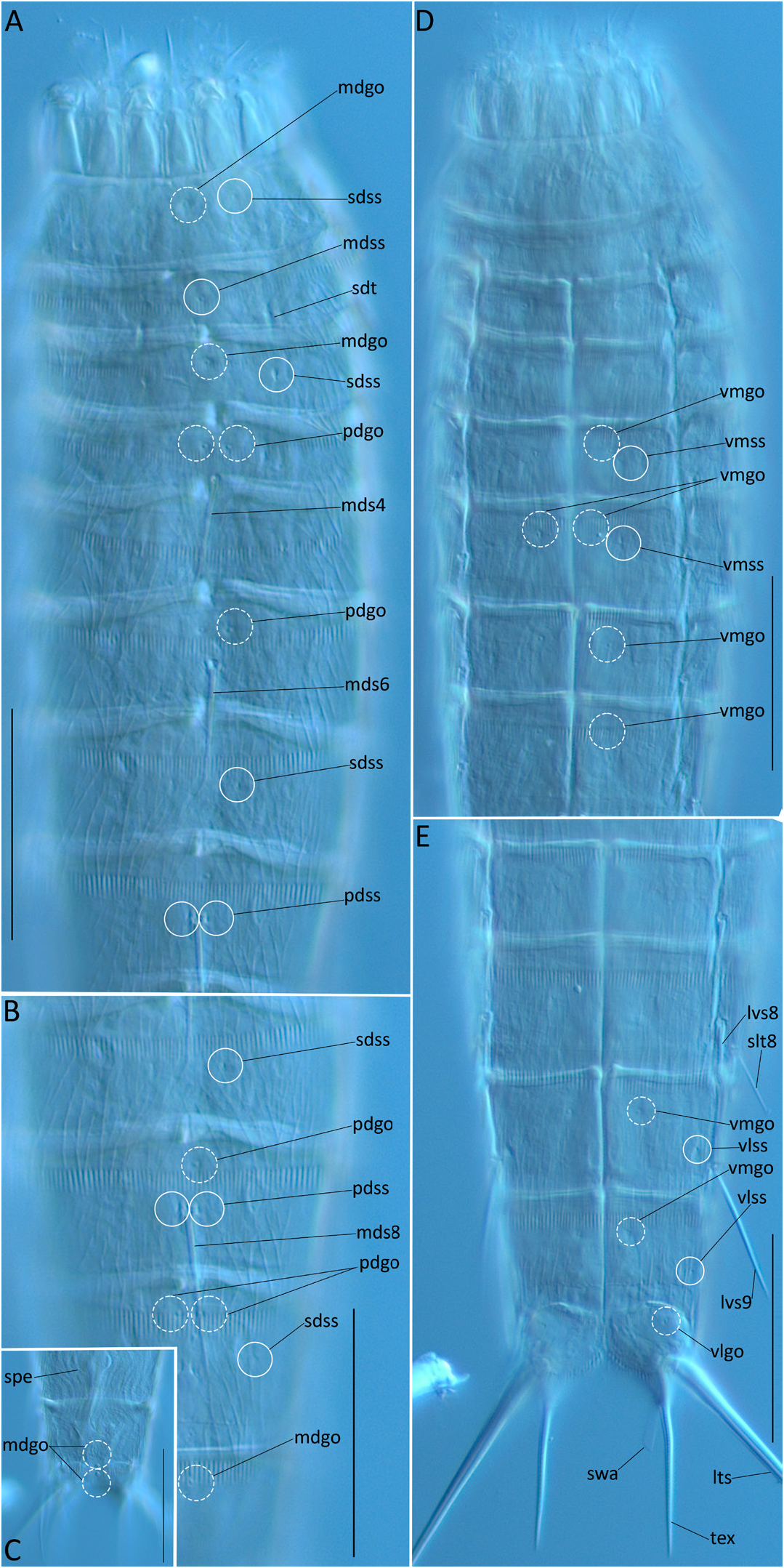
Holotype ( Fig. 2A–E View Fig ) RUSSIA • 1 ♂; Sea of Japan; 44.7942° N, 137.2550° E; depth 1530 m; 14 Aug. 2010; SoJaBio cruise; stn A3-7; multiple corer; oxidized brown clay; MIMB 41347 View Materials GoogleMaps .
Paratypes ( Fig. 3A–B View Fig ) RUSSIA • 4 adult ♂♂; same locality and station as for holotype; MIMB 41348 View Materials , MIMB 41349 View Materials , MIMB 41350 View Materials , MIMB 41351 View Materials GoogleMaps • 1 adult ♂; Sea of Japan ; 44.9439950° N, 137.2017717° E; depth 515 m; 13 Aug. 2010; SoJaBio cruise; stn A2-5; multiple corer; oxidized brown clay; MIMB 41352 View Materials GoogleMaps .
Other material
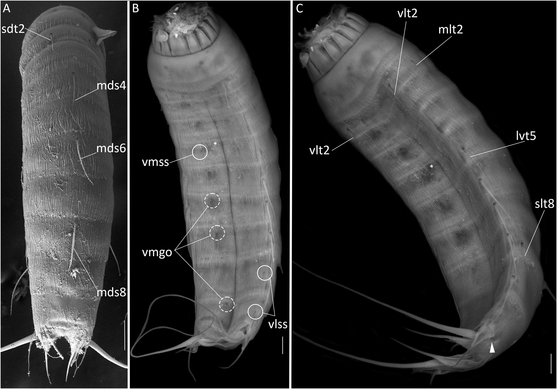
RUSSIA • 2 adult ♂♂, 2 adult ♀♀ (mounted for SEM); same locality and station as for holotype GoogleMaps .
Description
BODY. Adult specimen consists of head, neck and 11 trunk segments. Measurements (µm) and ratios (%) are provided in Table 1 View Table 1 . Head consists of retractable mouth cone and eversible introvert. Inner oral styles are arranged in two circlets (5 large tooth-like outer styles + 10 minute innermost styles). External mouth cone bears nine articulated outer oral styles. Head bears seven rings of spinoscalids and one ring of six trichoscalids. Neck consists of 16 placids. All placids trapezoid in shape, distinctly articulating with the first trunk segment. Midventral placid widest; remaining ones narrower. Two ventral and four dorsal trichoscalid plates present, ventral ones being broader than four dorsal ones.
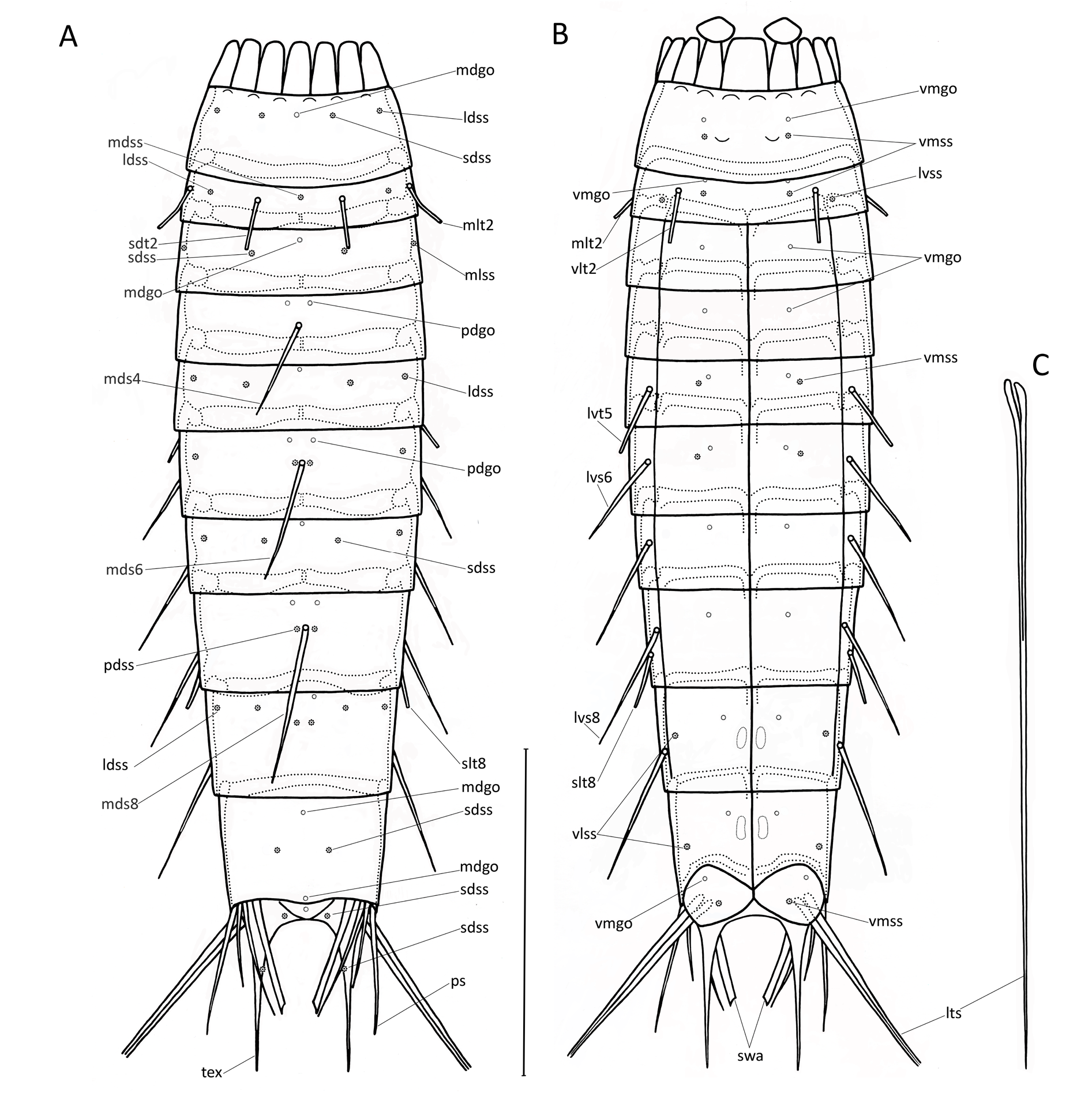
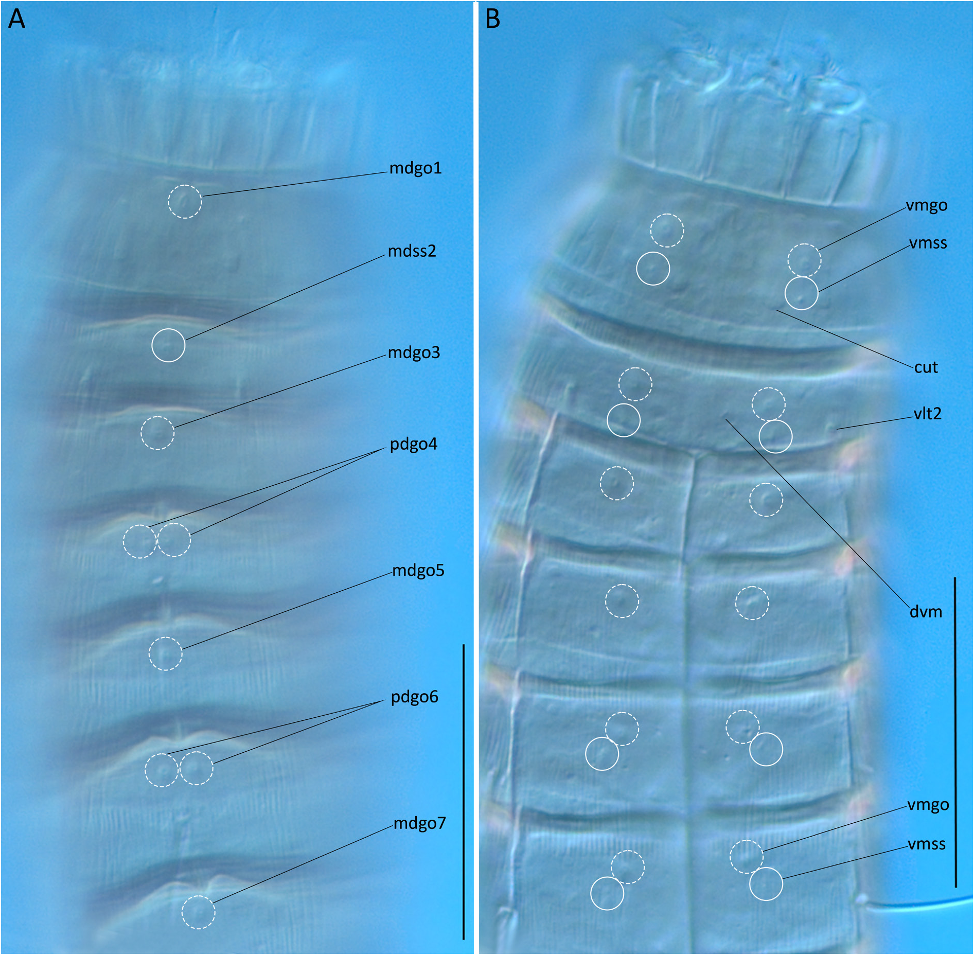
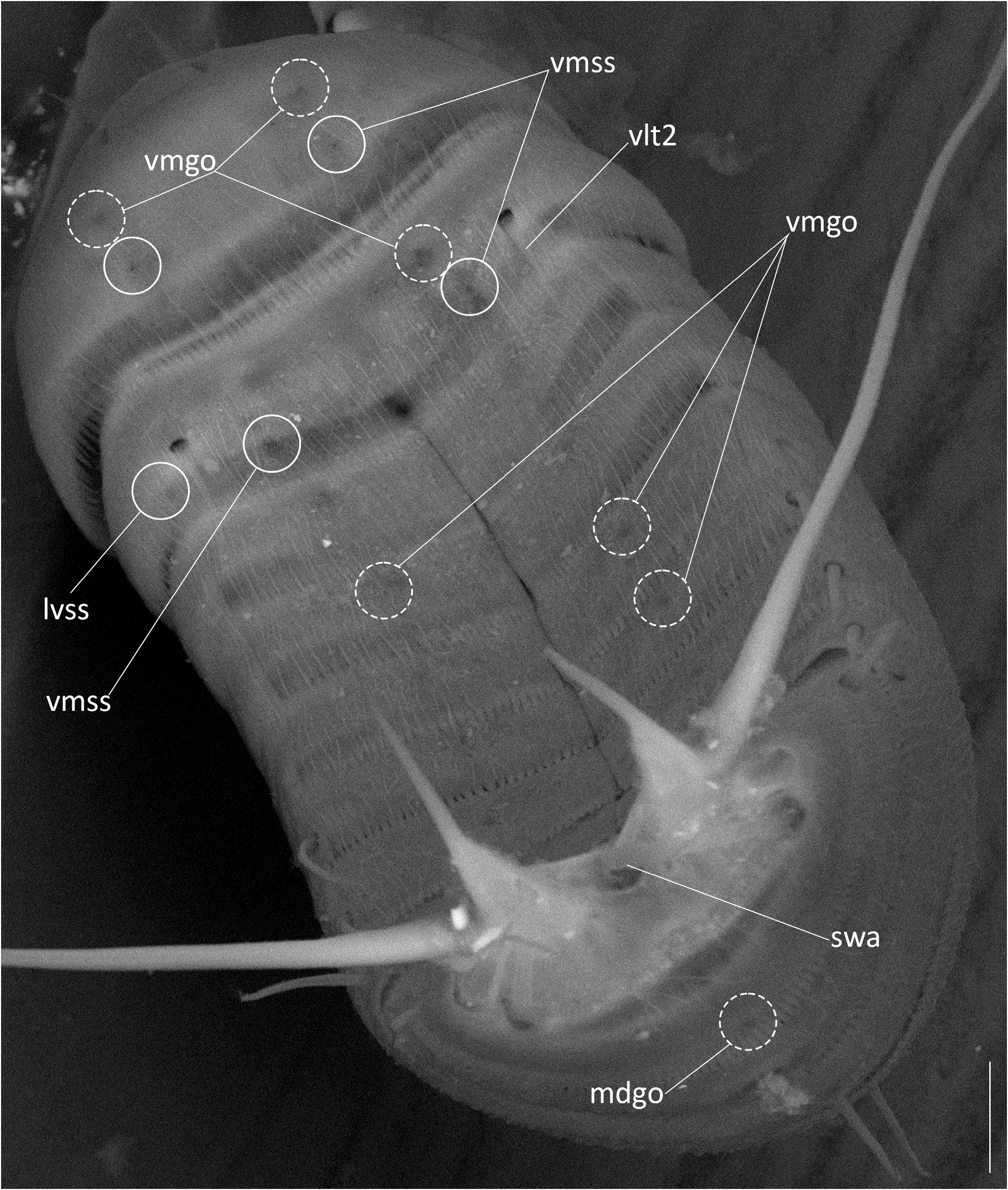
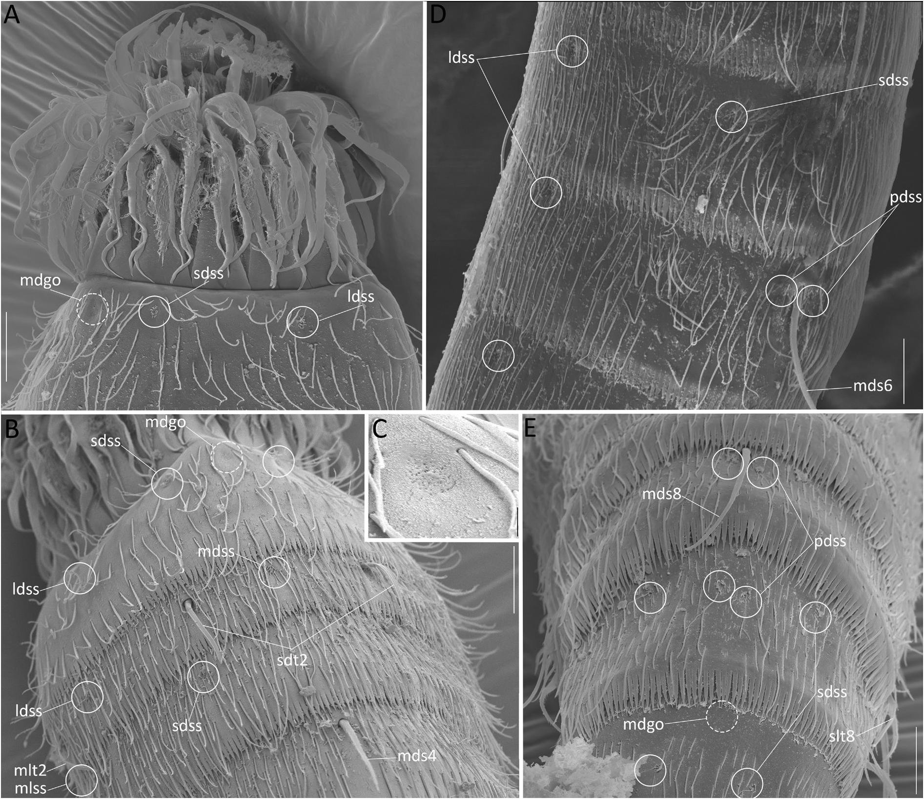
TRUNK SEGMENT 1. Closed cuticular ring, with three pairs of sensory spots located close to the anterior segment margin in subdorsal and laterodorsal and more posteriorly in ventromedial position ( Figs 1A–B View Fig , 2A View Fig , 3B View Fig , 5 View Fig , 6A–B View Fig , 7A View Fig ); sensory spots rounded in shape, with 15–20 very short petals and one or two pores ( Fig. 6A View Fig ); single glandular cell outlet of type 1 in middorsal position and two outlets in ventromedial position ( Figs 1A–B View Fig , 2A View Fig , 3A–B View Fig , 5 View Fig , 6A–C View Fig ); glandular cell outlets of type 2 absent as in all trunk segments; posterior margin of segment with pectinate fringe with fringe tips from midventral to ventrolateral position being shorter than others ( Fig. 7A View Fig ); long cuticular hairs on dorsal surface regularly arranged forming a continuous belt of 3–4 distinct transverse rows and irregularly arranged on ventral side ( Figs 6A–B View Fig , 7B View Fig ).
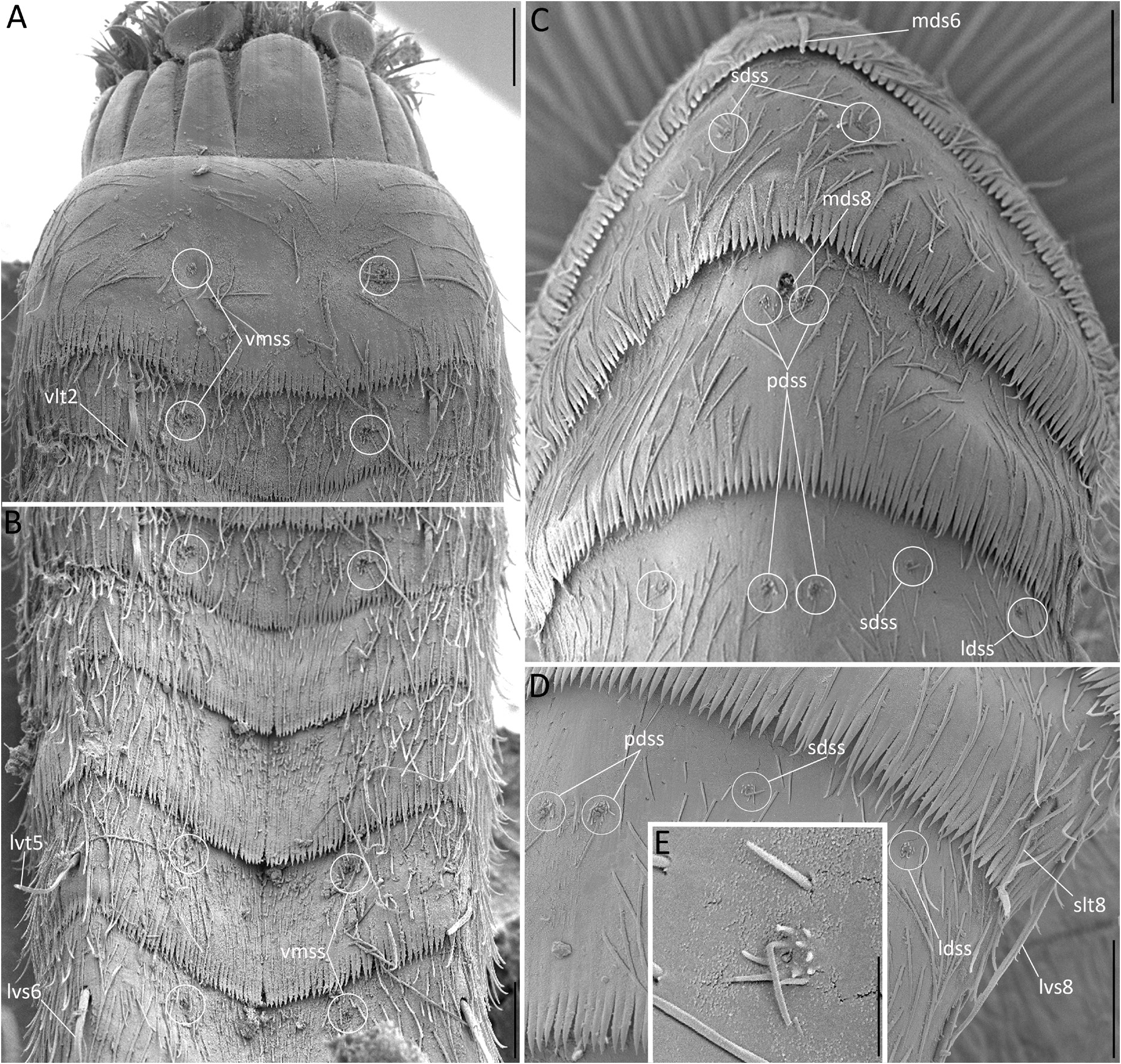
TRUNK SEGMENT 2. Closed cuticular ring, without any partial intra- or extracuticular fissures; three pairs of tubes in subdorsal, midlateral and ventrolateral positions; each tube with short and smooth basal part and longer distal part with two wing-like lateral projections ( Fig. 6B View Fig ); one sensory spot in middorsal position and three pairs in laterodorsal, lateroventral and ventromedial position, sensory spots on this and following segments with 10–15 very short petals and 1–2 very long posteriormost petals similar to cilia ( Figs 6B View Fig , 7C–E View Fig ); one pair of glandular cell outlets of type 1 in ventromedial position anterior to ventromedial sensory spots ( Fig. 3B View Fig ); ventral hairs on the trunk segment 2 seem to be more regularly arranged than those on trunk segment 1; pectinate fringe as in the preceding segment. It should be noted that glandular cell outlets at the anterior margin of tergal and sternal plates are usually hidden beneath posterior cuticular fringe of previous segment and seen more easily with DIC (see Figs 2–3 View Fig View Fig ).
TRUNK SEGMENT 3. One tergal and two sternal plates, with distinct tergosternal and midventral articulations, as in all remaining trunk segments; with two pairs of sensory spots in subdorsal and midlateral position ( Figs 1A–B View Fig , 2A View Fig , 3B View Fig , 6B View Fig ); single glandular cell outlet of type 1 in middorsal position located close to the anterior segment margin and hidden under preceding segment; with a pair of glandular cell outlets in ventromedial position; long cuticular hairs absent in ventromedial position, being replaced by much shorter cuticular hairs ( Fig. 7B View Fig ); other cuticular hairs and pectinate fringe as in the preceding segment.
TRUNK SEGMENT 4. With acicular spine in middorsal position ( Figs 2A View Fig , 4A View Fig ); with two pairs of glandular cell outlet of type 1 in paradorsal and ventromedial positions, paradorsal outlets usually hidden under previous segment ( Figs 2A, D View Fig , 3 View Fig ); long cuticular hairs absent in middorsal position beneath acicular spine; other cuticular hairs and pectinate fringe as in the preceding segment.
TRUNK SEGMENT 5. No acicular spine in middorsal position; one pair of tubes in lateroventral position ( Figs 4C View Fig , 7B View Fig ); three pairs of sensory spots in subdorsal, laterodorsal and ventromedial position; single glandular cell outlet of type 1 in middorsal position located close to the anterior segment margin and usually hidden under previous segment; with one pair of glandular cell outlets of type 1 in ventromedial positions; pectinate fringe and cuticular hairs as in the trunk segment 3.
TRUNK SEGMENT 6. With acicular spine in middorsal position ( Figs 2A View Fig , 4A View Fig , 6D View Fig ); with a pair of acicular spines in lateroventral position ( Fig. 4B View Fig ); with three pairs of sensory spots in paradorsal, laterodorsal and ventromedial position; with two pairs of glandular cell outlets of type 1 in paradorsal and ventromedial position, paradorsal outlets usually hidden under previous segment; pectinate fringe and cuticular hairs as in the trunk segment 4.
TRUNK SEGMENT 7. No acicular middorsal spine; with two pairs of sensory spots in subdorsal and laterodorsal position; with single glandular cell outlets of type 1 in middorsal position (usually hidden under previous segment) and one pair of outlets in ventromedial position; other spines, pectinate fringe and cuticular hairs as in the preceding segment.
TRUNK SEGMENT 8. With acicular spine in middorsal position ( Figs 2A View Fig , 4A View Fig , 6E View Fig ); with a pair of tubes in sublateral position ( Figs 2E View Fig , 4C View Fig , 7D View Fig ); with one pair of sensory spots in paradorsal position ( Figs 6E View Fig , 7C View Fig ); with two pairs of glandular cell outlets of type 1 in paradorsal and ventromedial position ( Fig. 2B, D View Fig ); other spines, pectinate fringe and cuticular hairs as in the preceding segment.
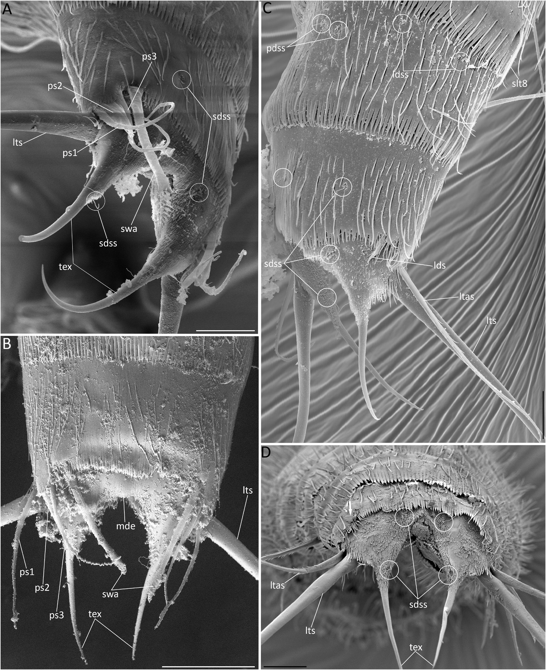
TRUNK SEGMENT 9. No acicular middorsal spine; no tubes; with four pairs of sensory spots in paradorsal, subdorsal, laterodorsal and ventrolateral position ( Figs 2E View Fig , 6E View Fig , 7C–E View Fig , 8C View Fig ); with two pairs of glandular cell outlets of type 1 in paradorsal and ventromedial positions ( Fig. 2B, E View Fig ); pair of excretory sieve plates in lateral accessory position; other spines, pectinate fringe and cuticular hairs as in the preceding segment.
TRUNK SEGMENT 10. No acicular spines; with two pairs of sensory spots in subdorsal and ventrolateral position ( Figs 2E View Fig , 6E View Fig , 8C View Fig ); with single glandular cell outlets of type 1 in middorsal position and one pair in ventromedial position ( Figs 2B, E View Fig , 5 View Fig ); males possess a pair of very long and flattened sword-like appendages (similar in shape to Japanese swords katana or wakizashi) in laterodorsal position, dorsally to penile spines, only slightly shorter than tergal extensions (about 80% of TE) ( Fig. 8A–B View Fig ); females with a pair of short tubes/ spines in laterodorsal position corresponding to sword-like appendages of male ( Fig. 8C–D View Fig ); other characters as in the preceding segment.
TRUNK SEGMENT 11. One pair of long lateral terminal spines ( Fig. 1C View Fig ); tergal extensions very long, about 15–16% of TL ( Figs 4–5 View Fig View Fig , 8 View Fig ); tergal plate with nearly rounded middorsal elevation ( Fig. 8B, D View Fig ); with two pairs of sensory spots in subdorsal and ventromedial position, and additional pair of sensory spots present at the base of tergal extensions ( Fig. 8A, C–D View Fig ); with two glandular cell outlets of type 1 in middorsal position and one pair ventrolateral position ( Figs 2C, E View Fig , 5 View Fig ); males with three pairs of penile spines, first and third pairs thin and flexible, spine of second pair shorter and cone-shaped with terminal tuft of cuticular hairs ( Figs 5 View Fig , 8A–B View Fig ); female with a pair of long and thin lateral terminal accessory spines; sexes can be also recognized by observation of gonads.
No known copyright restrictions apply. See Agosti, D., Egloff, W., 2009. Taxonomic information exchange and copyright: the Plazi approach. BMC Research Notes 2009, 2:53 for further explanation.
|
Kingdom |
|
|
Phylum |
|
|
Class |
|
|
Order |
|
|
Family |
|
|
Genus |