
Lamprohiza splendidula ( Linnaeus, 1767 )
|
publication ID |
https://doi.org/ 10.11646/zootaxa.4378.4.4 |
|
publication LSID |
lsid:zoobank.org:pub:D13D3271-9B08-4D35-85E4-21B1F69BA992 |
|
DOI |
https://doi.org/10.5281/zenodo.5952901 |
|
persistent identifier |
https://treatment.plazi.org/id/03D7BE5C-FFF6-6803-FF69-ED01FBE3F3F5 |
|
treatment provided by |
Plazi |
|
scientific name |
Lamprohiza splendidula ( Linnaeus, 1767 ) |
| status |
|
Lamprohiza splendidula ( Linnaeus, 1767) View in CoL
Material examined. Prague ( Czech Republic), ten higher-instar larvae out of sixteen collected at the end of August 2013, one male and one female pupa were reared from eleven specimens collected at the end of August 2014.
Diagnosis. Larvae flat, laterally explanate, brown and ochre towards the lateral edges of tergites with pairs of lighter pigmented spots on abdominal tergites I–VI; protergum with narrow emargination anteromedially; lightcoloured spot posteriorly behind each stemma; antennal sensorium with distinct basal constriction; retinaculum absent; mandibular channel opening covered by long feather-like or rounded-trapezium hyaline appendage; pretarsal claw ventrally bearing two long setae; photic organ of variable pattern, usually consisting of paired larger spots ventrolaterally on abdominal segments II and VI, sometimes with additional smaller spots ventrolaterally on abdominal segments III–V.
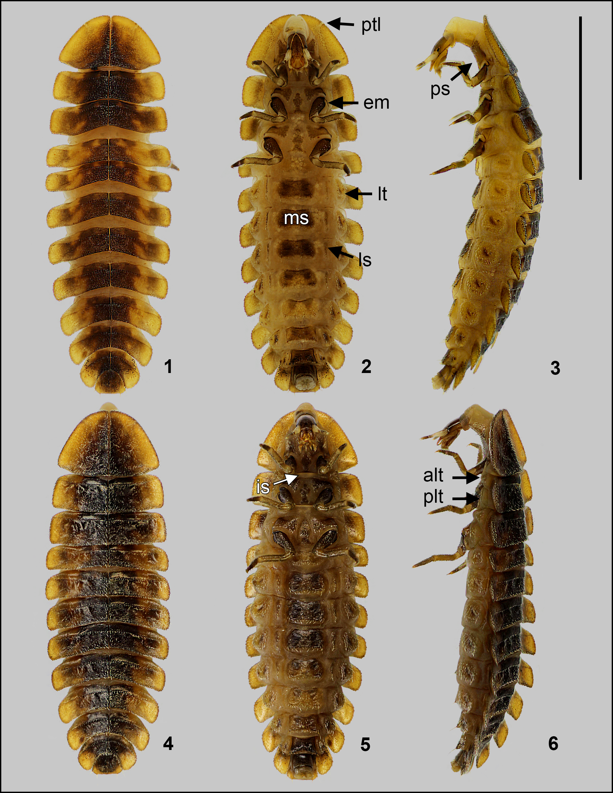
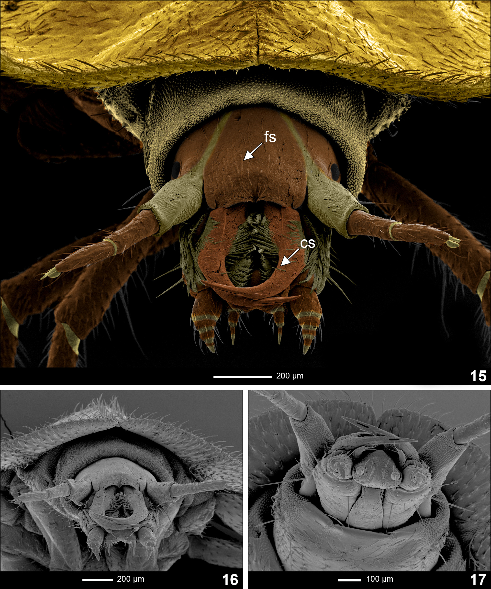
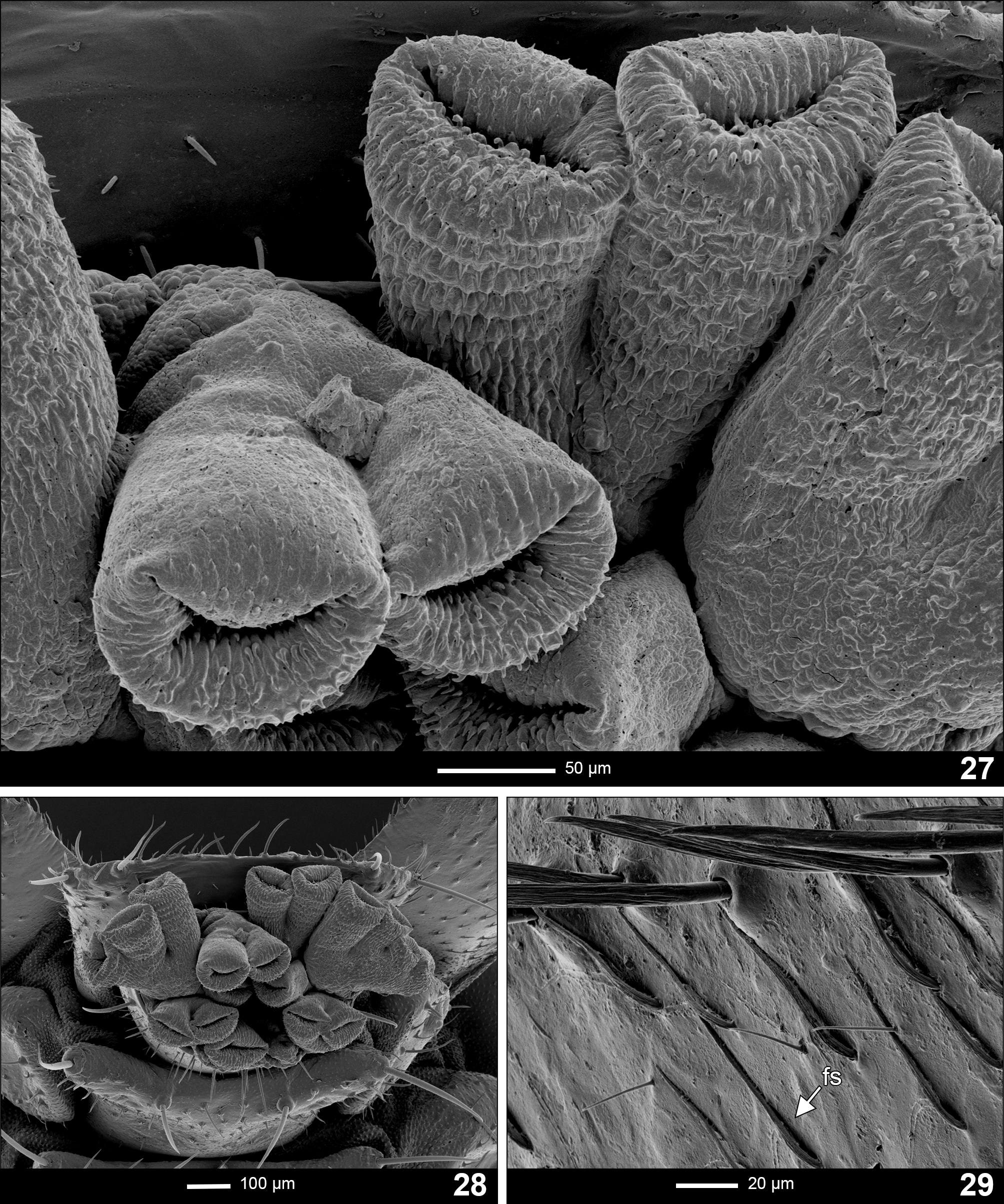
Description of mature larva ( Figs 1–6 View FIGURES1–6 ). Elongate and onisciform; dorsoventrally flattened, tergites of thorax and abdomen finely setose on the edges ( Figs 4 View FIGURES1–6 , 15 View FIGURES15–17 , 28 View FIGURES 27–29 ), and strongly laterally explanate ( Figs 1, 2, 4, 5 View FIGURES1–6 ). Body length ca. 11–12 mm (from the anterior margin of protergum to the apex of caudal segment); with 3 thoracic and 10 abdominal segments. Tergites from protergum to abdominal segment IX divided by sagittal line in dorsal view ( Fig. 1 View FIGURES1–6 ). Colouration: dorsally brown and ochre towards the lateral edges of tergites; with pairs of lighter pigmented spots on abdominal tergites I–VI ( Fig. 1 View FIGURES1–6 ), under which a variable number of localized photic organs may occur. Ventral region much lighter than dorsal, with ochre to light brown colouration, except central parts of sternal sclerites which are darker and more sclerotized. Spiracles on pleural plates are of dark brown colouration.
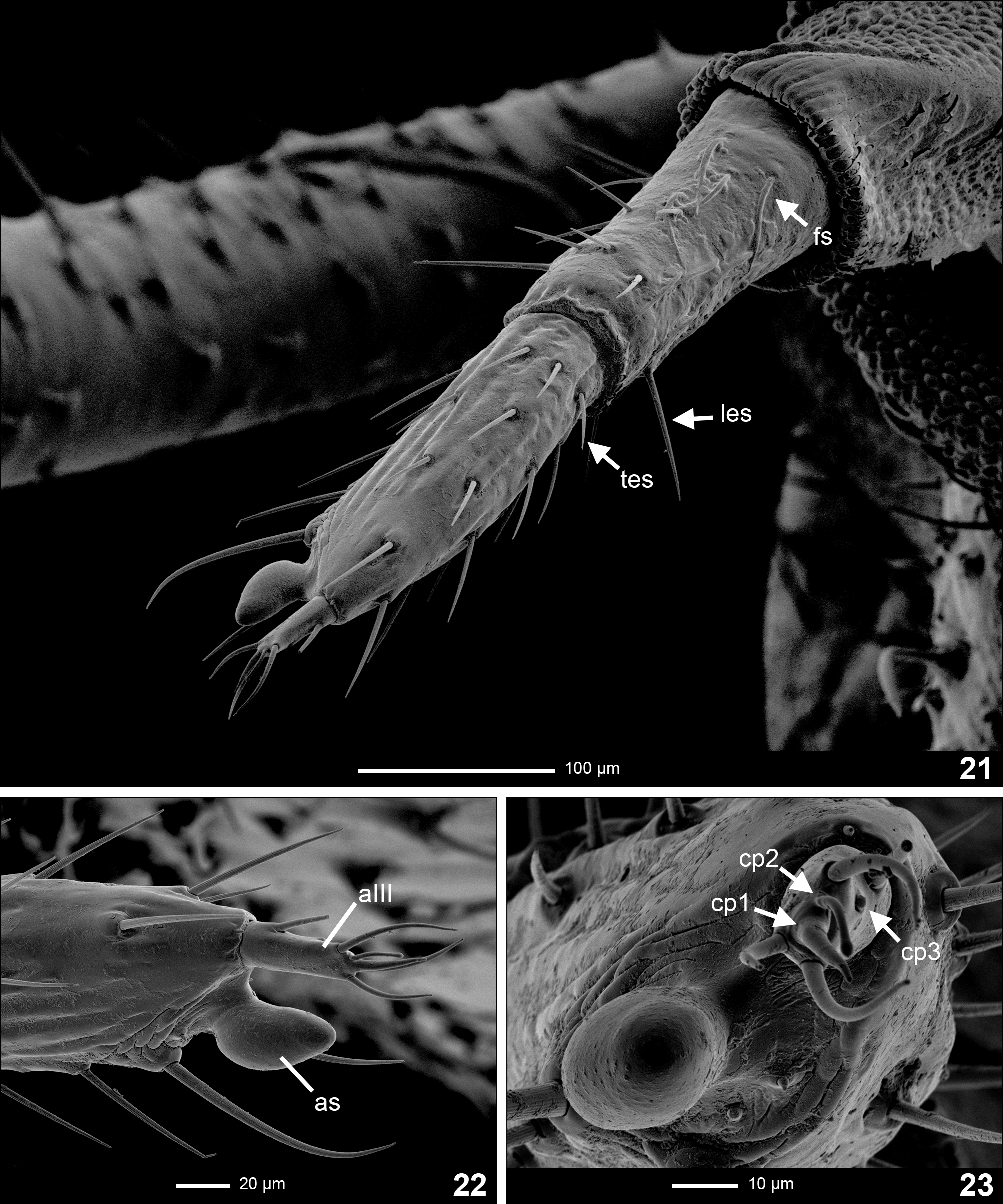
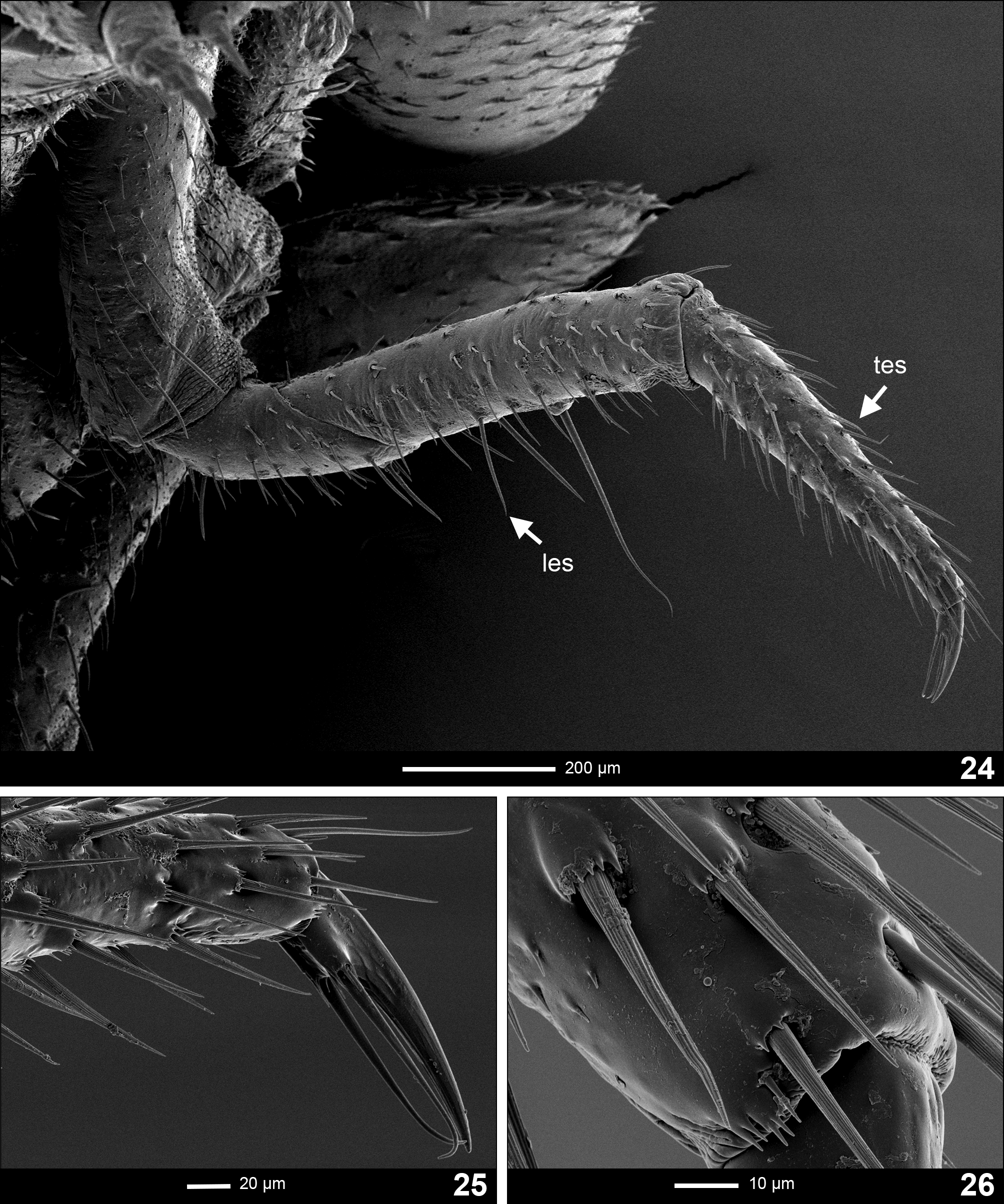
Types of general cuticular outgrowth observed. 1. Stout, long, erect setae ( Figs 21, 22 View FIGURES 21–23 ; les); 2. thin, short, erect setae ( Figs 21, 22 View FIGURES 21–23 ; tes); 3. flat setae, lying on/adjacent to the surface ( Figs 15 View FIGURES15–17 , 21 View FIGURES 21–23 , 29 View FIGURES 27–29 ; fs).
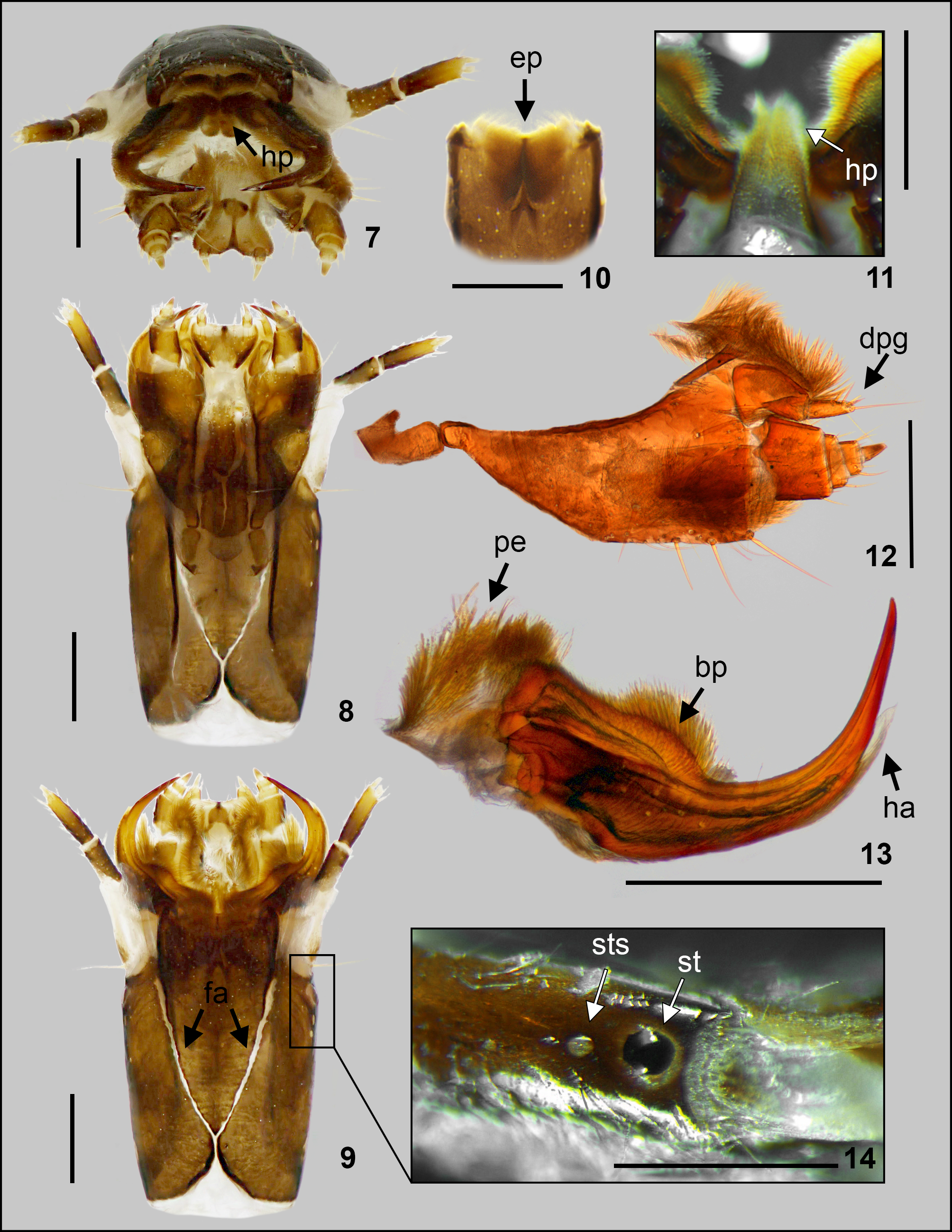
Head capsule ( Figs 7–9 View FIGURES7–14 , 15–17 View FIGURES15–17 ). Prognathous; retractable within prothorax, extensible neck membrane covered in extremely short spines and forming a two-layered envelope around retracted head; longer than wide, slightly tapering posteriorly. Epicranial plate laterally about 1.2 times longer than head capsule at its longest width, with one stout seta anterolateraly close to the base of antennae. Head capsule dorsally covered with long setae lying on surface ( Fig. 15 View FIGURES15–17 ; fs). Epicranial suture of light colouration, Y-shaped, with a very short epicranial stem, frontal arms V-shaped ( Fig. 9 View FIGURES7–14 ; fa). Gula not present ( Fig. 8 View FIGURES7–14 ). One stemma on each side of the head, with a light-coloured spot placed posteriorly behind the stemma ( Fig. 14 View FIGURES7–14 ; sts), possibly being a sensory organ. Labrum fused with clypeus forming clypeolabrum, covering the base of mandibles in dorsal view. Clypeolabrum double-arched in anterior view ( Figs 15, 16 View FIGURES15–17 ), with no distinguishable setae on lateroapical margins. Epipharynx ( Fig. 10 View FIGURES7–14 ; ep) formed by two plates and an anterior brush of long setae, which project centrally past anterior margin of the head. Hypopharynx ( Figs 7, 11 View FIGURES7–14 ; hp) with long setation.
Antenna ( Figs 21–23 View FIGURES 21–23 ). Trimerous, inserted on lateral distal margin of epicranial plate; partially retractable within membranous socket. Basal antennomere slightly wider than second antennomere, unsclerotized on posterolateral margin, bearing long flat setae lying on surface and erect setae lengthening towards the apical region. Second antennomere slightly narrower and longer than basal; bearing only erect setae equally spread across the antennomere, and with two longer setae on the outer apical region, next to sensorium. Sensorium of second antennomere ( Figs 22, 23 View FIGURES 21–23 ; as) oblong, potato-shaped, with distinct basal constriction as a connection with the second antennomere; with no visible surface pattern. Third antennomere ( Fig. 22 View FIGURES 21–23 ; a3) of similar size to sensorium of the second antennomere, bearing three setae on the apex, one seta on its body, and three cuticular projections ( Fig. 23 View FIGURES 21–23 ); first longer and thick (cp1), second longer and thin (cp2), third one placed on the body of antennomere forming a small bulge (cp3).
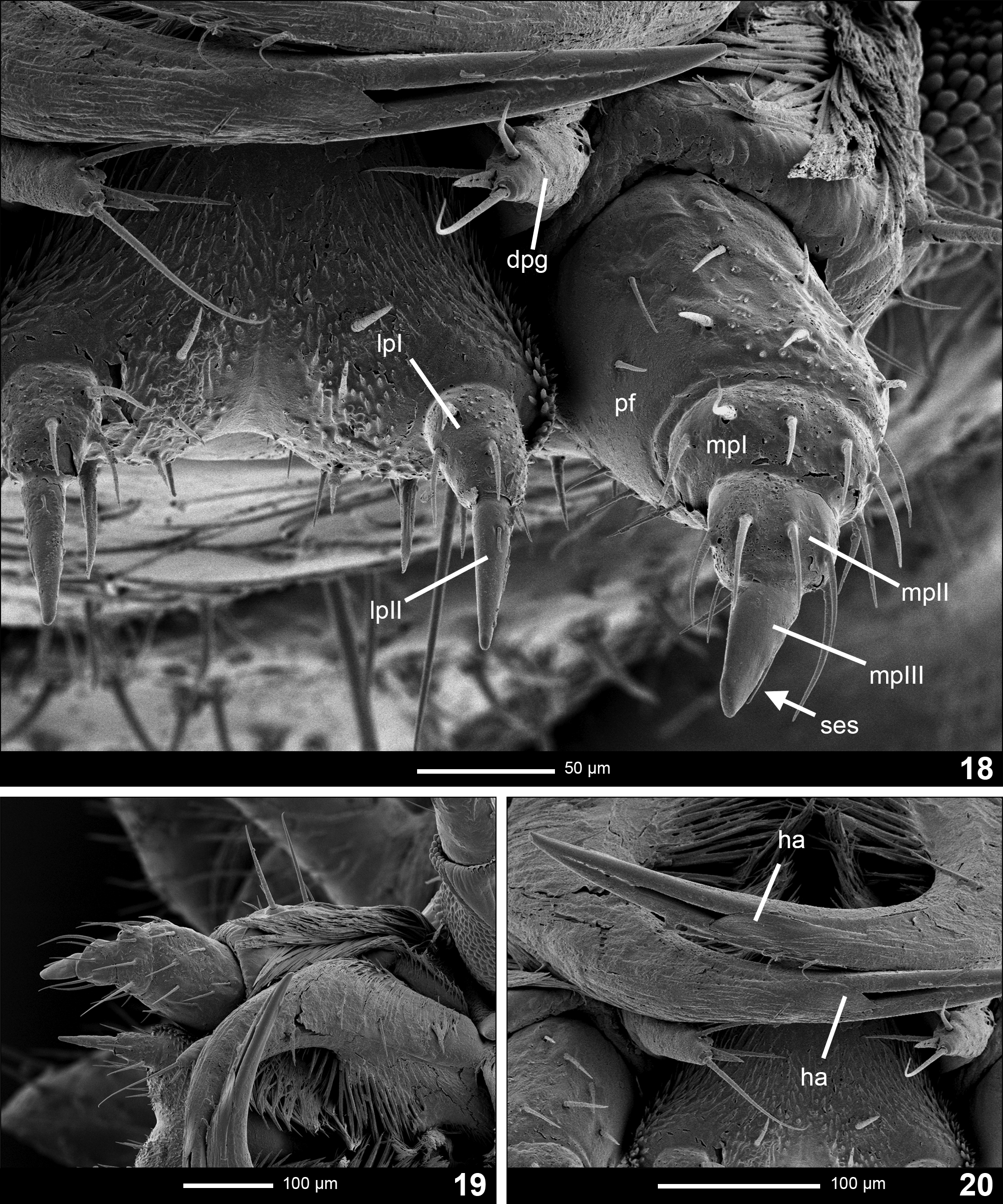
Maxilla ( Fig. 12 View FIGURES7–14 ). Consisting of five parts, attached to lateral margins labium forming a maxillo-labial complex. Cardo vertical, subrectangular, about twice as long as wide. Stipes elongated, ventrally covered with erect setae, with three long stout setae placed radially on the ventral apical region. Outer dorsolateral area covered with long dense setation reaching the base of maxillary palpus ( Fig. 19 View FIGURES18–20 ). Galea bimerous, with basal part larger than distal; distal part subcylindrical, pointing centrally, with short setae and one apical seta longer than body of the distal part and a blade-like flat cuticular projection on the apex ( Figs 12 View FIGURES7–14 , 18 View FIGURES18–20 ; dpg). Lacinia covered with brush of long setae on outer lateral margin ( Fig. 12 View FIGURES7–14 ). Maxillary palpifer ( Figs 18, 19 View FIGURES18–20 ; pf) large, subrectangular, of similar length and width. Maxillary palpus trimerous ( Figs 18, 19 View FIGURES18–20 ; mp1, mp2, mp3), basal and second palpomeres short and wide. Palpifer and palpomeres I–II covered with setae; palpomere III subconical, narrow, sharp, bare, with outer lateral longitudinal depression, possibly a sensory slot, covered with thick blunt seta ( Fig. 18 View FIGURES18–20 ; ses).
Labium ( Figs 7, 8 View FIGURES7–14 , 17 View FIGURES15–17 , 18 View FIGURES18–20 ). Closely attached to maxilla, formed by a short and strongly sclerotized prementum, mentum and mostly membranous submentum ( Fig. 8 View FIGURES7–14 ). Glossae absent. Prementum subtriangular, slightly heartshaped in ventral view; covered with brush of short setae and bearing several pairs of longer setae along sagittal line of the apex, shortening towards ventral region and with one pair of longer, stout setae on central regions of ventral part. Labial palpus bimerous ( Fig. 18 View FIGURES18–20 ; lp1, lp2); basal palpomere rectangular, longer than wide, bearing several setae; distal palpomere conical, longer and narrower than basal, bearing one short, thin seta lying on surface placed dorsally on basal part. Mentum elongated and subtriangular, unsclerotized on lateral margins, bearing numerous long setae lying on surface ventrally and a pair of long, erect setae posteromedially.
Mandible ( Figs 7-9, 13 View FIGURES7–14 , 15-17 View FIGURES15–17 , 20 View FIGURES18–20 ). Symmetrical, falcate, with an internal channel opening subapically on outer edge ( Fig. 20 View FIGURES18–20 ). Penicillus well developed ( Fig. 13 View FIGURES7–14 ; pe). Retinaculum featureless, present only as a blunt protuberance on basal third of the mandible ( Fig. 13 View FIGURES7–14 , bp). Basal half on inner margin of mandible covered with a brush of stout setae, being longest on the retinaculous protuberance. Basal two-thirds of mandible ventrally with dense setation adjacent to the surface aimed centrally. Dorsal part of mandibles with several stout setae aiming centrally and a strong distinct seta aimed centrally ( Fig. 15 View FIGURES15–17 ; cs), approximately in the central dorsal region of mandible. Lateral margin covered by brush of short setae lying on surface of basal two-thirds. Channel opening is partly covered by a feather-like or rounded-trapezium hyaline appendage with longer trapezoidal base situated ventrally ( Figs 13 View FIGURES7–14 , 20 View FIGURES18–20 ; ha).
Thorax ( Figs 1–6 View FIGURES1–6 ). Three-segmented, thoracic tergites divided by sagittal line in dorsal view. Protergum subtriangular, wider than long, rounded at posterolateral corners, with narrow emargination anteromedially; ventrally with two light-pigmented lines, leading diagonally from the margin towards anterolateral corners of prosternum ( Fig. 2 View FIGURES1–6 ; ptl). Meso- and metatergum subrectangular with round corners, ca. 4 times wider than long. Venter of prothorax composed of subrectangular, longer than wide prosternum ( Fig. 3 View FIGURES1–6 ; ps), subdivided into three well sclerotized areas; lateral ones extending above and to the sides of coxae fusing with episterna; medial area arrow-shaped (tip aiming anteriorly). Epimera forming thin sclerotized strands ( Fig. 2 View FIGURES1–6 ; em). Lateral areas of meso- and metathorax poorly sclerotized, composed of two laterotergites ( Fig. 3 View FIGURES1–6 ; alt, plt); anterior one bearing a well developed bilabiate spiracle in mesothorax. Anterior ventral area of meso- and metathorax formed by membranous intersternite ( Fig. 5 View FIGURES1–6 ; is) margined by paired anterior laterotergites ( Fig. 3 View FIGURES1–6 ; alt). Posterior ventral area subrectangular, wider than long, subdivided into three well sclerotized areas; lateral ones extending anteriorly and laterally to coxae, joining episterna; medial area hourglass-shaped. Epimera forming thin sclerotized strands.
Legs ( Figs 24–26 View FIGURES 24–26 ). Pentamerous, all pairs similar in shape and size. Coxa large, stout, bearing stout setae. Coxal-trochanteral membrane reaching ca. 1/3 of coxal longitudinal length ( Fig. 2 View FIGURES1–6 ). Trochanter smaller, elliptical in lateral view, shorter than femur, bearing shorter setae inclining to its surface and long stout setae, lengthening towards distal apex. Femur narrow and cylindrical in lateral view, bearing shorter setae inclining to its surface and long stout setae, lengthening ventrally, with one very long stout seta ventrally ( Fig. 24 View FIGURES 24–26 ). Tibiotarsus as long as femur, narrower, tapering towards distal end, bearing stout setae ( Fig. 26 View FIGURES 24–26 ). Pretarsus composed of a claw with fine ridges, ventrally bearing two long setae hooked apically towards each other, reaching the apex of the claw ( Fig. 25 View FIGURES 24–26 ).
Abdomen ( Figs 1–6 View FIGURES1–6 ). Ten-segmented, tapering towards posterior end, segments I to VIII subdivided by fine sagittal line in dorsal view. Tergites of segments I to VII subrectangular, similar in shape and colouration, ca. 4 times wider than long; tergite of segment VIII subcrescentiform; tergite of segment IX subsemicircular; segment X forming a narrow, incompletely sclerotized dark ring, bearing the holdfast organ—pygopod ( Figs 27, 28 View FIGURES 27–29 )—with several eversible processes. Segments I to VIII have single laterotergites ( Fig. 2 View FIGURES1–6 ; lt) on each side with poorly sclerotized plates bearing bilabiate spiracles; the venter of segments I to VIII consists of median sternite ( Fig. 2 View FIGURES1–6 ; ms), on segments I to V margined by paired narrow laterosternites ( Fig. 2 View FIGURES1–6 ; ls). Median sternites of segments I to VIII subrectangular, wider than long, bearing a pair of long stout setae posterolaterally; median sternite and laterosternites of segment I membranous. Sternite of segment IX well sclerotized, rectangular and dark. Sternites of segment V and VI less sclerotized. Functioning photic organs do not follow a solid pattern; composed of paired or single spots placed ventrolaterally on abdominal segments II to VI in variable number; most common configuration consisting of larger, usually paired spots on abdominal segments II and VI; possible additional smaller spots, if present, paired or single, on abdominal segments III—V.
Notes on larval behaviour. During the collecting of larvae, specimens were, thanks to their glow, frequently spotted from several metres away. While searching between bushes, the collecting team caused noise and vibrations, which were probably perceived by the larvae. However, when collected from the ground and handled, larvae often substantially reduced their glow, thus sometimes making the actual collecting almost impossible without a flashlight.
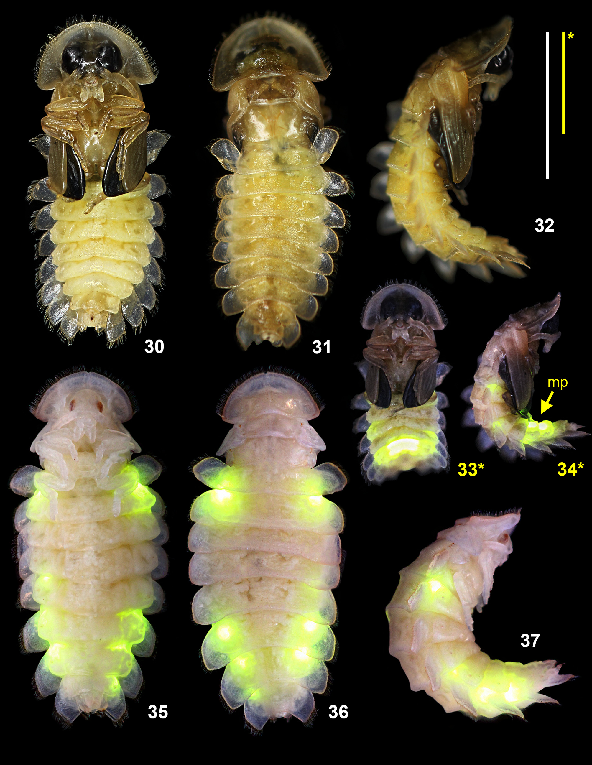
Description of female pupa ( Figs 35–37 View FIGURES 30–37 ). Type pupa adectica exarata libera. Curved, ventrally concave. Length 11–12 mm. Colouration: light yellow, with translucent protergum and abdominal tergites. Surface covered in short setae.
Head capsule. Completely covered by protergum in dorsal view. Eyes small, on sides of the head. Antennae short, extending laterally towards mid-femur of the prothoracic legs. Mouthparts visible in ventral view.
Thorax. Protergum similar in shape to larva, with long setae on the anterior edge. Meso- and metatergum smaller, subrectangular, bearing vestigial wing pads. All pairs of legs free, visible in ventral view. Spiracles present on pleural areas of mesothorax.
Abdomen. Abdominal segments subrectangular, wider than long. Tergites on segments I–IX covered with long setae on the edges, on segments I–VIII strongly laterally overlapping the body. Tergal plates on segments I–VII bearing pairs of depressions in cuticle, placed laterally around the sagittal line. Spiracles present on abdominal pleural areas of segments I–VIII. Segments II–VI bearing functioning larval photic organs with additional female photic organs on segments VII and VIII ( Figs 35–37 View FIGURES 30–37 ).
Description of male pupa ( Figs 30–32 View FIGURES 30–37 ). Type pupa adectica exarata libera. Curved, ventrally concave. Length 10–11 mm. Colouration: light beige on head, thorax and elytra, light yellow on abdomen, with distinctively black eyes and membranous wings ( Fig. 30 View FIGURES 30–37 ) and translucent protergum and abdominal tergites. Surface covered in fine short setae.
Head capsule. Completely covered by protergum in dorsal view. Eyes distinctly large, on sides of the head. Antennae short, extending laterally towards the distal end of femur of the prothoracic legs. Mouthparts visible in ventral view.
Thorax. Protergum similar in shape to larva, with long setae on the anterior edge. Meso- and metatergum smaller, subrectangular, bearing black wing pads covered externally with shorter elytra; wing pads reaching distal end of second abdominal segment when pupa is relaxed. Pro- and mesothoracic legs free, visible in ventral view; metathoracic legs almost completely covered by wing pads except for distal segments of tarsi, which extend past second ventrite. Spiracles present on pleural areas of mesothorax.
Abdomen. Abdominal segments subrectangular, wider than long. Tergites on segments I–IX covered with long setae on the edges, on segments I–VIII strongly laterally explanate, on segments I–VII bearing pairs of depressions in cuticle, placed laterally around the sagittal line. Spiracles present on abdominal pleural areas of segments I–VIII. Segment II–VI bearing functioning larval photic organs with additional male photic organs on ventrites of segments VI and VII ( Figs 33, 34 View FIGURES 30–37 ).
Notes on prepupae. The prepupal period begins when larva becomes inactive, lying curled up in a semicircle either on its side or back. Nevertheless, it still responds to any disturbance by activating its light organ and contracting its abdomen, a behaviour typical for pupae. Stage of prepupa in observed specimens lasted 3 days in female and 7 days in male.
Notes on ontogenetic changes in pupae. In the male, the pupa is generally indistinguishable from the female pupa ( Figs 35–37 View FIGURES 30–37 ) during the first three days. Moreover, as in the female, it glows from the same places as the larva. On the fourth day the wings begin forming and darkening in colour, signalling the beginning of the period when male and female pupae can be distinguished. On the fifth day the glow intensity of the larval lights is weaker than in the previous days. On the sixth day the male light organ activates and begins to glow ( Fig. 34 View FIGURES 30–37 ; mp). During this time larval lights still function and their glow intensity is back to normal. By this time the membranous wings, protruding under incipient elytra, are completely black ( Fig. 30 View FIGURES 30–37 ). The thorax starts to sclerotize, slowly darkening in colour. At the same time, the pupal eyes are turning into large male eyes, but their development is not yet complete, since they resemble a half-inflated balloon at this stage ( Fig. 30 View FIGURES 30–37 ). On the seventh day the male emerges. Its larval lights still operate and their glow is visible through the still incompletely sclerotized cuticle (this was also observed by Schwalb 1961). On the eighth day the cuticle of the adult male finally hardens and the larval lights are no longer visible.
The female pupa does not go through such obvious changes as the male does. It appears more or less similar throughout the whole pupal period ( Figs 35–37 View FIGURES 30–37 ), with the exception of slowly darkening small eyes and overall shift of the body colour towards a darker shade of yellow. The reason for this is neoteny as well as the paler pigmentation of the female. As in the male, the female emerges on the seventh day.
Notes on behaviour observed in pupae. The pupa is inactive, either lying on its side or back, responding only to disturbance. A response consisting of a ca. 10-second glow with quick intensification and gradual fading can be induced by handling the animal or even by vibrations. In the male pupa with the male photic organ already developed ( Figs 33, 34 View FIGURES 30–37 ), the response is similar, with exception of the newly developed lantern, which can still glow feebly at least three times longer than the larval lights. When under a strong light source, the pupa reacts in the same manner as pupa of Lampyris noctiluca , as described in Novák (2017).
No known copyright restrictions apply. See Agosti, D., Egloff, W., 2009. Taxonomic information exchange and copyright: the Plazi approach. BMC Research Notes 2009, 2:53 for further explanation.