
Disphaerobius svenhedini (Verhoeff, 1934) Verhoeff, 1934
|
publication ID |
https://doi.org/ 10.11646/zootaxa.4258.2.2 |
|
publication LSID |
lsid:zoobank.org:pub:949BAF3D-D8DF-4BC5-98CB-1F2D946EF802 |
|
DOI |
https://doi.org/10.5281/zenodo.6022943 |
|
persistent identifier |
https://treatment.plazi.org/id/03D7CE78-FFE1-FFE4-32F9-FF16FBE0FD2C |
|
treatment provided by |
Plazi |
|
scientific name |
Disphaerobius svenhedini (Verhoeff, 1934) |
| status |
comb. nov. |
Disphaerobius svenhedini (Verhoeff, 1934) comb. nov.
Figs 1–21 View FIGURES 1 – 5 View FIGURES 6 – 9 View FIGURES 10 – 15 View FIGURES 16 – 20 View FIGURES 21 – 24 , 29 View FIGURES 25 – 29
Pterygotergum svenhedini View in CoL — Verhoeff, 1934b: 29, Taf. 4, Abb. 1a, 2a, 3a, 4a, 5a, 6a, 7a (♂, ♀).
Material examined: Type material: Lectotype male (ZSM, in four slides labelled as No. A20030926. Lectotype 1. Pterygotergum svenhedini Verh. ♂ Kopf. Thian-Schan; No. A20030927. Lectotype 2. Pterygotergum svenhedini Verh. ♂ 1, 2 B. Thian-Schan; No. A20030928. Lectotype 3. Pterygotergum svenhedini Verh. ♂ 3.–8. B. Thian- Schan; No. A20030929. Lectotype 4. Pterygotergum svenhedini Verh. ♂ 9.–15. B. Thian-Schan).
This male was labeled as lectotype, but since this designation seems to have never been published, it becomes validated only with the present paper.
Other material examined: Kazakhstan: 1 male, 1 female ( ZMUM), 2 females ( PSU, No. 555), Almaty Region, Zhambyl District, 15 km NNW of Karabastau village, Tyrnakty , N 43˚53′, E 75˚30′, stony mountain steppe with rocks, 850–950 m a.s.l., 20–22.IV.2016, leg. A.A. Fomichev . Mongolia: 1 male ( PSU, No. 554), Baitag Bogd Uul Mt. Range , Gushoot-Shineetijn-Gol river terrace, N 45˚16′, E 91˚04′, stony terrain with sparse Achnatherum splendens growth, 1900 m a.s.l., 23–26.V.2015, leg. A.A. Fomichev .
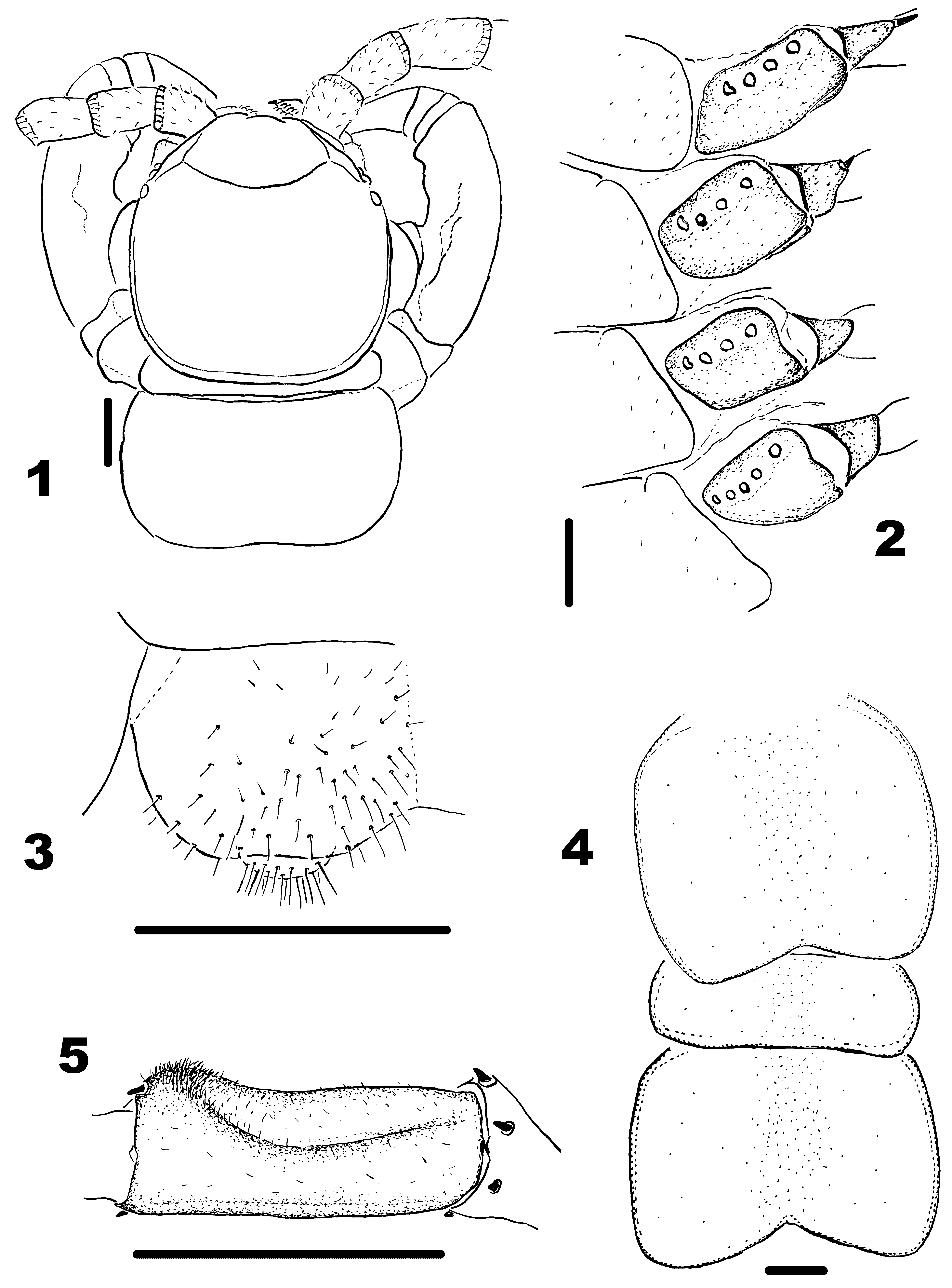
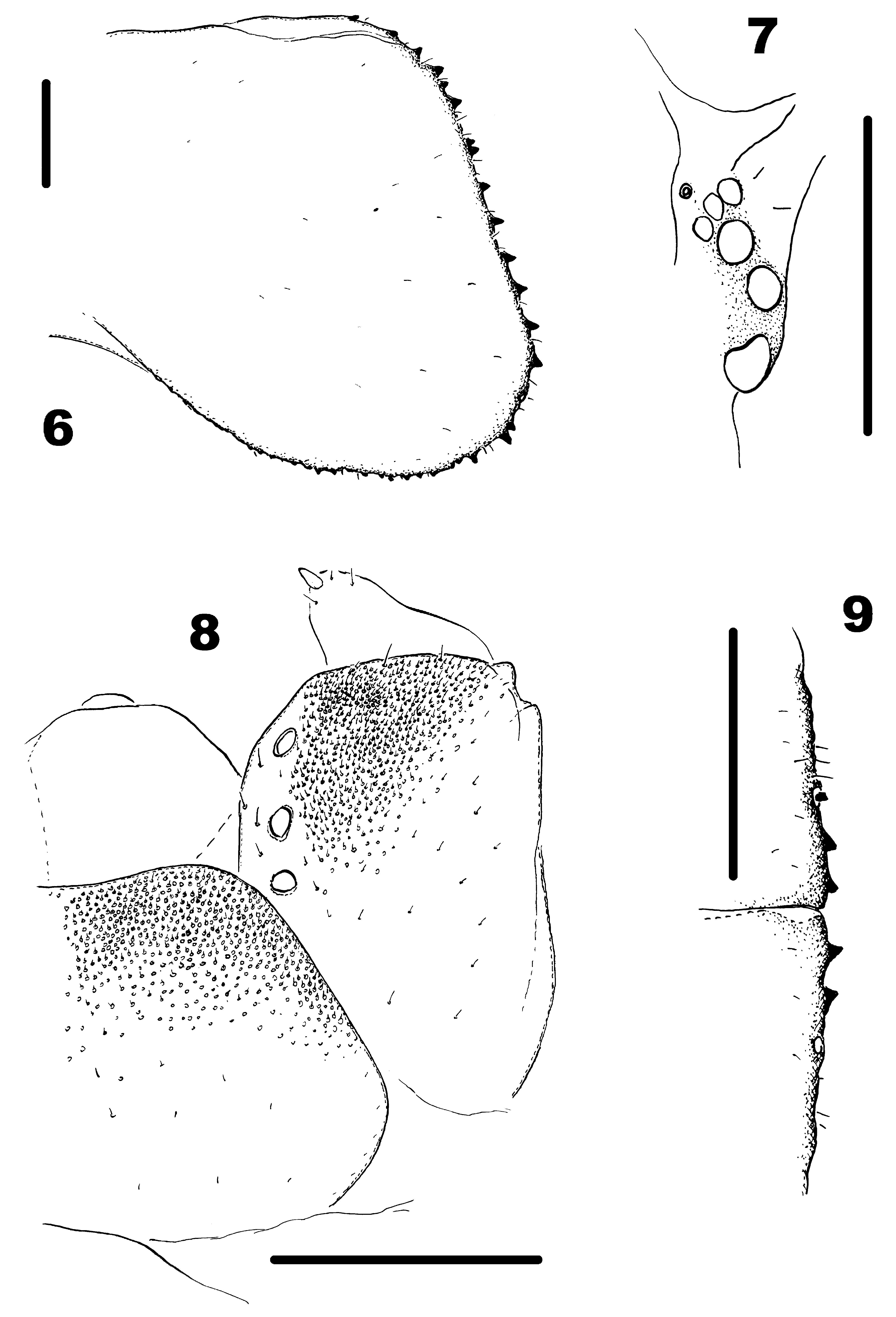
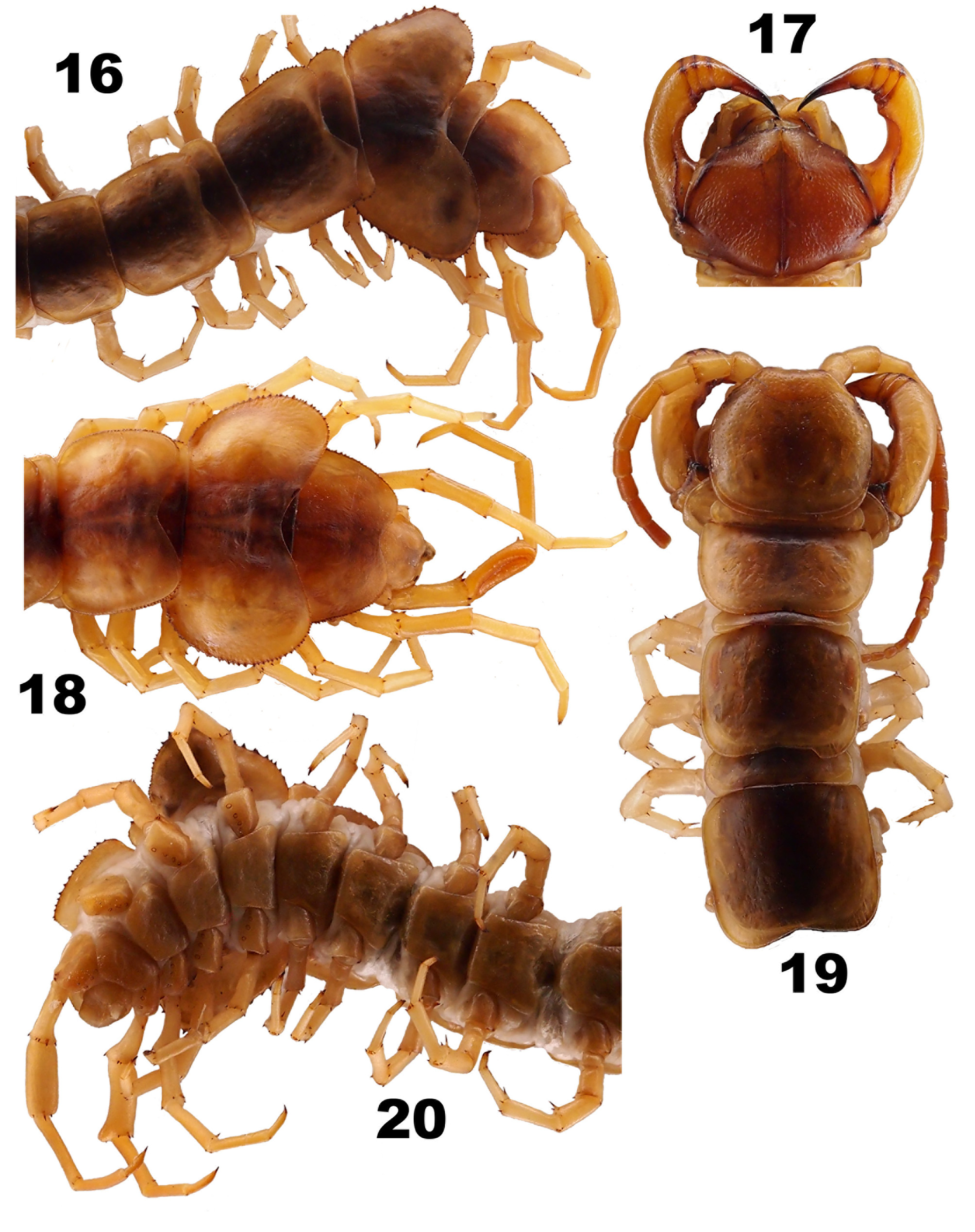
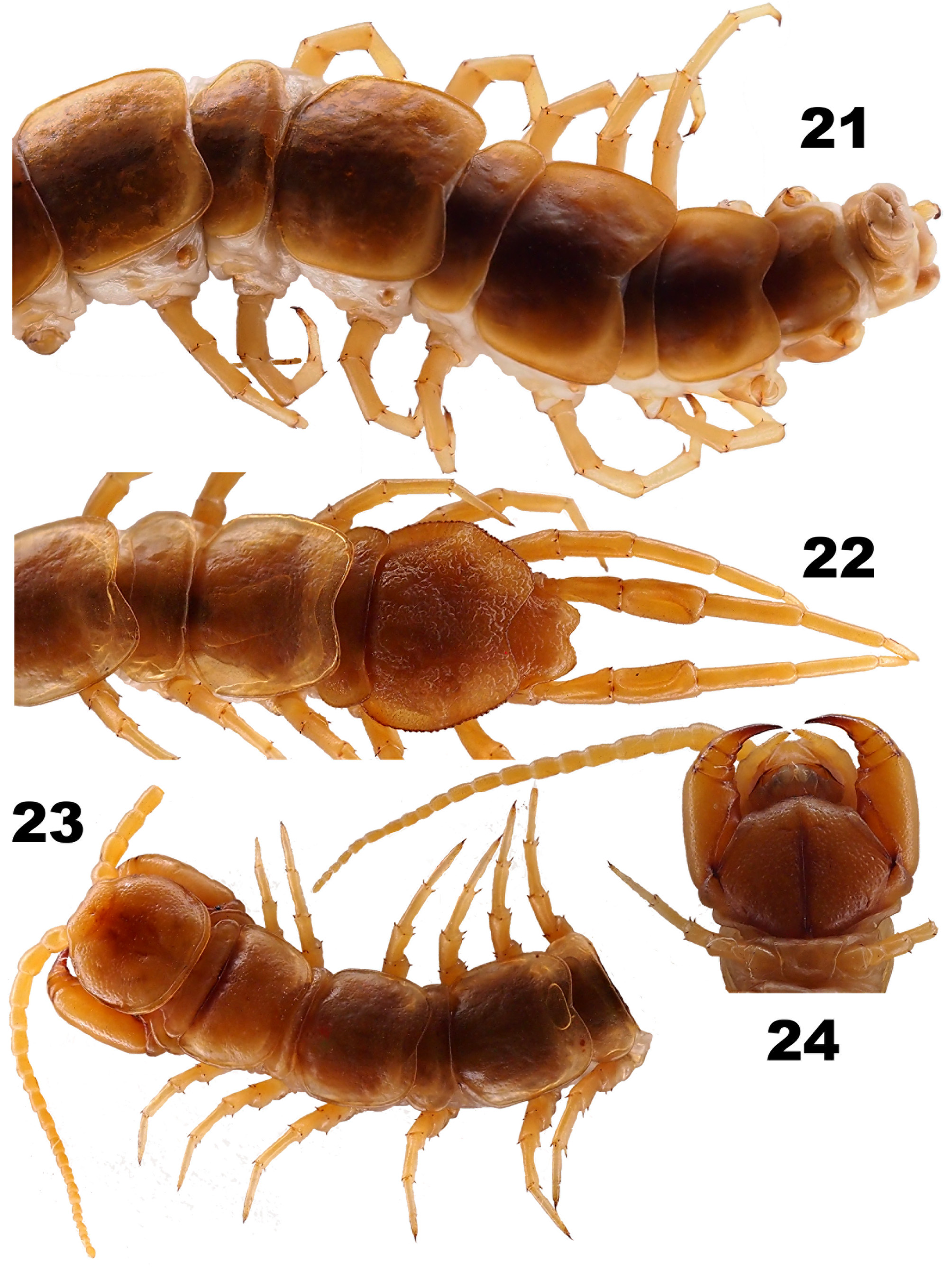
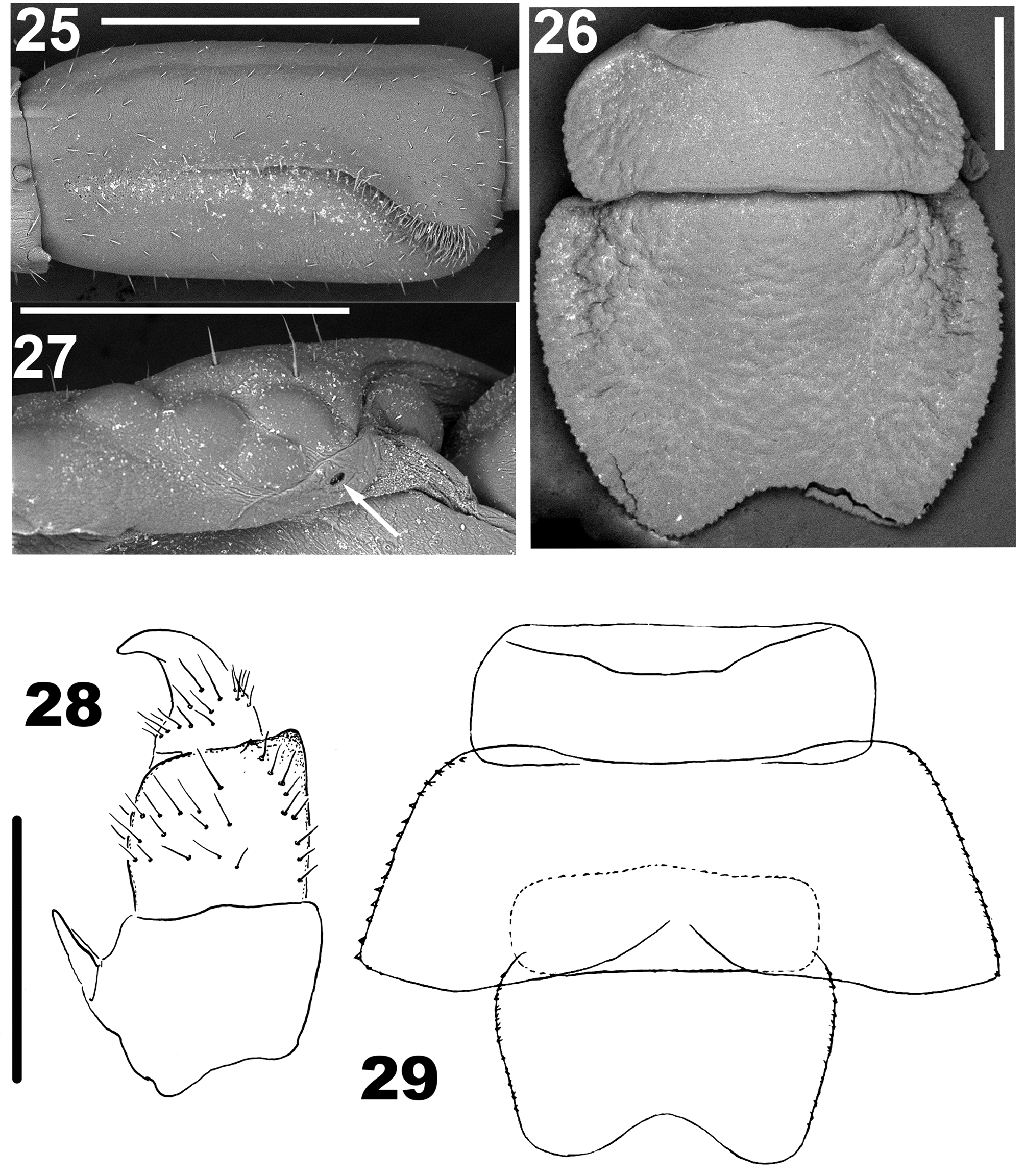
Diagnosis. Male D. svenhedini shows the most strongly broadened T 12 divided into two lobes and serrate at the lateral margin ( Figs 6 View FIGURES 6 – 9 , 16, 18 View FIGURES 16 – 20 & 29 View FIGURES 25 – 29 ). In the female, T 12 is noticeably, but not so much broadened as in the male, strongly notched at the rear edge to form lobes, and the lateral margin is devoid of serration ( Figs 4 View FIGURES 1 – 5 & 21 View FIGURES 21 – 24 ).
Description. Material examined: Male. Body 28.0 ( Mongolia) to 35.5 mm long ( Kazakhstan), poorly and sparsely punctate.
Coloration in alcohol yellow-brownish, with a vague, darker, broad, axial stripe on tergites. Ocellar area dark ( Fig. 7 View FIGURES 6 – 9 ).
Forcipular tergite slightly narrower than cephalic plate, with a ratio of 7.8:7.3. TT 10–Tim broadened, serrate and infuscate at lateral or all margins ( Figs 16 &18 View FIGURES 16 – 20 ). T 12 very strongly broadened so that its lateral parts forming rounded wing-like outgrowths, strongly serrate at edges. Serration irregular, strongly expressed on sides, gradually decreasing towards rear edge, fully lacking towards the middle of posterior edge of T 12. Short setae visible at high magnification between serrations ( Fig. 6 View FIGURES 6 – 9 ).
Axial length of T 12, 3.1 mm in both specimens, width 7.6 mm or 8.7 mm. Tergites 10–Tim broadened, serrate and infuscate at all margins ( Figs 16 &18 View FIGURES 16 – 20 ). Tergite 12 especially strongly broadened so that its lateral parts forming rounded wing-shaped projections distinctly serrate at edges; serration irregular, better developed laterally than caudally, totally disappearing towards the middle of caudal edge; short setae visible between teeth at high magnification ( Fig. 6 View FIGURES 6 – 9 ). Hind margin of Tim strongly emarginated in front considerably covered by a broadened T 14.
Punctation of cephalic plate denser than of tergites. Cephalic plate slightly broadened, 3.85 mm long, 3.90 mm wide in both specimens ( Figs 1 View FIGURES 1 – 5 & 19 View FIGURES 16 – 20 ).
Antennae composed of 18+9 and 20+17 segments, respectively (damaged in both specimens; according to the original description, antennae 20-segmented, but the lectotype has broken antennae), short, barely reaching the middle of T 5. Antennomeres elongated, first three covered with sparse and very short setae. Starting with basal 4– 5 antennomeres, antennae bright brown ( Fig. 19 View FIGURES 16 – 20 ), densely covered with very small and erect setae.
Ocelli 5–7, posteriormost ocellus always the largest.
Tömösváry’s organ very small, less than half the size of adjoining ocelli ( Fig. 7 View FIGURES 6 – 9 ).
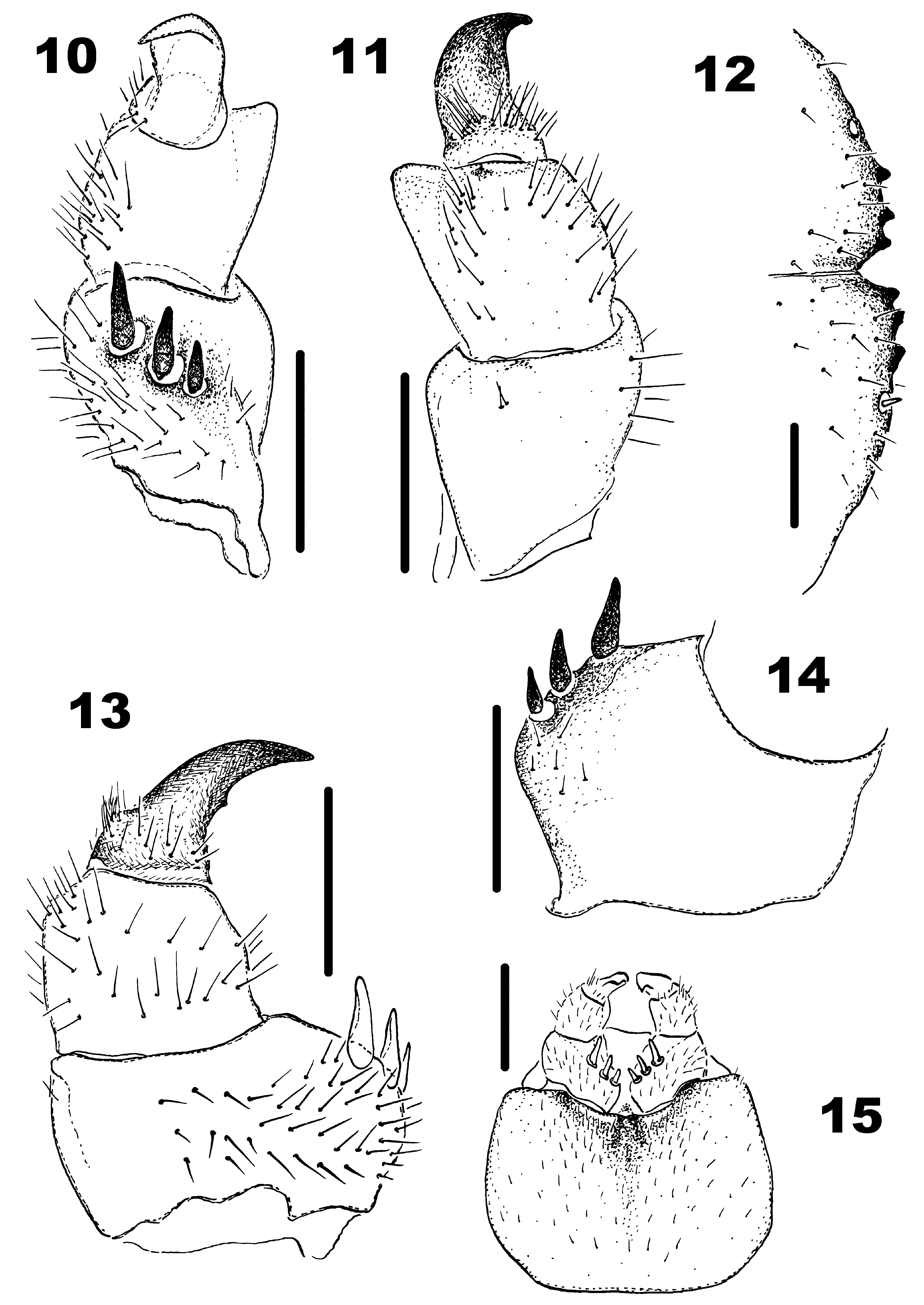
Forcipules dark brown, especially at joints. Lateral edges of trochanteroprefemur and part of coxosternite extended beyond cephalic plate, inner part of trochanteroprefemur concave, with a ventrally bent chitinous rib ( Fig. 17 View FIGURES 16 – 20 ). Forcipular coxosternite broad, with 2+2 obtuse and short teeth, shoulders of coxosternite varying from almost straight to slightly sloped, porodonts stout and strong, about as long as teeth; median notch from weak to wanting ( Figs 9 View FIGURES 6 – 9 & 12 View FIGURES 10 – 15 ).
Legs: 14–15P slightly incrassate. 15F incrassate, with two distinct sulci, i.e. a poorly expressed dorsolateral sulcus and a deep dorsal sulcus, the latter forming apically a round tubercle supporting a cluster of thick and short setae ( Fig. 5 View FIGURES 1 – 5 ). Length of legs: pair 1—t+P 1.00–1.05, F 0.90–0.95, Ti 1.05–1.15, Ts1 0.85–0.95, Ts2 0.45–0.50; pair 14—t+P 1.60–2.05, F 1.85–2.05, Ti 2.10–2.30, Ts1 1.80–1.85, Ts2 0.80–0.85; pair 15—t+P 1.85–2.20, F 2.25– 2.30, Ti 2.40–2.60, Ts1 2.05–2.20, Ts2 0.80–1.05. Leg plectrotaxy as in Table 2.
Leg pairs ventral dorsal All sternites sparsely punctate. SS 13–15 and coxae of the same legs covered with very small and dense setae in rear parts, especially so coxae 15. Attachment points of setae pigmented, therefore fields of setae resembling brown spots.
Genital sternite short and broad, covered with longer setae. Gonopod 1-segmented, with numerous setae (about 14–17) ( Fig. 3 View FIGURES 1 – 5 ).
Coxal pores present on legs 12–15, small, rounded, separated from one another by distances 2–2.5 times greater than their own diameter; formula 5,4,4,4.
Female. Body 37, 39 and 41 mm long. Diagnostic characters and plectrotaxy as in male, but all tergites and legs unmodified.
Head broadened, its length and width 3.60 (3.40–3.90) mm and 3.78 (3.50–4.25) mm, respectively.
Width of forcipular tergite 3.85 mm.
T 12 non-serrate, but as in male broadened and lobe-shaped at rear edge, lateral sides subparallel ( Figs 4 View FIGURES 1 – 5 , 21 View FIGURES 21 – 24 ); width 4.4 (3.90–5.15) mm, length in the middle 2.78 (2.60–3.10) mm. Hind margin of Tim strongly notched ( Fig. 21 View FIGURES 21 – 24 ).
Genital sternite broadened, sometimes pigmented ( Fig. 15 View FIGURES 10 – 15 ).
Antennae with 20+20 segments, not reaching the middle of T 5.
Ocelli 5–6 on each side of head, varying in size as in Fig. 7 View FIGURES 6 – 9 .
Forcipules as in male, coxosternite with 2+2 teeth (in one female 3+3) ( Fig. 12 View FIGURES 10 – 15 ). Inner face of forcipular trochanteroprefemur less strongly concave than in male and slightly shorter in length.
Length of legs: pair 1, t+P 1.13 (1.00–1.25), F 0.90 (0.80–1.00), Ti 1.05 (0.95–1.15), Ts1 0.85, Ts2 0.50 (0.45– 0.55); pair 14, t+P 2.01 (1.80–2.20), F 1.75 (1.55–1.90), Ti 1.97 (1.85–2.05), Ts1 1.80 (1.70–1.85), Ts2 0.83 (0.80– 0.85); pair 15, t+P 2.25 (2.05–2.45), F 2.21 (2.10–2.35), Ti 2.46 (2.40–2.55), Ts1 2.17 (2.15–2.20), Ts2 1.05 (1.05–1.10). Plectrotaxy as in Table 2.
Coxal pores as in Fig. 2 View FIGURES 1 – 5 .
Gonopods with thin setae and 3+3 ( Figs 10, 13 & 14 View FIGURES 10 – 15 ), 2+3 and 3+4 unequal spurs (usually obviously 3+3). First segment with numerous setae on external and, slightly, internal surfaces ( Figs 13, 14 View FIGURES 10 – 15 ). Second segment with a small distodorsal outgrowth shifted mesad ( Figs 10, 11 View FIGURES 10 – 15 ). No dorsolateral setae, but 1–2 short setae on first segment (absent from one specimen), two irregular rows of thin setae on second segment, and a group of very thin and dense setae on third segment ( Figs 11, 13 View FIGURES 10 – 15 ). Gonopodal claw simple, but in two females with a very small, additional denticle observed on external face of claw closer to its base, probably due to just a slightly uneven outer margin of claw ( Fig. 13 View FIGURES 10 – 15 ).
Remarks. The males at our disposal are differing from the original description ( Fig. 29 View FIGURES 25 – 29 ), as well as from the lectotype (despite the latter’s deformation during mounting on microscopic slides) by showing less sharp rear edges of T 12 ( Figs 6 View FIGURES 6 – 9 , 16 & 18 View FIGURES 16 – 20 ) and a different plectrotaxy pattern (pair 1: 221/333; pair 2: 222/333; pair 12: 00222/ 00333; pair 15: 10310/01432). The presence of four ventral spines on 15P in the original description is given without disposition details, which may mean either the presence of additional spines or as a misinterpretation. Lectotype spinulation is much more similar to our specimens (pair 1: 322/233, pair 15: 10311/01333), although spines are missing and often visible only as insertion points. In addition, both of the new specimens differ each other in the shape of TT 12 and 14 ( Figs 16 & 18 View FIGURES 16 – 20 ). The deep sulcus on 15F in the individual from Mongolia is brown at the bottom. However, all these minor variations in secondary sexual characters seem to be intraspecific, rather depending on age and condition ( Fusco et al. 2015).
The female described by Verhoeff (1934b) differs significantly from our specimens, but we have failed to access the female paralectotypes. First of all, the female he mentioned showed the gonopods with 2+2 spurs: “Die Gonopoden des ♀ sind denen der Abb. 11 ziemlich ähnlich, also die zwei Genitalklauen kegelig und schräg gegen einander gestellt, etwa um die eigene Breite von einander getrennt. Genitalklaue am Ende mit einer scharfen Spitze, aber schräg abgeschnitten, wodurch ein stumpfer Winkel und damit die Andeutung einer 2. Spitze entsteht…” [Gonopods of the ♀ are very similar to those depicted in fig. 11, so that the two gonopodal spurs are tapered and obliquely juxtaposed, separated by their own width. Gonopodal claw at the end with a sharp point, but cut obliquely, creating an obtuse angle and thus the forming as if a 2nd tip…] ( Verhoeff 1934b: 31). Despite this, Verhoeff did not mention a distodorsal outgrowth on the second gonopodal segment. Moreover, Verhoeff, when describing the female gonopod, referred to a figure of a different species, i.e. Lithobius giganteus (= Lithobius mongolicus Verhoeff, 1934 ) ( Verhoeff 1934b: Taf. 5, Abb. 11a).
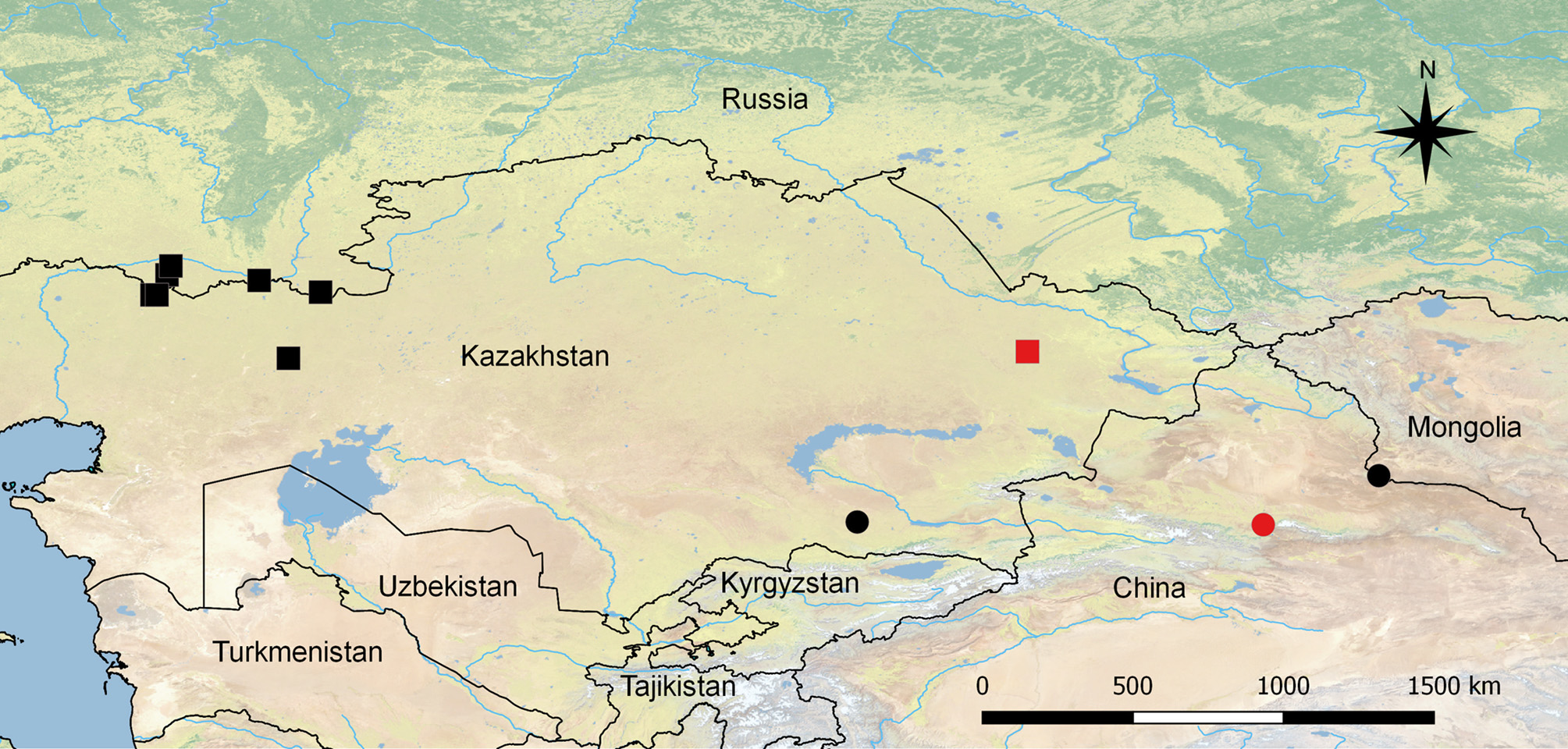
Distribution ( Fig. 30 View FIGURE 30 ). China: Xinjiang Uygur Autonomous Region, Urumqi (Tian Shan) ( Verhoeff 1934b). Kazakhstan: Almaty Region, Zhambyl District. Mongolia: Baitag Bogd Uul Mt. Range.
No known copyright restrictions apply. See Agosti, D., Egloff, W., 2009. Taxonomic information exchange and copyright: the Plazi approach. BMC Research Notes 2009, 2:53 for further explanation.
|
Kingdom |
|
|
Phylum |
|
|
Class |
|
|
Order |
|
|
Family |
|
|
Genus |
Disphaerobius svenhedini (Verhoeff, 1934)
| Farzalieva, Gyulli Sh., Nefediev, Pavel S. & Tuf, Ivan H. 2017 |
Pterygotergum svenhedini
| Verhoeff 1934: 29 |