
Odontesthes yucuman, Wingert, Juliana M., Ferrer, Juliano & Malabarba, Luiz R., 2017
|
publication ID |
https://doi.org/10.11646/zootaxa.4250.6.1 |
|
publication LSID |
lsid:zoobank.org:pub:3258EAD0-ED76-4B29-8F7B-2637013FA169 |
|
DOI |
https://doi.org/10.5281/zenodo.6030173 |
|
persistent identifier |
https://treatment.plazi.org/id/03D88787-0562-062A-8FCC-F90DFACCF5A5 |
|
treatment provided by |
Plazi |
|
scientific name |
Odontesthes yucuman |
| status |
sp. nov. |
Odontesthes yucuman , new species
( Figs. 4 View FIGURE 4 b, 5b, 7b, 8b, 9b, 10b, 11b, 16, 17, 18, 22)
Odontesthes aff. perugiae View in CoL . Zaniboni et al., 2004: 13 (photo, upper rio Uruguay).
Holotype. UFRGS 21310 View Materials (ts), 159.0 mm SL, Rio Grande do Sul State, Nonoai, rio Passo Fundo , downstream of the Monjolinho Hydroelectric Power Plant , 27°20’44”S 52°43’52”W, 1 Sep 2010, R. Angrizani. GoogleMaps
Paratypes. All from Brazil. MCN 19970 , 2, 125.3– 154.6 mm SL ; MCP 49447, 2, 146.0—161.0 mm SL, UFRGS 17840, 7 (1 xr, 1 sk, 7 ts), 118.2—161 mm SL, collected with holotype. MCP 40026 , 3, 41.7—76.8 mm SL, Santa Catarina State, Mondaí , locality of Capela, rio Uruguay , 27°11’52”S 53°38’42”W, 26 Jan 2006, C. A. S. Lucena, E. H. L. Pereira, J. P. Silva & V. A. Bertaco GoogleMaps . UFRGS 20536 , 1, 153.1 mm SL, Santa Catarina State, Itá, rio Uruguay at reservoir of the Itá Hydroelectric Power Plant , 27°17’10”S 52°20’26”W, 10 May 2015, R. Guereschi GoogleMaps . UFRGS 20537 , 1, 147.2 mm SL, Santa Catarina State, Concórdia, rio Uruguay at reservoir of the Itá Hydroelectric Power Plant , 27°18’32”S 52°06’05”W, 20 May 2015, R. Guereschi GoogleMaps . UFRGS 20538 , 1, 162.7 mm SL, Rio Grande do Sul State, Maximiliano de Almeida, rio Uruguay downstream of the Machadinho Hydroelectric Power Plant , 27°31’37”S 51°47’06”W, 22 May 2015, R. Guereschi GoogleMaps . UFRGS 10304 , 3 (1 c&s), 56.3—129 mm SL, Rio Grande do Sul State, Nonoai, rio Passo Fundo , Monjolinho Hydroelectric Power Plant , 27°20’44”S 52°43’52”W, R. Angrizani, 11 Jul 2008 GoogleMaps . UFRGS 11671 , 1 (ts), 156.6 mm SL, Rio Grande do Sul State, Nonoai, rio Passo Fundo , Monjolinho Hydroelectric Power Plant , 27°20’44”S 52°43’52”W, R. Angrizani, 11 Jul 2008, 14 May 2009. GoogleMaps
Non-type. UFRGS 19389, 1 (xr, ts), 168.7 mm SL, Santa Catarina State, Itá , rio Uruguay at reservoir of the Itá Hydroelectric Power Plant, 27°16’59”S 52°22’50”W. GoogleMaps
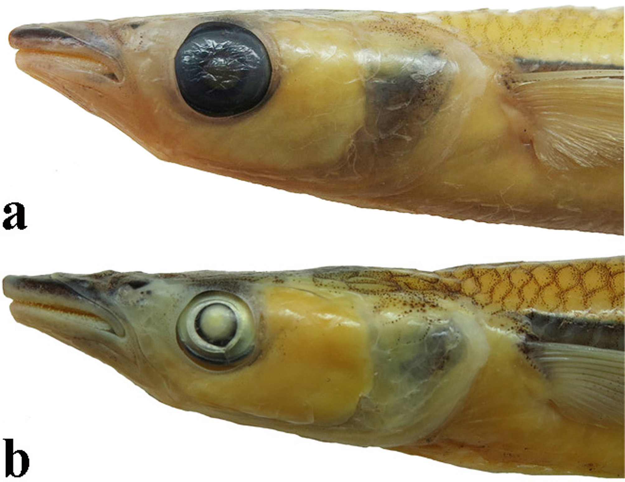
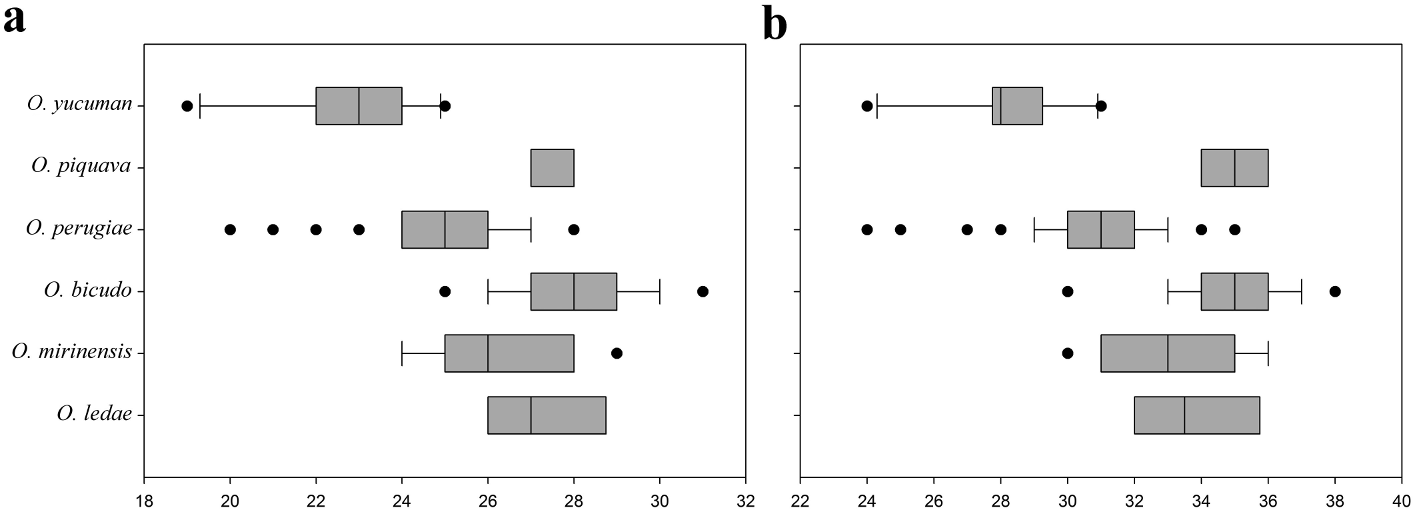
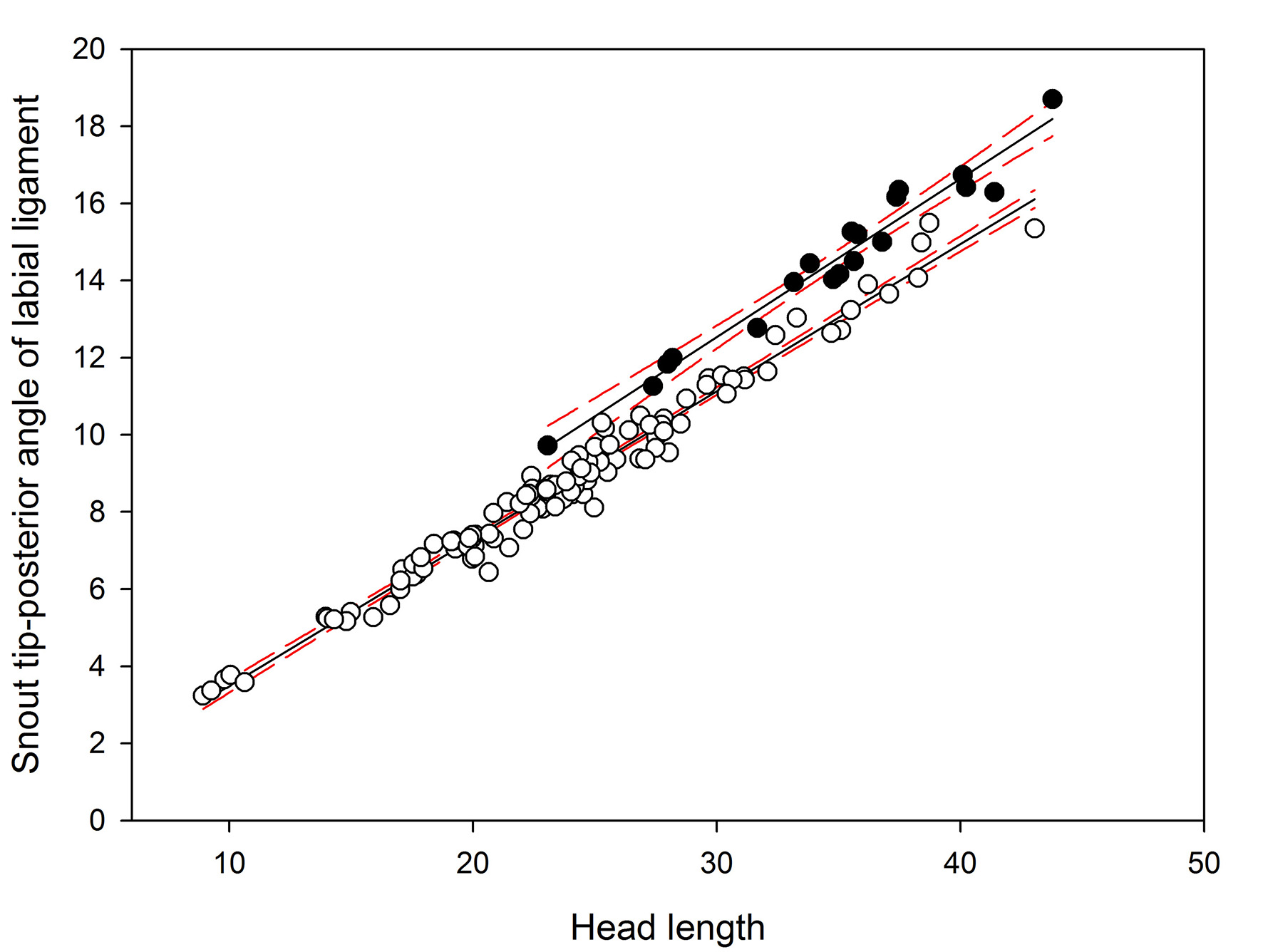
Diagnosis. Odontesthes yucuman is distinguished from congeners, with the exception of the species included in the O. perugiae species-group ( O. bicudo , O. ledae , O. mirinensis , O. piquava and O. perugiae ) by the possession of the following combination of characters: teeth of the outer row on dentary and premaxilla shorter than those in the inner row ( Fig. 18 View FIGURE 18 ; vs. teeth of outer row larger than those of inner row or teeth of outer and inner rows of equal size), origin of dorsal fin positioned over or posterior to anus ( vs. anterior to anus), and 7 to 9 longitudinal scale rows above left and right lateral bands. Odontesthes yucuman is distinguished from O. bicudo , O. ledae , O. mirinensis and O. piquava by the smaller number of lower branch gill rakers (19-25, vs. 26-29 in O. ledae , 24-29 in O. mirinensis , 25-31 in O. bicudo , and 27-28 in O. piquava , Fig. 19 View FIGURE 19 ). It is distinguished from O. perugiae by differences in the snout length ( Fig. 5 View FIGURE 5 , 35.6–39.5, mean 37.7 vs. 28.6–37.4%, mean 32.2 of HL), and the distance between snout tip and posterior angle of labial ligament (31.3—37.0, mean vs. 23.6—33.1%, mean 28.2 Oƭ HL; Fig. 21 View FIGURE 21 ).
Description. Morphometric and meristic data in Tables 1—2. Body elongate and slender; dorsal profile straight from snout tip to caudal peduncle. Ventral body profile slightly convex from tip of snout to origin of anal fin; straight and ascending along anal fin and convex near end of caudal peduncle. Caudal peduncle longer than deep.
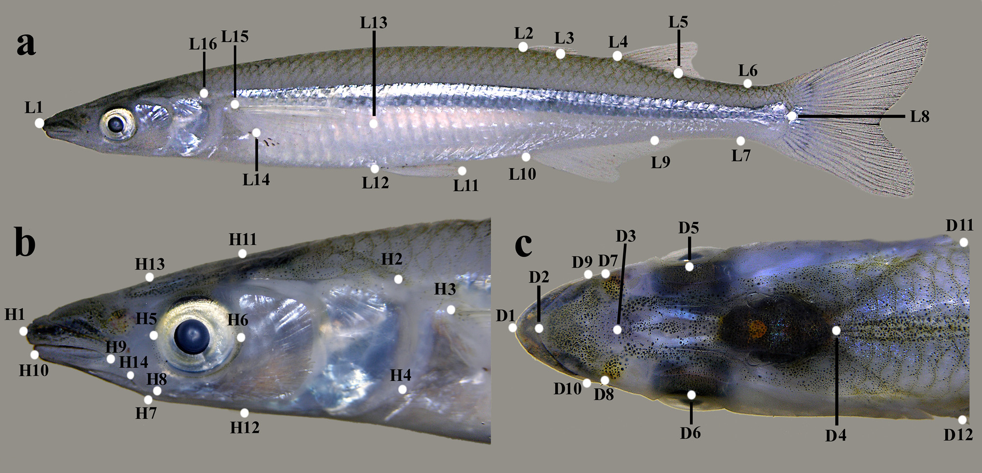
Head relatively large (22.1—24.8% of SL); dorsal profile straight from snout to posterior tip of supraoccipital bone; ventral profile of head straight or slightly concave from snout through region below anterior portion of eye. Snout relatively long ( Fig. 5 View FIGURE 5 b; 35.8—39.5 % of HL), pointed from dorsal and lateral views. Mouth terminal; protractile with large gape horizontal from lateral view and positioned in horizontal line crossing center of eye. Eyes rounded and well developed from lateral view, diameter distinctly smaller than snout length.
Pectoral-fin rays i+11—12 (i+11); unbranched and 1st to 4th branched rays longer, not reaching posterior to vertical through pelvic-fin origin. Pelvic-fin rays i+5 (i+5); unbranched and branched rays of same length, not reaching anal-fin origin. Pelvic fin insertion close to each other with interpelvic membrane uniting them at base or half-length of rays. First dorsal-fin rays iv—v (iv) with origin at same line or slightly posterior to anus and anterior to anal-fin origin. Second dorsal-fin rays ii+7—8 (ii+7); origin in second half of anal-fin base. Anal-fin rays ii+13— 15 (ii+14) with origin in final insertion of first dorsal fin, distal border usually concave. Principal caudal-fin rays i+15+i (i+15+i), with scales at least up to half of its length.
Scales large and cycloid with posterior margin smooth; striae forming complete circles. Scales in longitudinal series above lateral band 49—53 (50). Scales in longitudinal series above lateral band before the first dorsal fin 22— 26 (24). Rows of scales between origin of first dorsal fin and origin of anal fin 9—10 (9). Rows of scales between origin of first dorsal fin and posterior insertion of anal fin 8—9 (9). Dorsal scales between posterior insertion of first dorsal fin and origin of second dorsal fin 7—9 (8). Longitudinal scale rows above left and right lateral bands 9—10 (10). Predorsal scales 21—26 (21). Rows of longitudinal scales in the opercle 6—10 (7). Rows of longitudinal scales below eye 2 (2). Rows of longitudinal scales around caudal peduncle 16—18 (18).
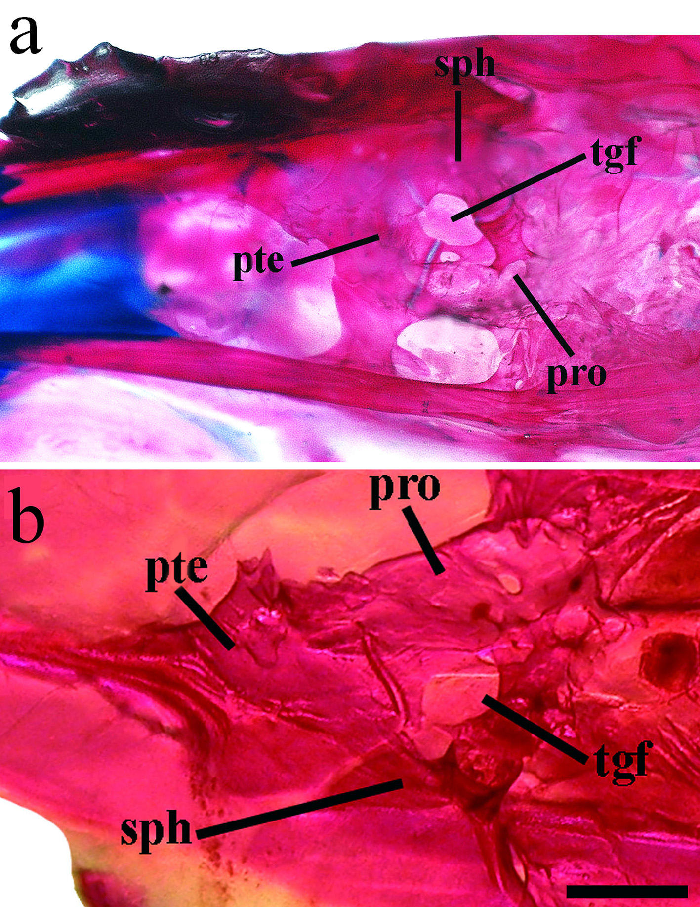
Osteology. Neurocranium. Vomer strongly concave from ventrolateral view; anterior margin pointed with two lateral condyles slightly rounded; three pointed posterior processes, two posterolateral and one central more elongate and extending to parasphenoid ( Fig. 7 View FIGURE 7 b). Vomer with three tooth patches with several teeth in some specimens, connected by single row of smaller similar teeth. Parasphenoid elongate and narrow with anterior portion wider; posterior portion with narrow lateral wings extending to basioccipital. Basioccipital rectangular, rough and covered dorsally by exoccipital. Exoccipital usually with two foramens and laterally expanded (exoccipital wing); connected dorsally with basioccipital and anteriorly with pterotic. Supraoccipital with forked laminar projection contacting epioccipital dorsally and parietal ventrally. Epioccipital elongate, flattened and with lateral extension; contacting supraoccipital ventrolaterally. Prootic large, trigeminofacial foramen large, in contact with prootic and sphenotic ( Fig. 8 View FIGURE 8 b). Sphenotic rectangular with anterodorsal process contacting frontal. Dermosphenotic slender with two pores connected dorsally to frontal and medial flange narrow covering all its extension ( Fig. 4 View FIGURE 4 b). Frontals rectangular, with anterior portion rounded and extending more than half skull length. Lateral ethmoid broad, not contacting parasphenoid. Nasal sensory canal detached anteriorly with ventral process pointed and contacting lacrimal subnasal shelf. Lacrimal expanded ventrally, almost contacting infraorbital 2.
Suspensorium ( Fig. 22 View FIGURE 22 ). Hyomandibula with dorsal head wide and robust and ventral portion narrow, presenting three articulating surfaces and foramen lodging hyomandibular nerve. Symplectic rectangular and narrow, its anterior portion thinner and posterior portion "v" shaped. Palatine without teeth, with distinct dorsal process rounded; anterior portion almost contacting quadrate. Ectopterygoid rod-shaped, located between anterior portion of palatine and posterior portion of quadrate. Quadrate contacting anguloarticular anteriorly and endopterygoid dorsally. Endopterygoid well developed with dorsal margin convex; contacting with palatine and quadrate. Metapterygoid rectangular and wide.
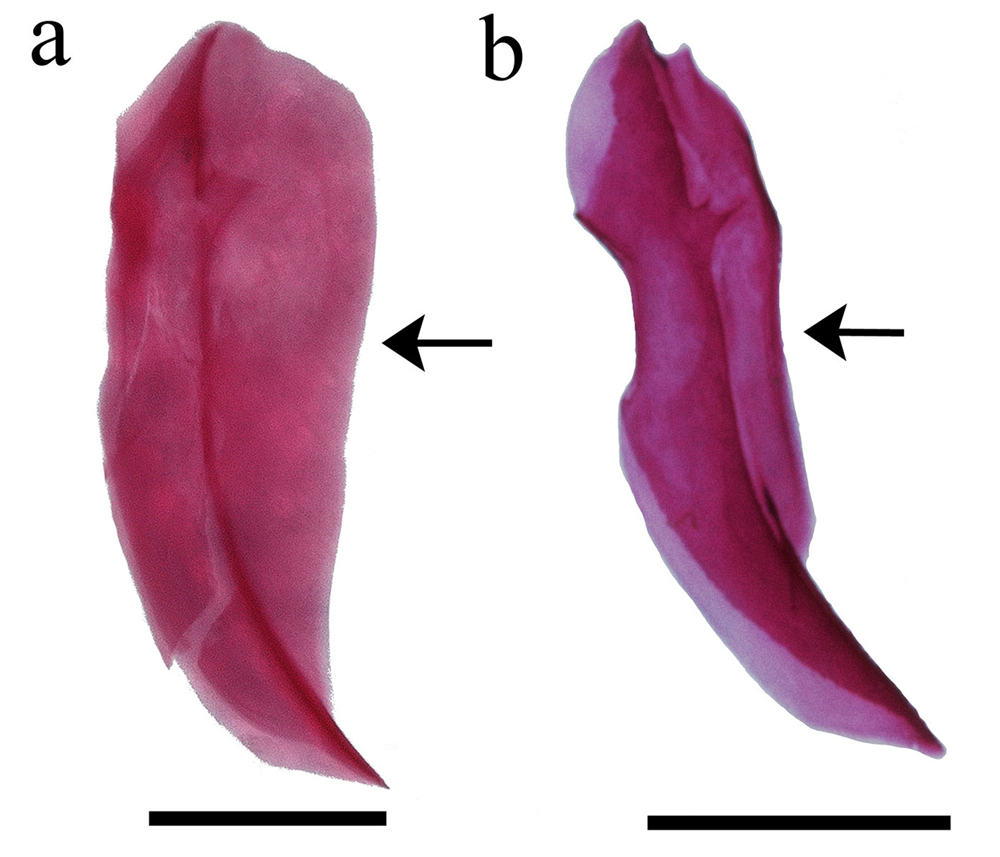
Jaws ( Fig. 22 View FIGURE 22 ). Maxilla curved, elongate with anterior groove and protruding process posteriorly. Premaxilla with two rows of pointed teeth; teeth of outer row with half length of teeth of inner row; inner row with teeth curved inward; dorsal margin of premaxilla with process ascending into maxilla groove; anterior process longer than dorsal process. Rostral cartilage long, almost contacting interpremaxillary ligament. Interpremaxillary ligament narrow, premaxillas close to each other. Dentary with two rows of pointed; teeth of outer row with half length of teeth of inner row; inner row with teeth curved inward; ventral margin concave. Dentary coronoid process broad and rounded, higher than anguloarticular coronoid. Meckel’s cartilage elongate and narrow. Anguloarticular coronoid pointed dorsoposteriorly and connected with retroarticular ventroposteriorly. Retroarticular with dorsoposterior portion pointed and connected to quadrate. Coronomeckelian elongate located over anguloarticular coronoid.
Opercular series ( Fig. 22 View FIGURE 22 ). Preopercle curved, with anterior and posterior portions elongated and pointed. Preopercular sensory canal with eight pores; pores 1, 2, 7 and 8 open and pores 3, 4, 5 and 6 enclosed by bone. Interopercle elongated with anterior portion pointed and posterior broad. Subopercle comma-shaped. Opercle broad with pointed process in superior margin and ventral margin over subopercle.
Branchial basket. Urohyal with broad posterior portion and two separate expansions in anterior portion. Lower branch gill rakers lower 19—25 (21); upper branch gill rakers 3—6 (4); total gill rakers 24—31 (25). Hyoid arch with six branchiostegals rays; four connected in anterior ceratohyal and two connected in posterior ceratohyal. Last three branchiostegal rays widest. Posterior ceratohyal and anterior ceratohyal separated by cartilage, with bony connection in medial portion. Interhyal rectangular and completely ossified connected through cartilage with ceratohyal. Ventral hypohyal rectangular, connected to ceratohyal by bony projections. Basihyal elongate, wider posteriorly. Ceratobranchials 1—4 narrow and long; fifth ceratobranchial enlarged in mid-length with several differently sized teeth; teeth of outer row longer. Epibranchials elongated and straight with cartilage at tips; epibranchials 1, 2 and 3 with pointed processes at anterior tip, longer in epibranchial 3; epibranchial 4 trapezoidal. Pharyngobranchial 1 long and ossified, pharyngobranchials 2 rectangular, wider than long with several teeth; pharyngobranchial 3 large and wide with several teeth; pharyngobranchial tooth plate well developed, triangular, with several teeth.
Paired fins. Pectoral girdle with supracleithrum narrow. Posttemporal connected ventrally to supracleithrum with two narrow and pointed processes; dorsal process largest ( Fig. 9 View FIGURE 9 b). Cleithrum with narrow dorsomedial wing not reaching scapular foramen, its anterior portion connected with coracoid. Coracoid partially separated from scapula by cartilage; scapula with rounded scapular foramen. Radials with ventral and dorsal borders cartilaginous; radial 1 and 2 largest. Extrascapular bone divided in two elements extending over to posttemporal ( Fig. 9 View FIGURE 9 b). Pelvic girdle with three pointed processes; dorsal plate well developed, dorsolateral process forming an angle less than 90 degrees with dorsal plate; median process smaller; medial plate small not reaching anterior tip of dorsal plate.
Medians fins. First dorsal fin with four pterygiophores; first one flat and longer than wide. Two interdorsal pterygiophores. Second dorsal fin with nine pterygiophores, first divided in two wide processes and last one rudimentary ( Fig. 10 View FIGURE 10 b). Anal fin with 16 pterygiophores, first one widest.
Axial skeleton. Precaudal vertebrae 24—27; caudal vertebrae 22—23; total vertebrae 46—49. Neural and hemal spines inserted anteriorly to vertebrae centrum. First to ninth neural spines extended laterally; seventh to ninth neural spines longer, remaining neural spines elongate and narrow. First haemal spine short and modified forming haemal-arch funnel; haemal-arch funnel beginning between second-fourth and third-five pterygiophores of anal fin; haemal-arch funnel with lateral expansion.
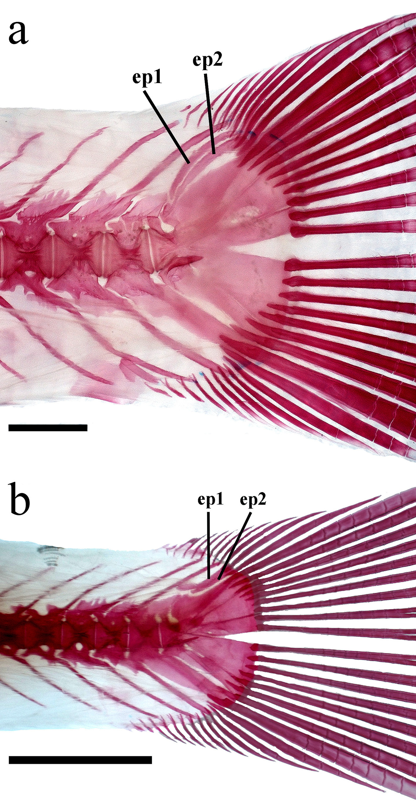
Caudal complex. (see Fig. 11 View FIGURE 11 b) Two narrow autogenous epurals, epural 1 with anterior process basally; parhypural near to first hypural. Lower caudal plate with hypurals 1 and 2 fused. Upper caudal plate with uroneural; hypural 3, 4 and 5 autogenous. Hypurapophysis narrow and pointed. Procurrent caudal fin rays 11 dorsally and 12 ventrally.
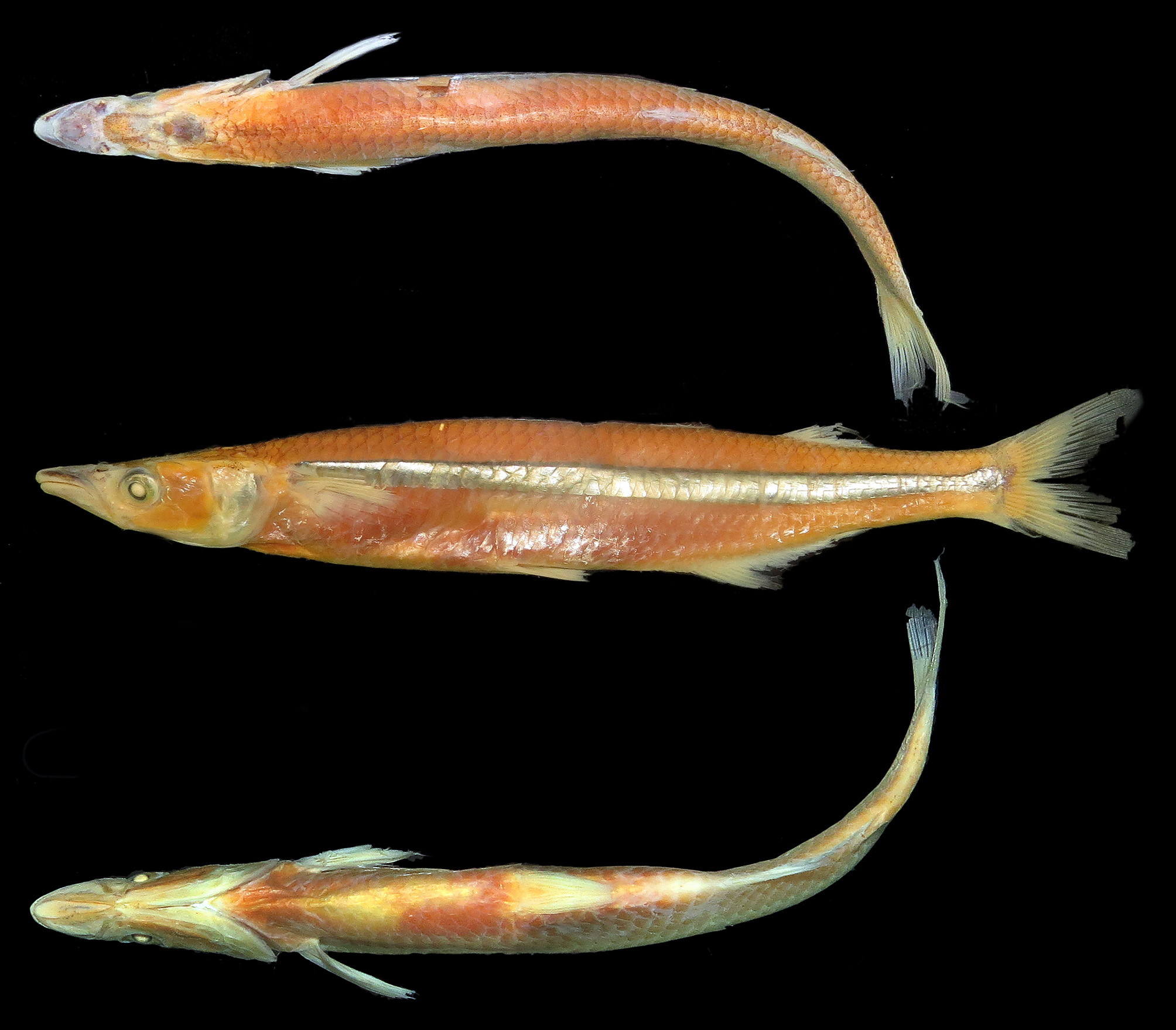
Color in alcohol. Lateral surface of body light yellow with conspicuous longitudinal silvery band in most of specimens extending to pectoral-origin to caudal-fin base and wider below dorsal fins; silvery band with thin black dorsal border ( Fig. 16 View FIGURE 16 ). Few specimens with silvery band darker or completely black after guanine degradation by formalin. Lateral surfaces of body above band presenting small black chromatophores on scales border. Dorsal surface of body yellow with small black chromatophores in predorsal region, more concentrated on scales border. Ventral surfaces of body yellow. Dorsal surface of head and temporal region with dark brown blotches larger than eye size, scattered from snout to nape region and two smaller blotches near each nostril. Small black chromatophores on snout, upper and lower lips, anteriormost portion of maxilla, and anteriormost portion of dentary. Fins translucent; pelvic and anal fin without marks. Second dorsal and anal fins with black chromatophores basally becoming inconspicuous towards tips. Caudal fin with black chromatophores at base and along middle portion of lobes.
Color in life. A photo taken of one specimen just after fixation shows overall color brighter than after preservation, a silvery lateral band, body light green above lateral band and white below lateral band ( Fig. 17 View FIGURE 17 ). The distal margins of second dorsal and anal fins are reddish and laterodorsal region of body reticulated due to conspicuous presence of black chromatophores in scale borders.
Sexual dimorphism. No clear sexual dimorphism could be observed in the specimens examined.
Size. Maximum length observed in types 168.7 mm SL.
Common names. Odontesthes yucuman is commonly known as “peixe-rei” in Brazil.
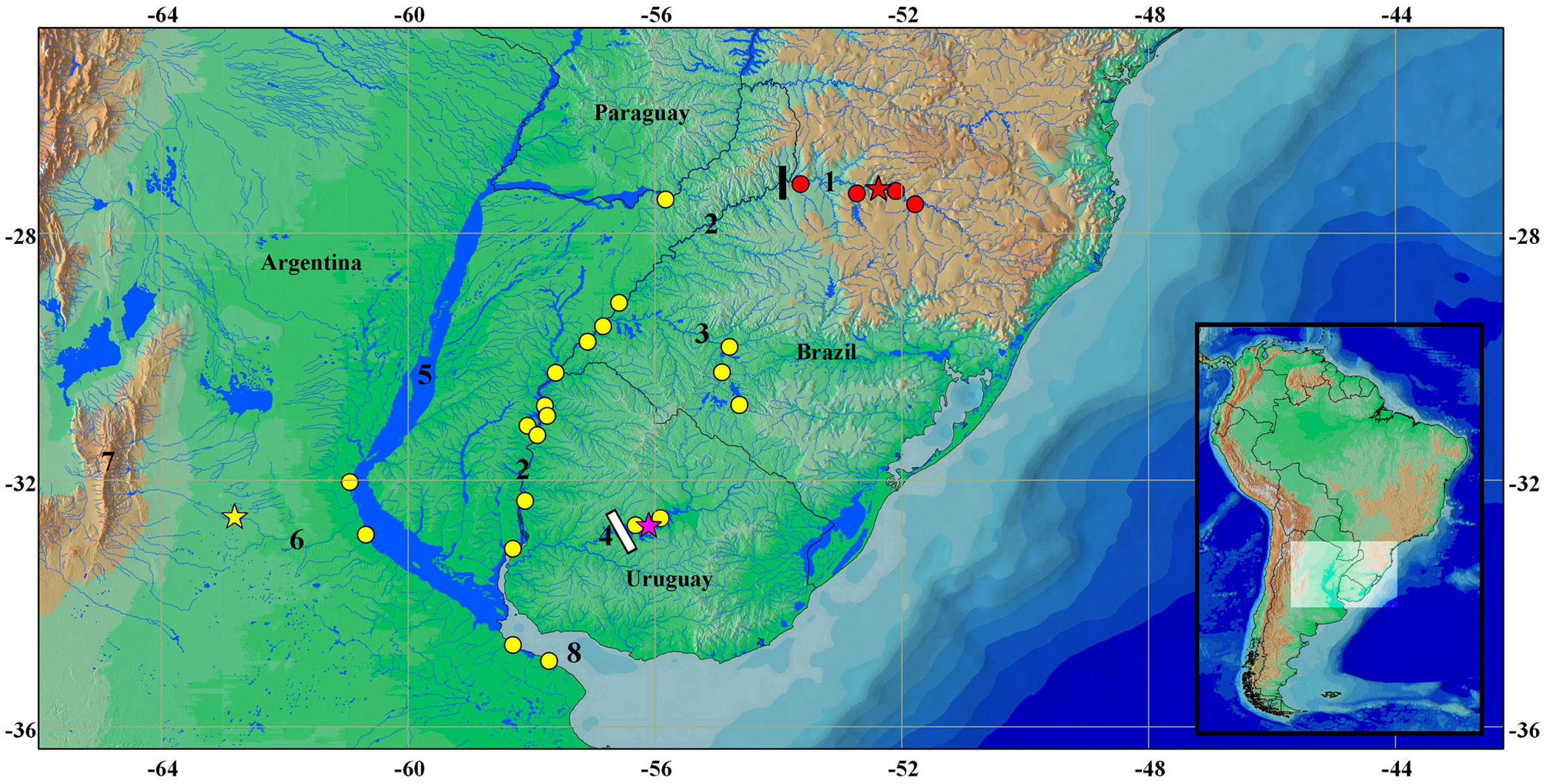
Distribution. Odontesthes yucuman is only known from the upper rio Uruguay and the rio Passo Fundo ( Fig. 12 View FIGURE 12 ), corresponding to the upper rio Uruguay ecoregion sensu Abell et al. (2008). All specimens were sampled at the reservoirs or immediately downstream of three Hydroelectric Power Plants of the rio Uruguay basin (Itá, Machadinho and Monjolinho) along fish monitoring programs. Only three of the examined specimens of O. yucuman were collected in a locality without influence of dams (MCP 40026). Other congeners occurring sympatrically with O. yucuman in the upper rio Uruguay are O. bonariensis (Valenciennes) and O. humensis de Buen.
A photo of a silverside identified as Odontesthes aff. perugiae published in an inventory of fishes from the upper rio Uruguay ( Zaniboni et al., 2004: 13) seems to correspond to Odontesthes yucuman . Zaniboni et al. (2004) inventory is based on fish collections made along seven years ( 1995—2002) in a stretch of upper rio Uruguay and some of its tributaries. Unfortunately, the sampled fishes were not preserved or catalogued in collections, precluding a definitive assessment of the identity of the depicted Odontesthes specimen.
Conservation status. Odontesthes yucuman has an extent of occurrence (EOO) smaller than 5,000 km ², and seems to be not frequently collected. Additionally, the species inhabits an area with a continued decline of habitat quality by the several hydroelectric dams built along the rio Uruguay and its main tributaries in the last years. However, there is no biological, ecological or behavioral data indicating the impact of the dams on the species, which could aid to infer more precisely its conservation status. Thus, O. yucuman is herein categorized as Near Threatened (NT) according to IUCN criteria (IUCN 2014).
Etymology. The specific epithet refers to the impressive longitudinal waterfall known as Salto do Yucumã in Brazil, which is the limit between upper and lower portions of the rio Uruguay basin. A noun in apposition.


| UFRGS |
Universidade Federale do Rio Grande do Sul |
No known copyright restrictions apply. See Agosti, D., Egloff, W., 2009. Taxonomic information exchange and copyright: the Plazi approach. BMC Research Notes 2009, 2:53 for further explanation.
|
Kingdom |
|
|
Phylum |
|
|
Class |
|
|
Order |
|
|
Family |
|
|
Genus |
Odontesthes yucuman
| Wingert, Juliana M., Ferrer, Juliano & Malabarba, Luiz R. 2017 |
Odontesthes aff. perugiae
| Zaniboni 2004: 13 |