
Agetocera taiwana
|
publication ID |
https://doi.org/ 10.5281/zenodo.194971 |
|
DOI |
https://doi.org/10.5281/zenodo.6204462 |
|
persistent identifier |
https://treatment.plazi.org/id/03D887C2-2150-FFA3-12CC-FB6ADB343821 |
|
treatment provided by |
Plazi |
|
scientific name |
Agetocera taiwana |
| status |
|
Agetocera taiwana species group
Diagnosis. General color (Figs 5–8) yellowish brown; antennomeres IX–XI or VIII–XI black; apical half of tibia, and tarsi darkened, two apical tarsomeres dark brown; elytra dark bluish-metallic. Pronotum transverse, about 0.6–0.7 times as long as wide. Elytra relatively longer, about 1.6–1.7 times as long as wide, widest at apical 1/3.
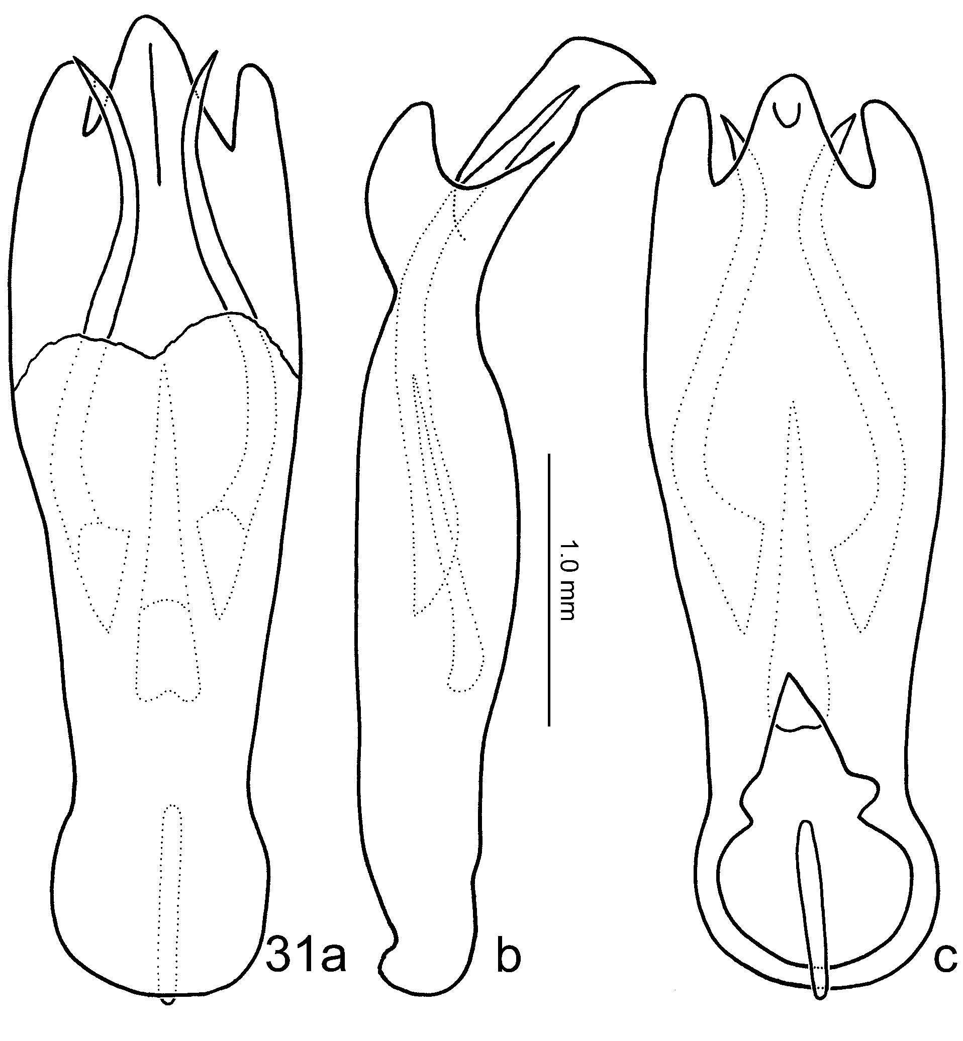
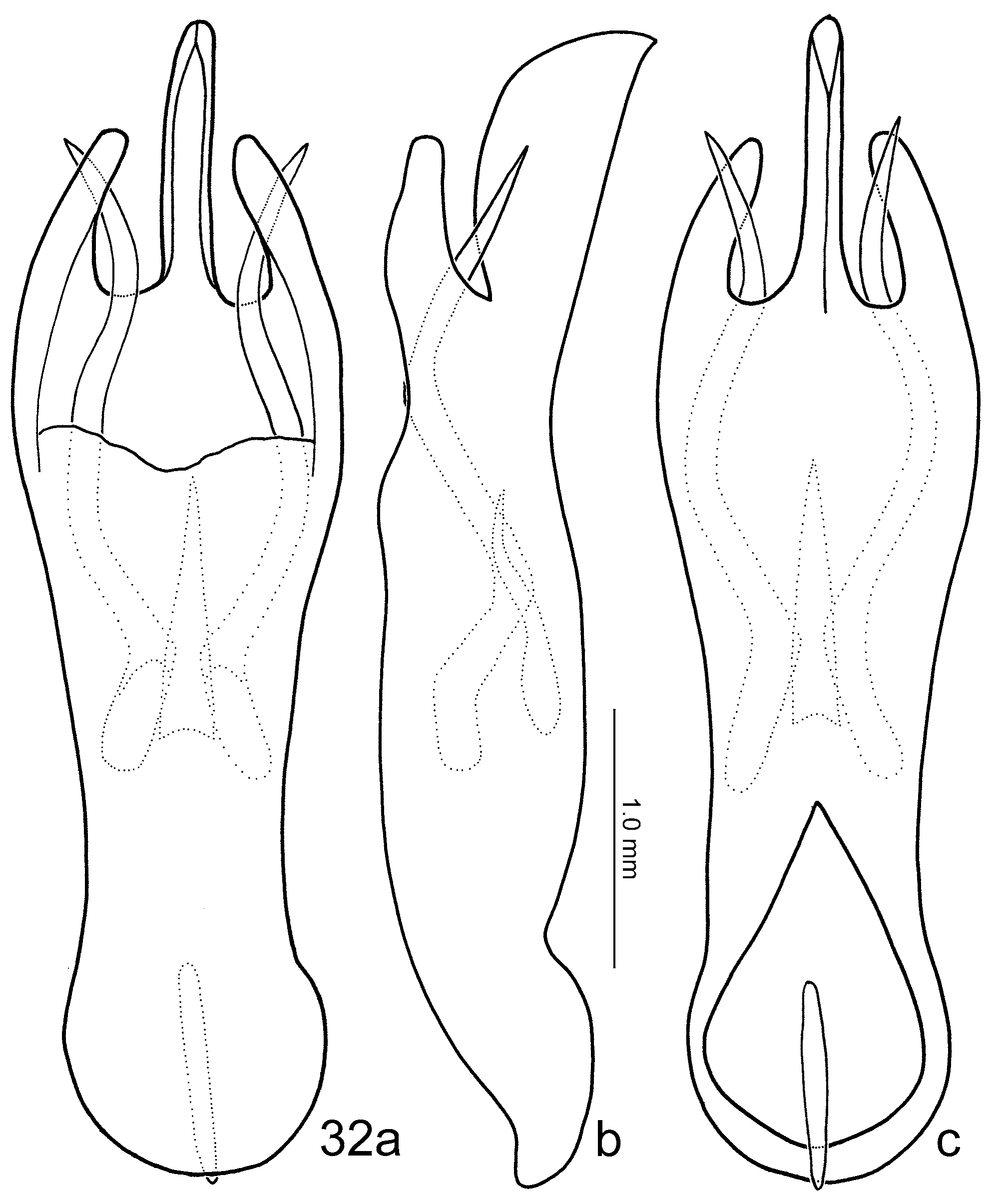
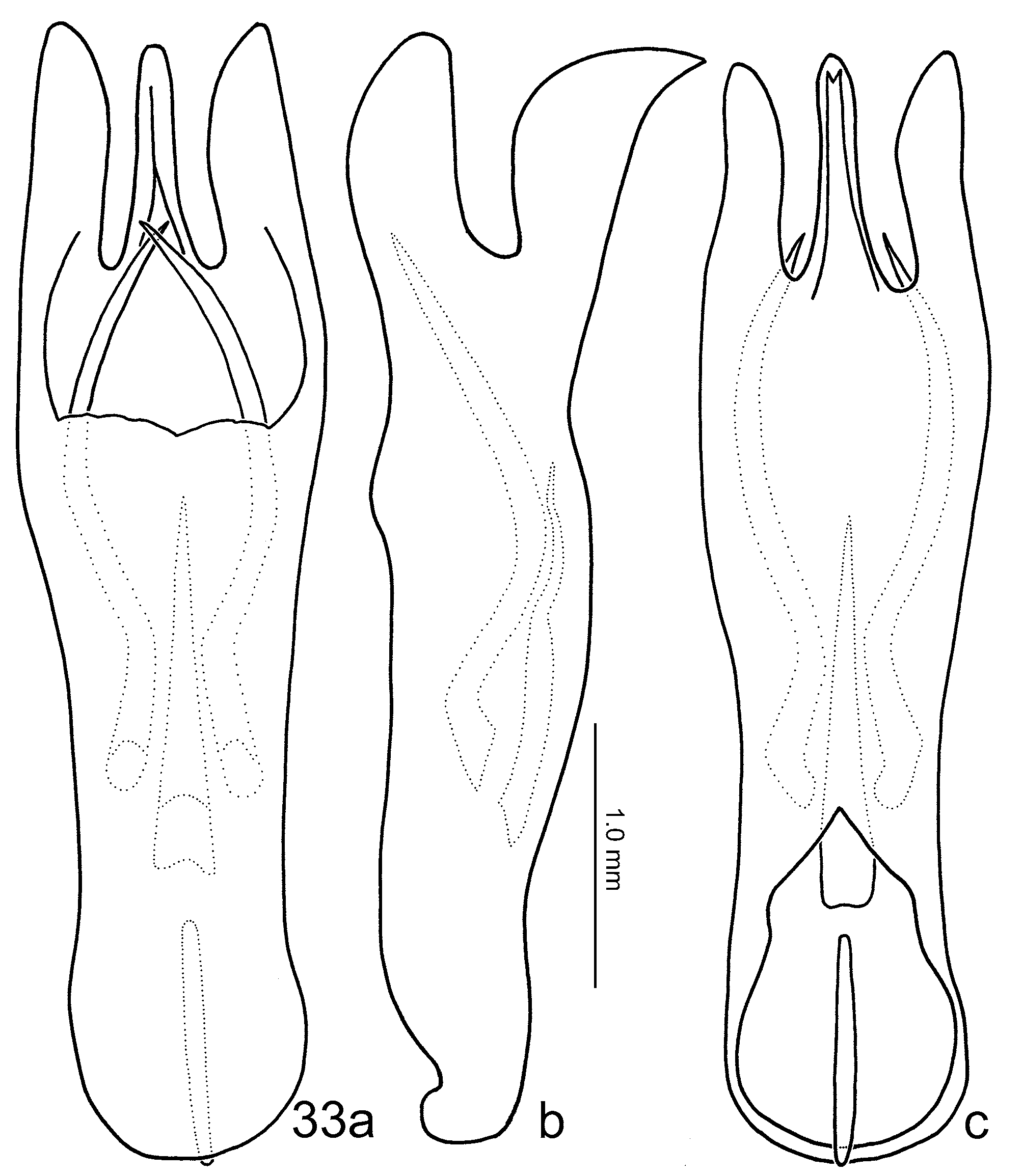
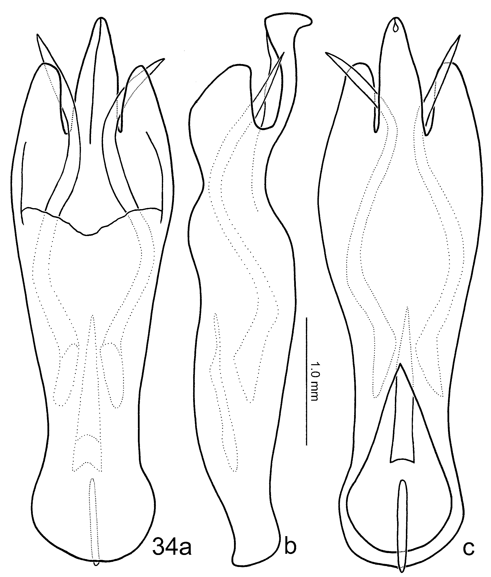
Male. Antenna long, extending to middle of elytra, antennomere VIII (Figs 11, 13) strongly swollen, with a short pointed process at antero-lateral angle, antennomere IX medially inserted on apex of VIII, moderately or strongly flattened dorsally (Figs 16, 17), antennomeres X and XI filiform. First pro- and mesotarsomeres relatively wider and shorter in males than in females. Abdominal ventrite V (Fig. 20) trilobed, apex of median lobe truncate, apices of lateral lobes rounded, apex of median lobe concave, with a median internal ridge from basal margin to base of median lobe. Genitalia ( Figs 31–34 View FIGURES 31 View FIGURES 32 View FIGURES 33 View FIGURES 34 ) symmetric, apex composed of a median process and a pair of lateral processes; endophallus composed of a pair of curved and horn-like lateral spiculae, and a median slender spiculae between lateral spiculae.
Female. Antenna filiform. Middle of apical margin of abdominal ventrite V (Fig. 21) widely rounded, slightly sinuate behind middle. Abdominal tergite VIII (Fig. 27) composed of two separated, weakly sclerotized sclerites, only antero-medial areas visible, with dense hairs. Receptacle of spermatheca (Fig. 29) shorter, and wider than pump; pump short and strongly curved. Gonocoxae (Fig. 30) separated, transverse and triangular, apex with dense setae, one of the setae being extremely long. Bursa-sclerites (Fig. 28) composed of a pair of sclerites, posteriorly widened, posterior margin rounded, anterior margin erected, surface convex near center and near posterior margin.
FIGURES 15–17. Antennomere IX, male, a: dorsal, b: lateral, c: horizontal. 15. Agetocera discedens ; 16. A. taiwana ; 17. A. yuae .
FIGURES 18–21. Abdominal ventrite V. 18. Agetocera discedens , male; 19, A. discedens , female; 20. A. taiwana , male; 21. A. taiwana , female.
Notes. After examination of females of all species belonging to this species group, we found that abdominal tergite VIII, spermatheca, bursa-sclerites, and gonocoxae are very similar and not diagnostic. These characters are excluded from the description of each species. The genitalia of two males of each species and one male from each locality were examined, the results show that male genitalia is diagnostic and shows no variation within a species.
Remarks. Paratypes of A. taiwana Chûjô, 1962 comprise three species. In addition to A. taiwana , A. yuae sp. nov. and A. choui sp. nov. were also found. They have been transferred and designated as paratypes of both new species.
No known copyright restrictions apply. See Agosti, D., Egloff, W., 2009. Taxonomic information exchange and copyright: the Plazi approach. BMC Research Notes 2009, 2:53 for further explanation.
|
Kingdom |
|
|
Phylum |
|
|
Class |
|
|
Order |
|
|
Family |
|
|
Genus |