
Microedus subtilis ( Sharp, 1889 ) Shavrin, 2024
|
publication ID |
https://doi.org/ 10.11646/zootaxa.5443.2.4 |
|
publication LSID |
lsid:zoobank.org:pub:173C52E1-C650-4B3A-92AF-AE7AC29E8D2C |
|
DOI |
https://doi.org/10.5281/zenodo.11060435 |
|
persistent identifier |
https://treatment.plazi.org/id/03D887ED-FF8B-815C-73E1-ACD371C2F677 |
|
treatment provided by |
Plazi |
|
scientific name |
Microedus subtilis ( Sharp, 1889 ) |
| status |
comb. nov. |
Microedus subtilis ( Sharp, 1889) comb. n.
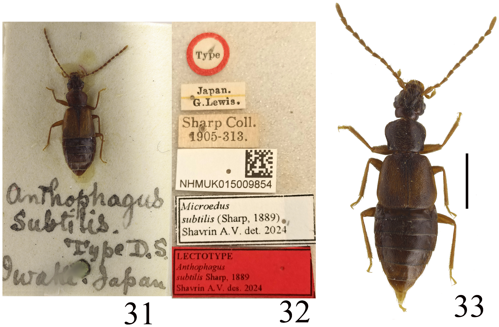
( Figs 31–42 View FIGURES 31–33 View FIGURES 34–42 )
Anthophagus subtilis Sharp, 1889: 471 View in CoL ; Bernhauer & Schubert 1910: 81
Philydrodes subtilis : Scheerpeltz 1933: 1067, Watanabe 1985: 268
Philydrodes (Lioplax) subtilis : Nakane & Sawada 1956: 184; Adachi 1957: 166; Shibata 1976: 123 Liophilydrodes subtilis View in CoL : Watanabe 1990: 302, Shibata et al., 2013: 75, Hayashi 2022: 412
Liophilydrodes subtilis iturupensis Lafer, 2004: 90 View in CoL syn. n.
Philydrodes (Lioplax) troglophila Nakane & K. Sawada, 1956: 184 View in CoL syn. n.
Philydrodes (Lioplax) troglophilus : Uéno & Watanabe 1966: 321, Watanabe 1982: 410, 1996: 9
Philydrodes (Liophilydrodes) subtilis : Watanabe 1986: 545
Liophilydrodes troglophilus View in CoL : Watanabe 1990: 306, Zerche 2003: 289, Shibata et al. 2013: 75, Hayashi 2022: 412
Type material examined. Lectotype (here designated) of Anthophagus subtilis Sharp, 1889 ♀ ( Figs 31–32 View FIGURES 31–33 ): ‘ Anthophagus | subtilis. | Type D.S. | Iwake. Japan’ <handwritten on a card with the specimen, in black India ink>, ‘Type’ <round label with red margin, printed>, ‘Japan [underlined by yellow] | G. Lewis.’ <printed>, ‘ Sharp Coll. | 1905-313.’ <printed>, ‘ NHMUK015009854 ’ <printed label, with BMNH barcode on rigth side of the label>, ‘LECTOTYPE | Anthophagus | subtilis Sharp, 1889 | Shavrin A.V. des. 2024’ <red, printed>, ‘ Microedus | subtilis ( Sharp, 1889) | Shavrin A.V., det. 2024’ <printed> ( BMNH).
Paralectotype ♀ (left apical antennomere, left middle tibia and tarsus and right elytron are absent): ‘ Anthophagus | subtilis. D.S.’ <handwritten on a card with the specimen, in black India ink>, ‘ Japan. [underlined by yellow] | G. Lewis | 1910-320.’ <printed>, ‘Iwake’ <handrwitten>, ‘ Philydrodes [handwritten] | subtilis Sh. [handwritten] Det. C.Koch’ <printed>, ‘ Microedus | subtilis ( Sharp, 1889) | Shavrin A.V., det. 2024’ <printed> ( BMNH).
Additional material examined: JAPAN: HONSHU: 7 ♂♂, 6 ♀♀: ‘JAPAN: Honshu B.M. 1980-492 P.M. Hammond’, ‘ Gumma Pref. Mt, Hotaka (foot) ca 1300 m 14-15.viii. [19]80’ ( BMNH) .
Redescription. Measurements (n=15): HW: 0.72–0.87; HL: 0.47–0.63; OL: 0.15–0.21; LT: 0.12–0.14; AL (lectotype): 2.96; PL: 0.67–0.78; PWmax: 0.90–1.03; PWmin: 0.75–0.87; ESL: 1.02–1.32; EW: 1.30–1.57; MTbL (lectotype): 1.05; MTrL (lectotype): 0.37 (MTrL 1–4: 0.17; MTrL 5: 0.20); AW: 1.35–1.68; AedL: 0.75–0.80; BL: 2.90–5.30 (lectotype: 4.70).
Habitus as in Fig. 33 View FIGURES 31–33 . Body reddish-brown, usually with darker head and abdomen and paler elytra (some specimens with darker mediobasal portion of elytra; lectotype and paralectotype somewhat paler); antennae brown, sometimes with paler antennoimeres 1–2; mouthparts and legs yellow-brown to brown; apical maxillary palpomere and tarsi yellowish. Head with very dense isodiametric microreticulation, more transverse in middle of vertex, finer and sometimes indistinct in middle, and coarser on infraorbital portions, mediobasal portion of head with dense and fine isodiametric meshes; neck with dense and coarse isodiametric sculpture; pronotum with very dense and coarse isodiametric microsculpture, distinctly finer and sometimes indistinct in middle; scutellum with very fine transverse or isodiametric meshes; abdominal tergites with very dense and coarse isodiametric microreticulation.
Head 1.3–1.5 times as broad as long; medioapical depression slightly or strongly narrowing posteriad toward level of apical margins of eyes; temples 1.2–1.5 times as long as longitudinal length of eyes. Ocelli located distinctly below level of posterior margins of eyes, distance between ocelli 1.3–1.6 times as long as distance between ocellus and posterior margin of eyes. Punctation of head sparse and irregular, sparser and finer (sometimes indistinct) in middle and slightly larger, deeper and denser on infraorbital portions. Antennomeres 4–10 elongate; basal antennomere slightly more than three times as long as broad, 2 slightly narrower and distinctly shorter than basal antennomere, 3–5 distinctly longer than 2, 6–7 slightly longer and indistinctly broader than 5, 8–10 slightly shorter than 7, apical antennomere 1.4–1.7 times as long as 10.
Pronotum 1.3 times as broad as long, 1.1–1.2 times as broad as head, from widest anterior portion slightly or strongly narrowed posteriad, laterobasal margins in front of hind angles sometimes straight and parallel-sided. Punctation dense and fine, sparser and sometimes indistinct in middle.
Elytra 1.1–1.2 times as broad as long, 1.5–1.6 times as long as pronotum. Punctation dense and regular, distinctly larger and deeper than that on pronotum, finer in parascutellar portion and along suture.
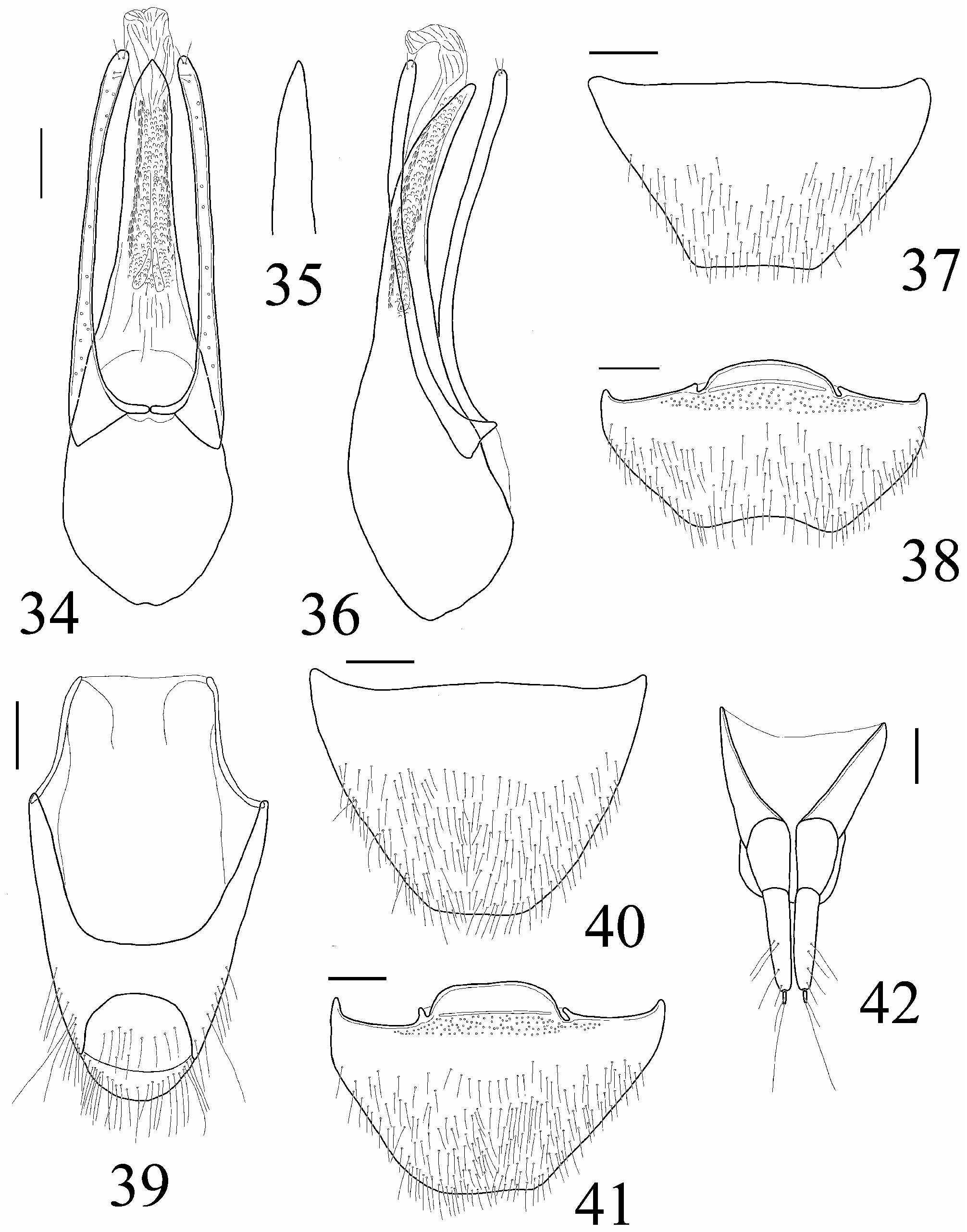
Male. Apical margin of abdominal tergite VIII truncate ( Fig. 37 View FIGURES 34–42 ). Apical margin of abdominal sternite VIII widely concave ( Fig. 38 View FIGURES 34–42 ). Genital segment as in Fig. 39 View FIGURES 34–42 . Aedeagus with moderately narrow basal portion, gradually narrowed toward long and narrow median lobe, slightly ( Fig. 34 View FIGURES 34–42 ) or strongly ( Fig. 35 View FIGURES 34–42 ) narrowed apicad; parameres narrow, distinctly exceeding apex of median lobe, sometimes sligthly broadened apically, with two short apical and two preapical setae; internal sac narrow and long, with two narrow elongate and sclerotized structures in middle (sometimes these structures invisible). Lateral aspect of the aedeagus as in Fig. 36 View FIGURES 34–42 .
Female.Apical margins of abdominal tergite VIII ( Fig. 40 View FIGURES 34–42 ) and sternite VIII ( Fig. 41 View FIGURES 34–42 ) somewhat straight. Genital segment as in Fig. 42 View FIGURES 34–42 .
Comparative notes. Based on the shape of the slightly flattened pronotum, the presence of coriaceous ground sculpture on head and pronotum, and the general shape of the aedeagus, M. subtilis is similar to the Japanese M. yamanakai Watanabe, 1990 , known from Honshu ( Watanabe 1990), from which it can be distinguished by the larger eyes, the finer punctation of head and pronotum, and the distinctly narrower median lobe of the aedeagus.
Distribution. Microedus subtilis is known from Japan (Honshu) and Russia (Iturup Island, Kurile islands).
Bionomics. Some specimens were collected under stones near streams and rivers. Detailed bionomical data are uknown.
Remarks. Anthophagus subtilis was originally descibed from “…Iwakisan…” based on “…two mutilated examples”. I studied two syntypes (females) from BMNH, and one of them, with additional label ‘Type’, I designated here as the lectotype in order to fix the name. Nakane & Sawada (1956) regarded it as a species of Philydrodes (Lioplax) . Watanabe (1990) redescribed it and transferred it to the genus Liophilydrodes , and provided additional material from northeastern Honshu and illustrations of the aedeagus.
Philydrodes (Lioplax) troglophila was originally described based on two specimens from “Nippara cave, Okutama, Tokyo ”. Watanabe (1990) redescribed it, transferred it to the genus Liophilydrodes , provided new material from Honshu and illustrations of the aedeagus. Based on the external morphology of the body, and the shape of the aedeagus, this species corresponds with other specimens of M. subtilis . I could not find sufficient morphological differences between the type and additional specimens. The apex of the median lobe is variable and can be narrow or somewhat wide ( Figs 34–35 View FIGURES 34–42 and Figs 407, 414 in Watanabe (1990)). It should be noted that the width of the apical portions of the median lobe and the parameres is variable in many species of Anthophagini (e.g. Shavrin 2022b). Thus, this species is synonymized with M. subtilis .
Liophiydrodes subtilis iturupensis was originally described based on one female from Iturup Island (“Konservnaya bay”), Kurile islands. Based on the original description, features of the body of this taxon correspond with morphological features of specimens of M. subtilis . Thus, L. subtulis iturupensis is synonymyzed with M. subtilis .
| BMNH |
United Kingdom, London, The Natural History Museum [formerly British Museum (Natural History)] |
No known copyright restrictions apply. See Agosti, D., Egloff, W., 2009. Taxonomic information exchange and copyright: the Plazi approach. BMC Research Notes 2009, 2:53 for further explanation.
|
Kingdom |
|
|
Phylum |
|
|
Class |
|
|
Order |
|
|
Family |
|
|
Genus |
Microedus subtilis ( Sharp, 1889 )
| Shavrin, Alexey V. 2024 |
Liophilydrodes subtilis iturupensis
| Lafer, G. Sh. 2004: 90 |
Liophilydrodes subtilis
| Hayashi, Y. 2022: 412 |
| Watanabe, Y. 1990: 302 |
Liophilydrodes troglophilus
| Hayashi, Y. 2022: 412 |
| Zerche, L. 2003: 289 |
| Watanabe, Y. 1990: 306 |
Philydrodes (Liophilydrodes) subtilis
| Watanabe, Y. 1986: 545 |
Philydrodes (Lioplax) troglophilus
| Watanabe, Y. 1996: 9 |
| Watanabe, Y. 1982: 410 |
| Ueno, S. - I. & Watanabe, Y. 1966: 321 |
Philydrodes (Lioplax) subtilis
| Shibata, Y. 1976: 123 |
| Adachi, T. 1957: 166 |
| Nakane, T. & Sawada, K. 1956: 184 |
Philydrodes subtilis
| Watanabe, Y. 1985: 268 |
| Scheerpeltz, O. 1933: 1067 |
Anthophagus subtilis
| Bernhauer, M. & Schubert, K. 1910: 81 |
| Sharp, D. 1889: 471 |