
Microlaimus porosus Bussau et Vopel, 1999
|
publication ID |
https://doi.org/ 10.11646/zootaxa.2096.1.11 |
|
DOI |
https://doi.org/10.5281/zenodo.5334904 |
|
persistent identifier |
https://treatment.plazi.org/id/03D8C423-FF8B-FFD0-FF77-0B27E776FB96 |
|
treatment provided by |
Felipe |
|
scientific name |
Microlaimus porosus Bussau et Vopel, 1999 |
| status |
|
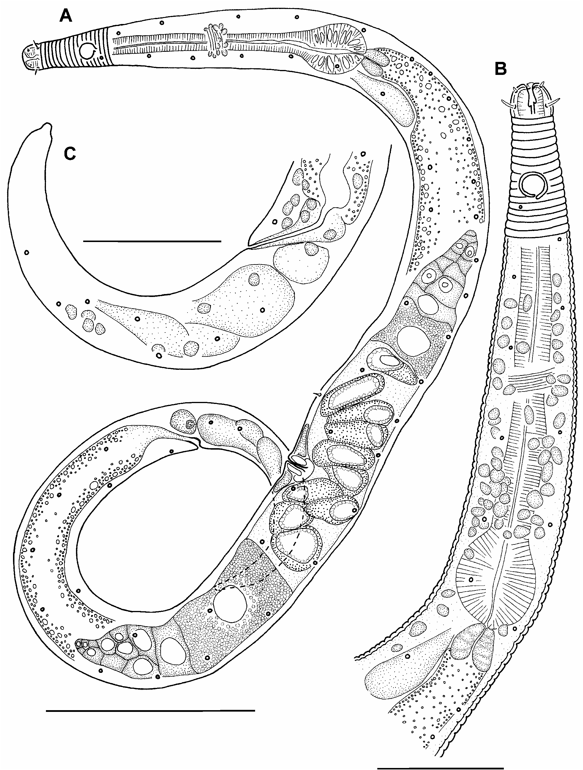
Microlaimus porosus Bussau et Vopel, 1999
( Figs 20–21 View FIGURE 20 View FIGURE 21 , Table 7)
Material examined: 4 females ( Table 7).
Locality: Tables 1, 7.
Measurements: Table 7.
Description of female: Body in shape of elongated spindle, with narrowed anterior end and conical tail. Non-annulated cephalic capsule present at anteriormost part of cephalic end. Length of cephalic capsule slightly more than its width. Cuticle of rest body annulated, annuli strongly pronounced, their width 0.9–1.1 µm. Cuticle thickness 0.7 µm at level of cephalic capsula and 1.0–1.1 µm along rest of body. Four submedian rows of round pores 1 µm in diameter situated along whole body. Number of pores in dorsolateral rows 1.2–1.5 times more than those in ventrolateral rows. Anteriormost pore of dorsolateral rows situated behind amphid at a distance approximately equal to amphidial c.b.d., and posteriormost one situated at posterior quarter of tail. Anteriormost pore of ventrolateral rows situated behind amphid close to it, and posteriormost one situated at a middle part of tail. Number of pores in rows varying in different specimens from 27 to 42 in dorsolateral rows and from 17 to 33 in ventrolateral rows. Pores located in rows regularly. Somatic setae very rare, irregularly situated, 1.5 µm long. Sensilla of cephalic end arranged in 2 rings: 6 short outer labial setae 1 µm long situated at anterior part of cephalic capsule and 4 submedian cephalic setae 2.5 µm long, situated at a distance of 2/3 of length of cephalic capsule from anterior end. Amphid monospiral, round, 40–50% of c.b.d. in diameter. There are 8–11 cuticular annules between anterior end of amphid and cephalic capsule. Stoma present, with a greater length than that of cephalic capsula, however its posterior end not clearly visible. Walls of stoma thicker than in posterior pharyngeal lumen. Two teeth visible in stegostoma. Pharynx thin but having a well-developed terminal oval bulb, constituting 70–80% of c.b.d. in width. Nerve ring situated at a level of middle of pharynx. Large cellular body of renetta located at level of beginning of intestine. Cardia quite large and round. Reproductive system didelphic, amphidelphic, with outstretched ovaries. It occupying approximately 40% of total body length. Anterior ovary lies to the right of intestine, posterior one lies to the left of intestine. Mature oocyte elongated, its size 15x27 µm. Large spermatozoa of different shape visible in uterus. Vulvar glands seen.
Abundance: The average density of this species was about 1.5 inds/ 10cm 2 and relative abundance within the nematode community was 1–2% at the stations where it was found.
Remarks: M. porosus was initially described by Bussau and Vopel (1999) from the South-Eastern Pacific area which is located about 5200 km from the area of the new finding. The specimens from the new area bear a good resemblance to the original description.
The presence of 2 teeth in the stegostoma is unusual feature in the genus Microlaimus , where three teeth are described as a rule. Meanwhile, Bussau and Vopel (1999) also mentioned two teeth in the original description of M. porosus . Perhaps, 2 subventral teeth are touched very closely and optically seem fused when viewedunder light microscope.

No known copyright restrictions apply. See Agosti, D., Egloff, W., 2009. Taxonomic information exchange and copyright: the Plazi approach. BMC Research Notes 2009, 2:53 for further explanation.
|
Kingdom |
|
|
Phylum |
|
|
Class |
|
|
Order |
|
|
Family |
|
|
Genus |