
Microlaimus parviporosus, Miljutin & Miljutina, 2009
|
publication ID |
https://doi.org/ 10.11646/zootaxa.2096.1.11 |
|
DOI |
https://doi.org/10.5281/zenodo.6486055 |
|
persistent identifier |
https://treatment.plazi.org/id/03D8C423-FF8E-FFED-FF77-0A82E682FF49 |
|
treatment provided by |
Felipe |
|
scientific name |
Microlaimus parviporosus |
| status |
sp. nov. |
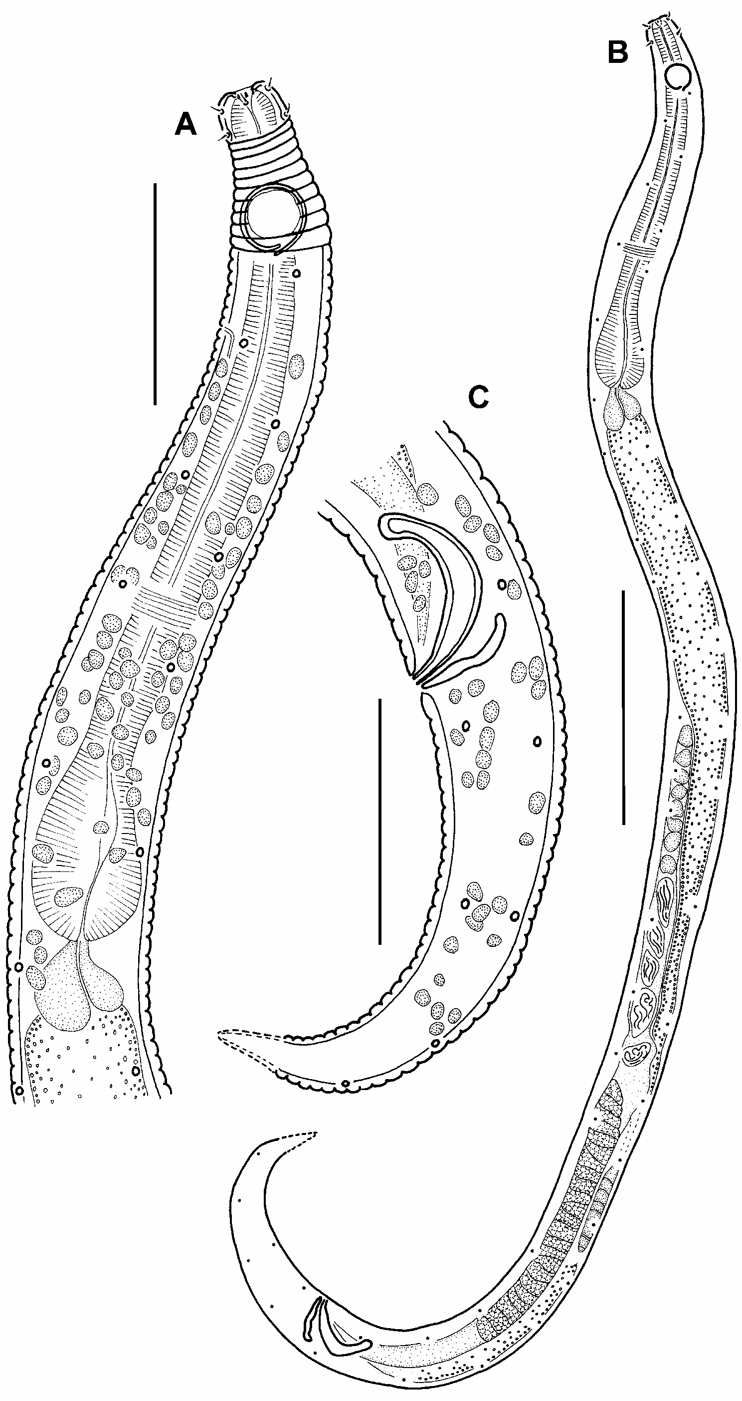
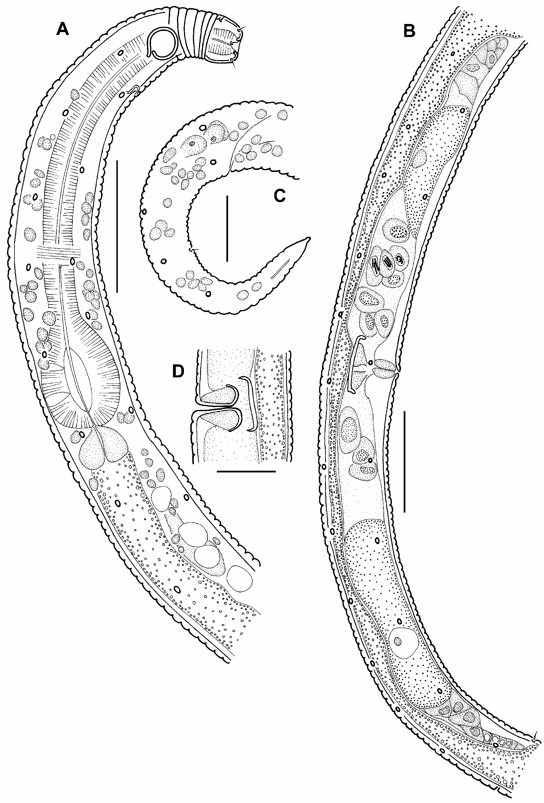
Microlaimus parviporosus sp. n.
( Figs 17–19 View FIGURE 17 View FIGURE 18 View FIGURE 19 , Table 6)
Type material: Collection number MNHN-BN492 . Holotype: one male. Paratype: 4 males, 3 females ( Table 6) .
Locality: Tables 1, 6.
Etymology: From Latin parvus (scanty, hidden) and porus (pore).
Measurements: Table 6.
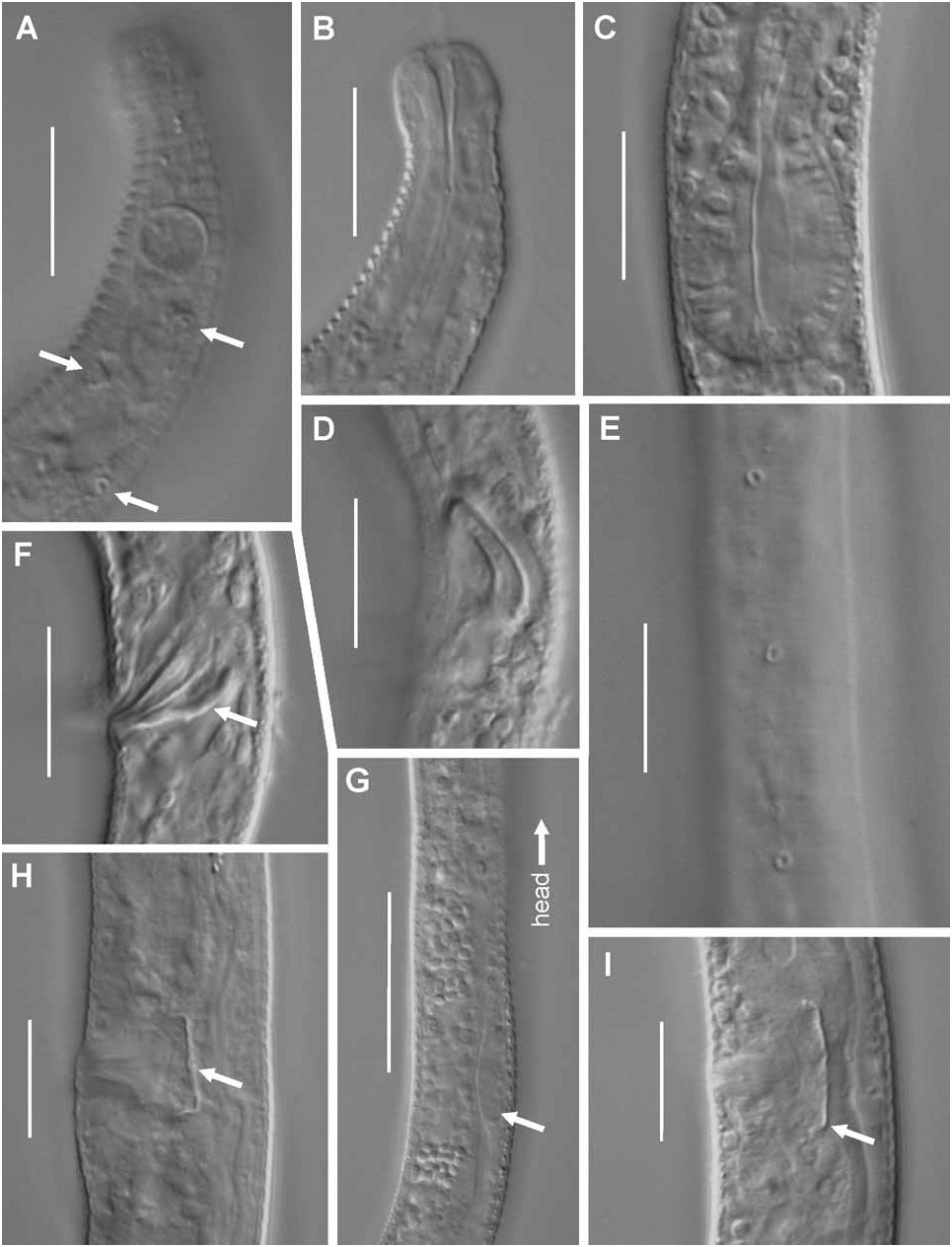
Description of males: Body cylindrical, with slightly narrowed anterior end and conical tail. Cuticle with strongly pronounced annuli, which beginning at posterior of cephalic capsule (width of every annulus 1.1–1.3 µm). Cuticle thickness 0.3 µm at level of cephalic capsula and 0.6–0.7 µm along rest of body. Four submedian rows of round pores 0.5 µm in diameter situated along whole body. Number of pores in dorsolateral rows 1.2–1.5 times more than in ventrolateral ones. Anteriormost pore of dorsolateral rows situated at a distance less than amphidial c.b.d. behind amphid, and posteriormost one situated at posterior third of tail. Anteriormost pore of ventrolateral rows situated at a distance of approximately 1.5 amphidial c.b.d. behind amphid, and posteriormost one situated at anterior third of tail. Number of pores in rows varying in different specimens from 21 to 28 in dorsolateral rows and from 17 to 21 in ventrolateral rows. Location of pores in rows not absolutely regular, as distance between pores varying. As a rule, pores situated more sparsely in ventrolateral rows in region of midbody. Somatic setae 1.2–1.5 µm long, rare and irregularly situated. Sensilla of cephalic end arranged in 2 rings: 6 short, outer, labial setae 1.2 µm long at level of cephalic tip and there 4 submedian cephalic setae of about the same length near posterior border of cephalic capsule. Amphidial fovea monospiral, round, 55–67% of c.b.d. in its diameter. There an amphidial aperture visible in holotype, which is less in diameter than amphidial fovea. From 5 to 7 cuticular annules visible between anterior end of amphid and cephalic capsule. Vestibulum short, cup-shaped, possessing 3 small sclerotized odontia. Small dorsal tooth visible in anterior quarter of stegostoma. Pharynx thin but has a well-developed terminal pear-shaped, or oval, bulb which being 61–86% of c.b.d. in its width. Nerve ring being 2/3 of pharyngeal length from anterior end. Renetta with large cellular body located at level of beginning of intestine and outlet opening at 21 µm (approximately 2 amphidial c.b.d.) from anterior end. Cardia large and triangular. Reproductive system diorchic, with outstretched anterior testis, and with reflected posterior one. Posterior testis shorter and thinner then anterior one, situated at dorsal side, and poorly visible. Anterior testis lying dorsally from intestine, and small posterior one being to the right from intestine. Two zones of spermatogenesis are visible in anterior testes: small roundish spermatogonia with fine-grained content, large oval spermatogonia with fibrillar content. Vas deferens thick, 29% of total body length, filled with puck-shaped spermatids approximately 6 µm in diameter with large-grained content. Spicules rather large, curved, 6 µm, with feebly marked knob at its distal end. Gubernaculum rod-like, slightly bent anteriorly. Supplementary organs not visible. Whole reproductive system occupying about a half of total body length. Tail conical. Three roundish cellular bodies of caudal glands visible close to anus.
Females: Females similar to males in most parameters, however amphids smaller, 44–56% of c.b.d. Reproductive system didelphic, amphidelphic, with outstretched ovaries, and occupying about 2/5 of total body length. Intestine pushed to dorsal side by reproductive system. Small part of dorsal wall of uterus opposite vulva being thicker and strongly sclerotized. Every ovary containing one mature elongated oocyte approximately 40 µm long. Spermatozoa 5.5x4.8 µm in size visible in uterus. Two pairs of vulvar glands seen.
Abundance: The density of this species was 0.8–2.9 inds/ 10cm 2 and relative abundance within the nematode community was 1–3% at the stations where it was found.
Differential diagnosis: The peculiar feature of M. parviporosus sp. n. is the occurrence of four submedian rows of pores spreading along the whole body length. Only three Microlaimus species possessing the same feature have been described before: M. cyatholaimoides de Man, 1922 (according to Jensen (1978), whereas Hopper and Meyers (1967) mentioned the presence of four rows of short, fleshy setae associated with hypodermal glands for this species), M. porosus Bussau and Vopel, 1999 , and M. discolensis Bussau and Vopel, 1999 .
M. parviporosus sp. n. differs from M. cyatholaimoides by the absence of preanal supplementary organs; by the total body length (360–415 µm vs 700–1000 µm in the latter species); by weakly cuticularized stoma with inconspicuous teeth vs. very sclerotized stoma with quite large teeth. The outer labial setae and the cephalic setae are of about equal length in the new species whereas the cephalic setae are much longer in M. cyatholaimoides .
The new species differs from M. porosus by its 2 rings of head setae similar in length, whereas the cephalic setae are much longer than the outer labial ones in M. porosus . Besides, it differs from M. porosus by the shorter spicule (16–18 µm vs. 24 µm) and by the shape of spicule and gubernaculum (more curved and thicker spicule and gubernaculum are observed in the new species). The aperture of the amphid of the new species is narrower than the fovea, whereas it is of similar diameter in M. porosus .
The new species differs from M. discolensis by its shorter head setae of two rings (1.2 µm vs. 6–9 µm); by the width of cephalic capsule (6–7 µm vs. 11–16 µm) by the number of pores on submedian rows (21–28 in dorsal rows and 17–21 in ventral rows vs. 35–45 and 9–15 respectively in M. discolensis ); by the shape of outlet openings of hypodermal glands (pores vs short tubes with pores on its distal ends); by the length of spicule (16–18 µm in arc vs. 28–33 µm); by the shape of gubernaculum (curved vs. straight); by the character of annulation (there are wide furrows between the annuli in M. discolensis , whereas M. parviporosus does not possess such a feature). The hypodermal glands associated with pores were not observed in M. parviporosus in spite of M. discolensis , where these glands are very big and noticeable.
No known copyright restrictions apply. See Agosti, D., Egloff, W., 2009. Taxonomic information exchange and copyright: the Plazi approach. BMC Research Notes 2009, 2:53 for further explanation.
|
Kingdom |
|
|
Phylum |
|
|
Class |
|
|
Order |
|
|
Family |
|
|
Genus |