
Microlaimus abyssalis, Miljutin & Miljutina, 2009
|
publication ID |
https://doi.org/ 10.11646/zootaxa.2096.1.11 |
|
persistent identifier |
https://treatment.plazi.org/id/03D8C423-FF9A-FFE6-FF77-0820E19AFD13 |
|
treatment provided by |
Felipe |
|
scientific name |
Microlaimus abyssalis |
| status |
sp. nov. |
Microlaimus abyssalis sp. n.
( Figs 9–12 View FIGURE 9 View FIGURE 10 View FIGURE 11 View FIGURE 12 , Table 4)
Type material: Collection number MNHN-BN491 . Holotype: one male. Paratype: 5 males, 4 females ( Table 4) .
Locality: Tables 1, 4.
Etymology: Greek abyssos (= precipice, abyss).
Measurements: Table 4.
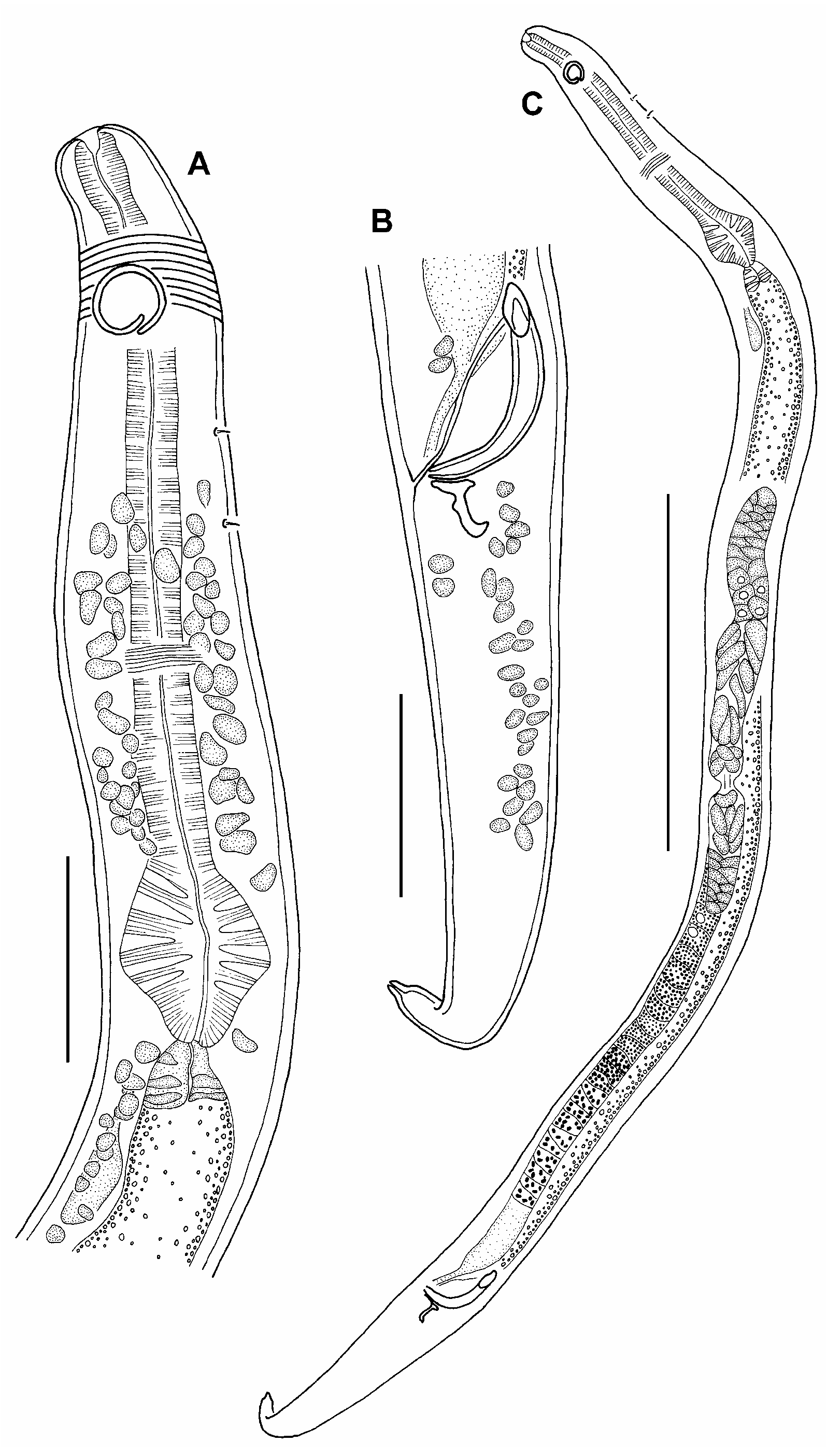
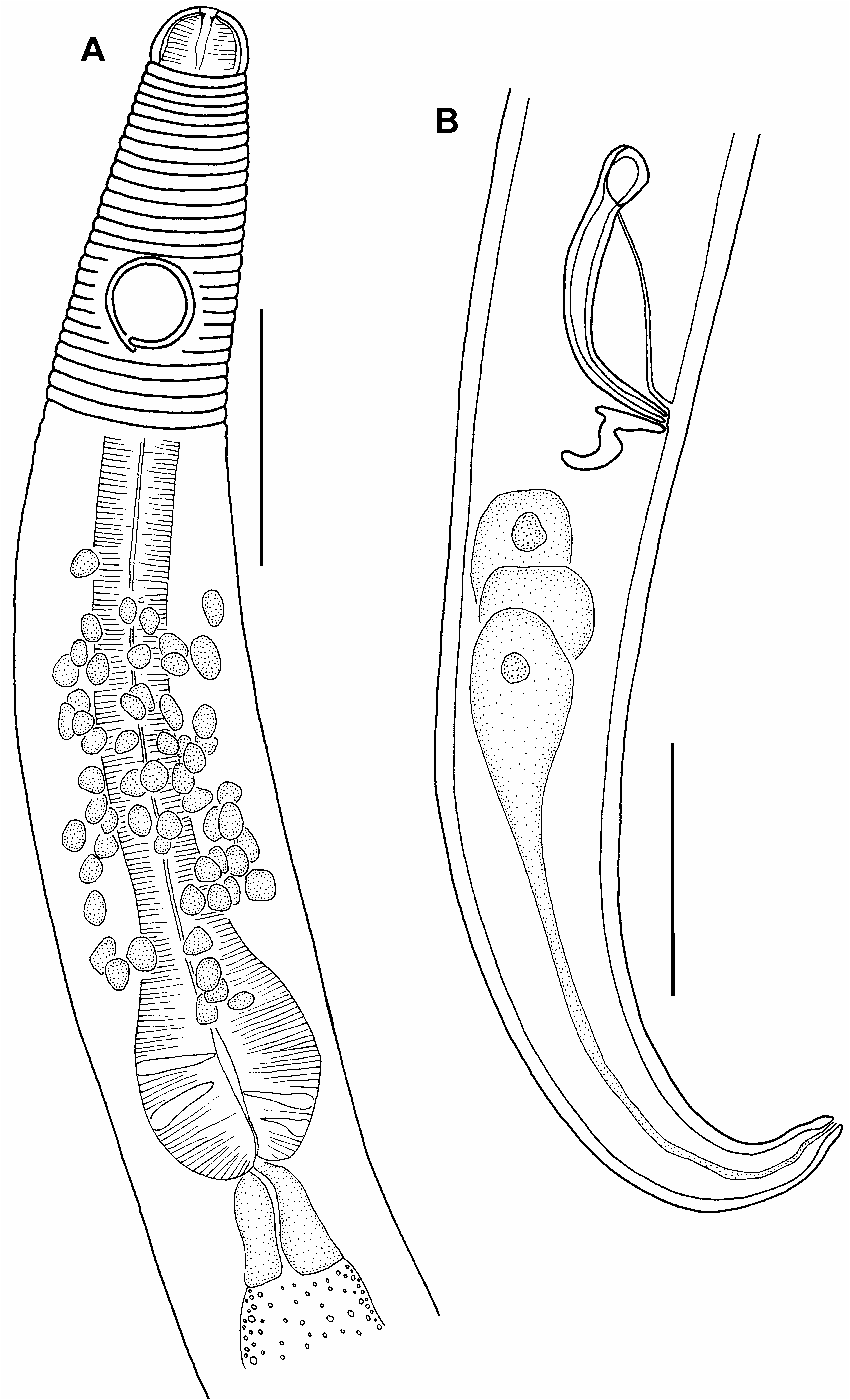
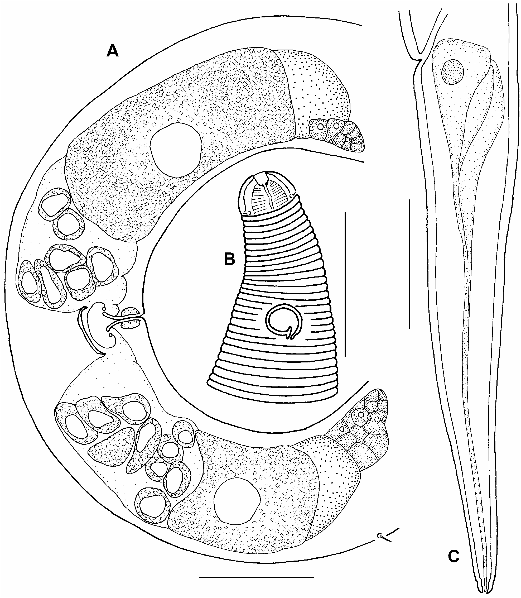
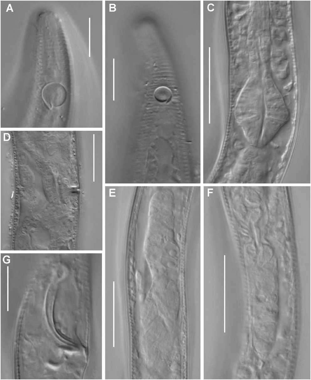
Description of male: Body cylindrical, with slightly narrowed anterior end and conical tail. Cuticle annulated along whole body except cephalic capsule. Cuticular annuli approximately 1 µm in width. Cuticle thickness approximately 0.6 µm at level of cephalic capsule, 0.8 µm at level of midbody and approximately 0.7 µm at level of tail. Holotype possessing two small somatic setae 1.5–2.0 µm long at pharyngeal part of the body between amphid and nerve ring. Cephalic end of holotype male was hard to discern, so the main characteristics of the apical end were described using paratypes. Paratype possessing 1 circle of 4 minute cephalic setae of approximately 1 µm length. Amphid monospiral, round, distinct cuticular edging, 58% (46–58%) of c.b.d. in its diameter. In holotype, there are 15 (15–17 in paratypes) cuticular annules between anterior end of amphid and cephalic capsule. Buccal cavity present, its cuticular walls looking thicker than in more posterior part of pharynx. Three small sclerotized teeth visible in anterior part of buccal cavity (one bigger dorsal one and two smaller subventral ones). Pharynx consisting of slender cylindrical anterior part with well developed muscular elements and posterior terminal muscular bulb with plasmatic interruptions. Bulb rhomboid with rounded corners or oval, 74% (74–95%) of c.b.d. in width with several plasmatic interruptions. Length of bulb 19 (16–19) µm. Nerve ring at a distance 2/3 of pharyngeal length from anterior end. Cellular body of renetta located at level of beginning of intestine. Cardia large, triangular, 43% of c.b.d. in its width. Holotype possesses plasmatic interruptions in cardia. Reproductive system diorchic, with outstretched anterior testis, and reflected posterior one. Anterior testis lies to the left of intestine. Posterior testis shorter than anterior one and lying to the right of intestine. Vas deferens thick, 36% of total body length, filled with big spermatids of approximately 9 µm in diameter with coarse-grained content. Spicules rather large, curved, with feebly developed capitulum at its distal end and thin velum. Gubernaculum in shape of a curved, thick rod. Thick prominent bent, hook-like apophysis present which liyng perpendicularly to gubernaculum to caudal direction in its basal part and than changing to be dorsal. Supplementary organs not visible. Whole reproductive system occupying 52% of total body length. Tail conical. Caudal glands of holotype not visible. Paratypes possess three cellular bodies of caudal glands located close to anus. Outlet of caudal glands visible in caudal tip.
Female: Females very similar to males in most parameters, however amphids being smaller, 36–38% of c.b.d. Reproductive system didelphic, with outstretched ovaries. Oocytes large, with granular cytoplasm. Anterior ovary lying to the right of intestine, posterior one lying to the left of intestine. Every ovary containing one mature elongated oocyte. Whole reproductive system occupying 29% of total body length. Several oval or roundish spermatozoa approximately 21x42 µm in size situated in uterus. Small part of dorsal wall of uterus located opposite vulva being thicker and strongly sclerotized. Middle part of uterus opposite vulva narrow. No vulvar glands seen.
Abundance: The density of this species was 1.0–3.4 inds/ 10cm 2 and relative abundance within the nematode community was 1–2% at the stations where it was found.
Differential diagnosis: M. abyssalis sp. n. is characterized by presence of large hook-like apophysis directed perpendicularly to gubernaculum in caudal direction in its basal part and than turning to dorsal direction. The presence of apophysis perpendicular to gubernaculum is a rare feature within Microlaimus species. Only M. crassiceps Gerlach, 1953 , M. undulatus Gerlach, 1953 , M. decraemerae (Muthumbi et Vinx, 1999) , and M. mnazi (Muthumbi et Vinx, 1999) possess similar apophyses. [ M. decraemerae and M. mnazi were transferred from Aponema to Microlaimus because of the presence of 2 testes in these species (Kovalyov and Miljutina 2008)].
M. abyssalis sp.n. may be distinguished from M. crassiceps and M. undulatus in several features. In the new species cephalic setae are very short, and outer labial setae are not visible, whereas cephalic setae of M. crassiceps and M. undulatus are quite long, and outer labial setae are discernible. The amphids in the new species are located far from the cephalic end (about in 2 amphidial c.b.d.), 15–17 annuli are present between the anterior rim of amphids and the posterior border of non-annulated cephalic capsule. The amphids of M. crassiceps are located at a level of cephalic setae, at the posterior half of the cephalic capsule. The amphids of M. undulatus are located close to the cephalic capsule (only 1 annule is between the amphidial anterior rim and the posterior border of cephalic capsule). Besides these features, the body length of M. abyssalis sp.n. is half the body length of M. crassiceps (427–568 µm vs. 1095–11120 µm).
M. abyssalis sp. n. differs from M. mnazi by being longer (427–568 µm vs. 253–328 µm in M. mnazi ); by shorter pharyngeal region (b = 5.0–5.9 vs. 4.0–4.7); by the shape of spicules (possessing distinct round capitulum it their distal end vs. edged distal end of spicula); by absence of supplemental precloacal organs (whereas M. mnazi possess 1 easily discernible one).
M. abyssalis sp. n. most resembles M. decraemerae . However the new species differs from M. decraemerae by its body length (427–568 µm vs. 296–378 µm in M. decraemerae ); by shorter tail (c = 6.5–7.3 vs. 5.1–6.0). The main difference is in the proportions and the shape of gubernaculum and apophysis: the length of the gubernaculum and the apophysis is approximately equal in the new species, whereas, in M. decraemerae , the apophysis is twice as long as the gubernaculum. The distal end of the apophysis is rounded in the new species in spite of the apophysis of M. decraemerae possessing an edged distal end.
No known copyright restrictions apply. See Agosti, D., Egloff, W., 2009. Taxonomic information exchange and copyright: the Plazi approach. BMC Research Notes 2009, 2:53 for further explanation.
|
Kingdom |
|
|
Phylum |
|
|
Class |
|
|
Order |
|
|
Family |
|
|
Genus |