
Eumastigonus waitahae, Korsós, Zoltán & Johns, Peter M., 2009
|
publication ID |
https://doi.org/ 10.5281/zenodo.186931 |
|
DOI |
https://doi.org/10.5281/zenodo.6225532 |
|
persistent identifier |
https://treatment.plazi.org/id/03D8DB74-3E1A-FFB9-FF61-4F38B310FEC2 |
|
treatment provided by |
Plazi |
|
scientific name |
Eumastigonus waitahae |
| status |
sp. nov. |
Eumastigonus waitahae View in CoL sp. n.
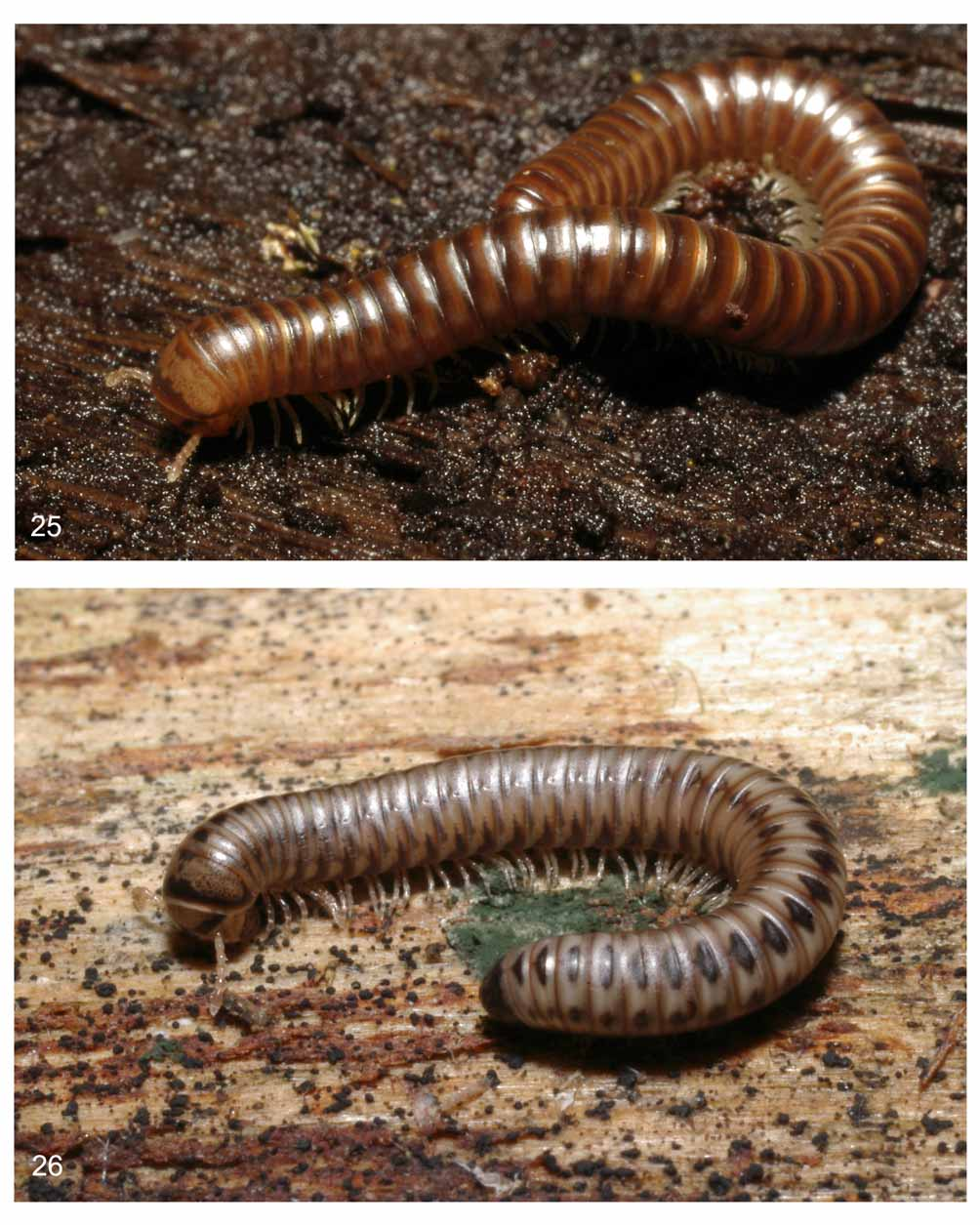
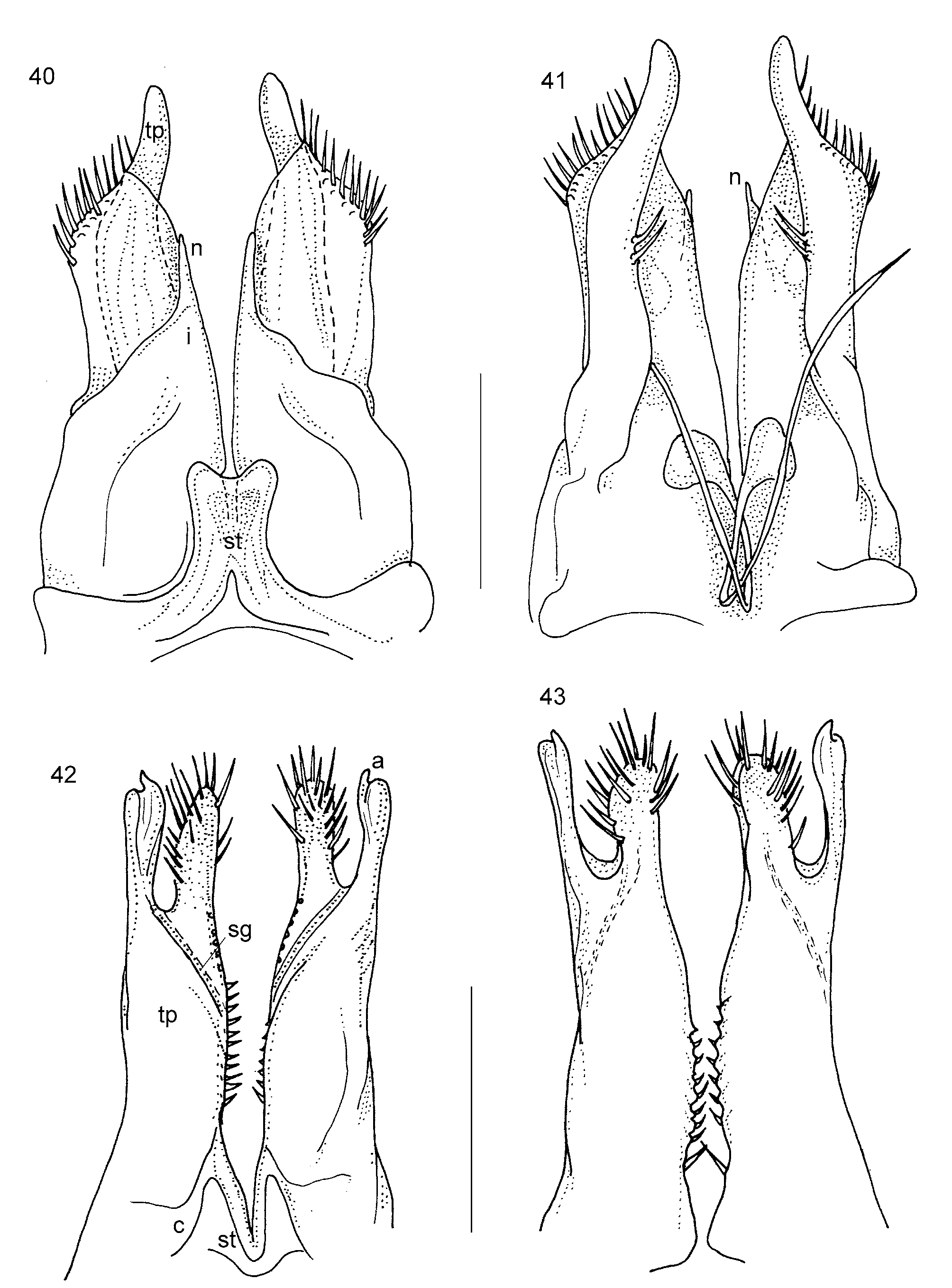
Figs. 26 View FIGURE 26. E , 33 –49, 51.
Type material: Holotype 3 ( CMNZ): New Zealand, South Island, Canterbury Region, Kaikoura, Ohau Point Scenic Reserve, 15 m, leg. P. M. Johns, 26 Aug. 2000, under rocks.
Paratypes (103 and 11Ƥ): 1Ƥ – Same locality and date ( CMNZ); 23, 2Ƥ – Kekerengu, coastal ngaio bush, 3-20 m, leg. P. M. Johns & W. Thomas, 19 July 1963; 43, 3Ƥ ( CMNZ: one male is dissected for SEM: head with 1st legpair, 2nd legpair with penis, gonopods, 7th ring) – Kaikoura, Kahautara River, 280 m, leg. P. M. & M. Johns, 23 March 1962; 2Ƥ ( HNHM, see Fig. 25, also vulva prep.) – Kaikoura, Mt. Fyffe Conservation Area, S42° 21’ – E173° 34’, 192 m a.s.l., leg. Z. Korsós & P. M. Johns, 3 June 2006, coastal broadleaf forest; 13 ( CMNZ: dissected for SEM: head, 1st legpair, 2nd legpair with penis, gonopods, anal ring). – Oxford, Okuku Pass, leg. P. M. Johns & Z. Korsós, 15 March 2008; 13 ( MNHN Paris) – Rangiora, Okuku Pass (lower stream flat) (420 m original labels), 370 m, leg. P. M. Johns, 27 Feb. 1972, stones, under Nothofagus ; 23, 3Ƥ ( ZMUC Copenhagen) – Rangiora, Okuku Pass (lower stream flat), 370 m (original label: 420 m), leg. P. M. Johns, 14 March 1970, stones, under Nothofagus .
FIGURES 35–39. E. waitahae male from Okuku Pass, Canterbury Region. 35: Gnathochilarium, ventral view (gp = gnathochilarial pit), 36: 2nd legpair, posterior view (p = penis), 37: first legpair, anterior view, 38: first legpair, posterior view, 39: body end, left lateral view. Scales 35, 39: 1 mm, 36-38: 0.5 mm.
FIGURES 44–49. E. waitahae paratype male from Okuku Pass, Canterbury Region, scanning electronmicrographs. 44: Gnathochilarium, ventral view (gp = gnathochilarial pit), 45: antennal tip, 46: 7th ring, right lateral view, 47: first legpair, anterior view, 48: 2nd legpair with penis (p), posterior view, 49: body end, left lateral view.
Other material studied: 1313, 201Ƥ, and 12 juveniles from 60 sites.
Diagnosis: A species of Eumastigonus , standing quite alone with its characteristic live colour pattern ( Fig. 26 View FIGURE 26. E , and described below), and diagnostic shape of gonopods: inner coxal process short, needle-like, telopodites with wide oblique lamella marginally strongly setose, and two long cylindrical processes almost crossing each other. Posterior gonopods with two subequal processes, one blunt and hairy, the other slim and with a slightly pointed tip.
Etymology: Named after the distribution of the species, eastern part (Waitaha) of the South Island, New Zealand.
Description: Slightly smaller than the previous species, length: 16–28 mm, max. midbody diameter: 1.4–2.4 mm, no. of rings: 30–45 podous + 1–4 apodous and telson.
Head rounded, glabrous, with typical pattern, no setae, gnathochilarium and antennae as in generic description (Figs. 35, 44–45). Ocelli in 3–4 rows, in less number (14–23) than the previous species, in a compact irregular, oval or subrectangular field.
Collum broad, rounded, covering caudal part of head until ocellarium, with a few (2–3) but distinct short striae at its corner. Prozonae of each ring with slightly reticulated surface, metazonae below ozopores with 10–14 longitudinal striae turning upwards along suture and melting into the sculpture of prozonae. Ozopores situated at about 1/2 metazonal length behind suture, openings distinct, easily visible. Telson smooth, without striae and/or projection, paraprocts with 2+2, rarely 1+1 marginal setae (Figs. 39, 49).
Colouration: The typical live colouration involves a light greyish-brown ground colour with conspicuous dark brown markings ( Fig. 26 View FIGURE 26. E ): a transversal stripe between ocellaria, broad anterior margin on collum, anterio-lateral edge of mandibular stipes, a series of dark brown side spots on the lower third of metazonae and at about the level of ozopores, and transversally oval or diamond-shaped patches along the dorsum. This colour pattern can fade, especially in populations going inland on the northeastern part of South Island; most typical pattern shown in the east coast specimens. Head pattern and dark brown telson with contrastingly light paraprocts are invariable, however.
Male sexual characters: Mandibular and gnathochilarial stipites, first legpair (Figs. 37–38, 47) as for Eumastigonus , coxa wide, flat, laterally with small group of setae; prefemur with strong triangular process (provided with short setae) directed frontally; femur, postfemur, tibia and tarsus normal, clawless. 2nd legpair (Figs. 36, 48) normal, coxae separate, elongated, deeply bifurcated penis (p) closely behind them, its tips tubelike with hairy openings. Prefemur subquadrate, caudally with 2+2 setae, one long and one short, femur and othe podomeres normal, claw long. 3–7th legs normal, without modifications, walking legs from 10th onwards with femoral pads.
Protrusion on 7th ring (Fig. 46) conspicuous, surrounding and protecting embedded gonopods behind.
Gonopods ( Figs. 33–34, 40–43 View FIGURES 40 – 43. E ): Anterior gonopods with wide, bilobed sternal plate (st), inner coxal process (i) short, reaching only to half of length of entire gonopod, with pointed, needle-like tip (n) closely clung to median lobe. Telopodites (tp) each with a lateral, obliquely cut shoulder marginally with a set of hairs, and a pair of long cylindrical processes bent mesad, almost like wanting to cross each other. Remnant of second telopodomere missing. Flagellum attached to coxae of anterior gonopods, long, pointed, tip not penicillate, in situ fits into seminal groove of posterior gonopods. Coxal parts of posterior gonopods (c) widely separated, telopodite (tp) parallel-sided, apically divided into two processes subequal in length, inner one blunt, densely setose, lateral part slimmer and thinner, its tip (a) small, tooth-like. Anterior side of posterior gonopods longwise split, with overlapping clefts housing flagellum in situ. Margin of cleft mediobasally strongly serrated, with small spikes sitting on each tooth. Seminal groove (sg) runs entirely on anterior side along cleft, and ends between the two terminal branches, perhaps opening at base of lateral one.
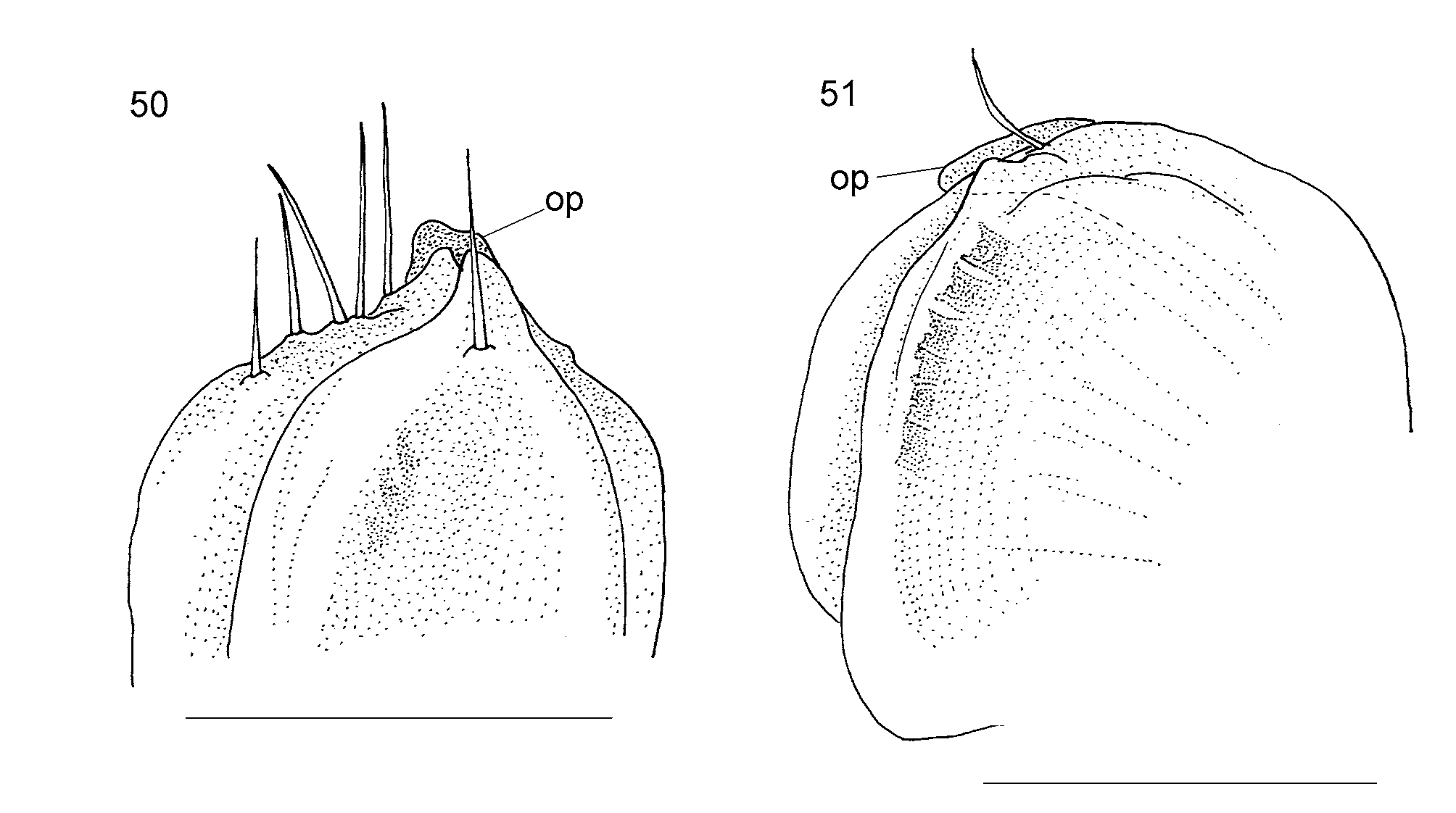
Female sexual characters: Vulva ( Fig. 51 View FIGURES 50 – 51 ) typical Eumastigonus -like, disc-shaped, compressed, opculum (op) very small. Posterior bursal valve with a single seta only, anterior hairless. Instead, in bursal opening, between valves a series (5–6) of peculiar small, quadratic objects whose fine structure, however, even with SEM could not be revealed.
Distribution: New Zealand, South Island, Canterbury and Marlborough Regions ( Fig. 53). All records are centered on low broadleaf forest or shrub vegetation. Some sites are quite dry with yearly rainfall being less that 700 mm. It is restricted to the north-eastern portion of the South Island from Dashwood Pass, Blenheim, to Kaikoura where it is very common in the steepland forest down to the tussocks close to the sea. In North Canterbury it is in podocarp and Nothofagus forests, and on Banks Peninsula it is in many types of vegetation, some of which has been modified over the years by farming.
No known copyright restrictions apply. See Agosti, D., Egloff, W., 2009. Taxonomic information exchange and copyright: the Plazi approach. BMC Research Notes 2009, 2:53 for further explanation.