
Dicyemennea tobaense, Furuya, 2018
|
publication ID |
https://doi.org/ 10.12782/specdiv.23.143 |
|
publication LSID |
lsid:zoobank.org:pub:82CD9349-810A-42F1-A602-343EBA1AE7A4 |
|
persistent identifier |
https://treatment.plazi.org/id/8A3CFAB1-B1C2-459C-9741-EE98553B008A |
|
taxon LSID |
lsid:zoobank.org:act:8A3CFAB1-B1C2-459C-9741-EE98553B008A |
|
treatment provided by |
Felipe |
|
scientific name |
Dicyemennea tobaense |
| status |
sp. nov. |
Dicyemennea tobaense sp. nov.
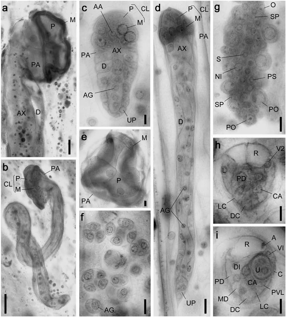
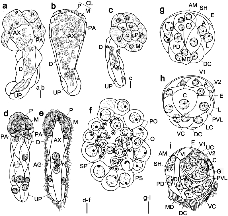
( Figs 20 View Fig , 21 View Fig ; Tables 2, 3)
Diagnosis. Large dicyemid; body length to 5,000 µm. Calotte wheel-shaped. Vermiform stages with 40–47 peripheral cells: 4 propolars+5 metapolars+2 parapolars+29–36 trunk cells. Infusoriform embryos with 37 cells; refringent bodies solid; and nucleus present in each urn cell.
Description. Nematogens ( Figs 20a, b View Fig , 21a, c, d View Fig ). Body length 500–4,960 µm and width 80–530 µm; widest in region of metapolars; trunk width narrower to posterior. Peripheral cell number 40–47 ( Table 3): 4 propolars+5 metapolars+2 parapolars+27–34 diapolars+2 uropolars. Calotte wheelshaped, cilia on calotte about 6 µm long, oriented anteriorly. Propolar cells and their nuclei larger in size than metapolar cells and their nuclei ( Fig. 21c, d View Fig ). Cytoplasm of metapolar cells more darkly stained by hematoxylin than cytoplasm of other peripheral cells ( Fig. 20a View Fig ). Verruciform cells absent. Axial cell cylindrical, enlarging at anterior part, and extending forward to propolar cells ( Fig. 20b View Fig ). About 50 vermiform embryos present per axial cell of large individuals.
Vermiform embryos ( Figs 20c, d View Fig , 21f, g View Fig ). Full-grown vermiform embryos length 106–224 (maximum 319) µm and width 28–40 µm. Peripheral cell number 40–47 ( Table 3); trunk cells arranged in opposed pairs. Anterior end of calotte rounded. Axial cell rounded anteriorly and extending to propolar cells. Anterior abortive axial cell present ( Figs 20c View Fig , 21g View Fig ). Axial cell of full-grown embryos with up to 23 agametes.
Rhombogens ( Figs 20e View Fig , 21e View Fig ). Body similar in length to nematogens but stuggy, length 500–3,190 µm, width 120– 385 µm. Peripheral cell number 41–45 ( Table 3). Calotte disc-shaped. Axial cell shape and anterior extent similar to nematogens. Verruciform cells absent. Usually, 6–10 (maximum 16) infusorigens present in axial cell of each parent individual. Usually, 100 (maximum 133) infusoriform embryos present per axial cell of large individuals. Accessory nuclei occasionally present in trunk cells.
Many spherical hollow cell masses, consisting of three to ten cells, present within the axial cell ( Fig. 20f View Fig ).
Infusorigens ( Figs 20g View Fig , 21h; n View Fig =10). Mature infusorigens large; composed of 60–117 (mode 76) external cells (oogonia and primary oocytes)+21–52 (mode 44) internal cells (spermatogonia, primary spermatocytes, and secondary spermatocytes)+8–51 (mode 21) spermatozoa. Four to 8 nuclei present within axial cell. Mean diameter of fertilized eggs 14.8 µm and that of spermatozoa 3.2 µm. Axial cell round or ovoid, diameter 15–27 µm.
Infusoriform embryos ( Figs 20h, i View Fig , 21i–k; n View Fig =50). Fullgrown embryos large, length 31.8±1.5 µm (mean±SD; excluding cilia); length-width-height ratio 1.0: 0.85: 0.84; shape ovoid, bluntly rounded to pointed posteriorly; cilia at posterior end 6 µm long. Refringent bodies present, solid, and occupying anterior 30% of embryo length when viewed laterally ( Fig. 20i View Fig ). Cilia projected from ventral internal cells into urn cavity ( Fig. 21k View Fig ). Capsule cells containing small granules. Mature embryos with 37 cells:33 somatic+4 germinal cells. Somatic cells of several types present: external cells covering large part of anterior and lateral surfaces of embryo (2 enveloping cells); external cells with cilia on external surfaces (2 pairs of dorsal cells+1 median dorsal cell+2 dorsal caudal cells+2 lateral caudal cells+1 ventral caudal cell+2 lateral cells+2 posteroventral lateral cells), external cells with refringent bodies (2 apical cells); external cells without cilia (1 couvercle cell+2 first ventral cells+2 second ventral cells+2 third ventral cells); internal cells with cilia (2 ventral internal cells); and internal cells without cilia (2 dorsal internal cells+2 capsule cells+4 urn cells). Each urn cell containing single nucleus and single germinal cell ( Fig. 21k View Fig ). All somatic nuclei appearing pycnotic in mature infusoriform embryos.
Remarks. Dicyemennea tobaense sp. nov. is morphologically very similar to D. trochocephalum in having a wheelshaped calotte but the former is clearly distinguished by the number of peripheral cells (40–47 vs. 25–29) and the cell number of infusoriform embryos (37 vs. 39) ( Furuya 1999). The new species is also distinguished by possessing the most peripheral cells of all species of dicyemids.
Dicyemennea tobaense sp. nov. has spherical hollow cell masses in the rhombogen. Similar hollow cell masses have been reported in the rhombogen of D. trochocephalum ( Furuya 1999; see present study, Fig. 21 View Fig ), but cells in the latter tightly adhere to morula like adjacent cells (vs. not present in D. tobaense sp. nov., Fig. 19f View Fig ).
Etymology. The species name tobaense refers to the localition of Toba Aquarium where the new species was firstly found.
Taxonomic summary. Type material: a syntype slide (NSMT-Me-56) collected at 30 November 2015; additional syntypes on slide series No. OT3308 (5 slides) in the author’s collection.
Type locality: off Minami-Ise (34°05′N, 136°50′E), Mie Prefecture, Honshu , Kumano-nada Sea, North Pacific Ocean, Japan, depth 260 m GoogleMaps .
Other materials examined: slide series No. OT1451 (5 slides) collected off Owase (33°54′N, 136°18′E), Mie Prefecture, Honshu , Kumano-nada Sea, North Pacific Ocean, Japan, depth 300 m, 9 June 2004; Nos GoogleMaps . OT3412–3418 (each 5 slides) collected off Kii-Nagashima (33°58′N, 136°28′E), Mie Prefecture, Honshu , Kumano-nada Sea, North Pacific Ocean, Japan, depth 300 m, 14 March 2016 in the author’s collection GoogleMaps .
Host: symbiotype, Octopus tenuicirrus ( Sasaki, 1929) (Mollusca: Cephalopoda: Octopoda ), male (mature), 75 mm ML (NSMT-Mo-85871).
Site : anterior ends (calottes) inserted into crypts of the renal appendages within the renal sacs.
Prevalence: in 15 of 48 host specimens examined (31.3%).

No known copyright restrictions apply. See Agosti, D., Egloff, W., 2009. Taxonomic information exchange and copyright: the Plazi approach. BMC Research Notes 2009, 2:53 for further explanation.