
Ascidia, Linnaeus, 1767
|
publication ID |
https://doi.org/10.11646/zootaxa.4387.3.3 |
|
publication LSID |
lsid:zoobank.org:pub:F0845057-D918-4693-8D80-E94E6CA6EE8C |
|
DOI |
https://doi.org/10.5281/zenodo.5967757 |
|
persistent identifier |
https://treatment.plazi.org/id/03D9A066-FFF3-FFB3-F2C1-1B70FB3FF862 |
|
treatment provided by |
Plazi |
|
scientific name |
Ascidia |
| status |
|
Ascidia sp.
Fig. 9 View FIGURE 9
Station AB 157, 14°29,9N–61°05,4W, 28m (MNHN P5 ASC.A 431)
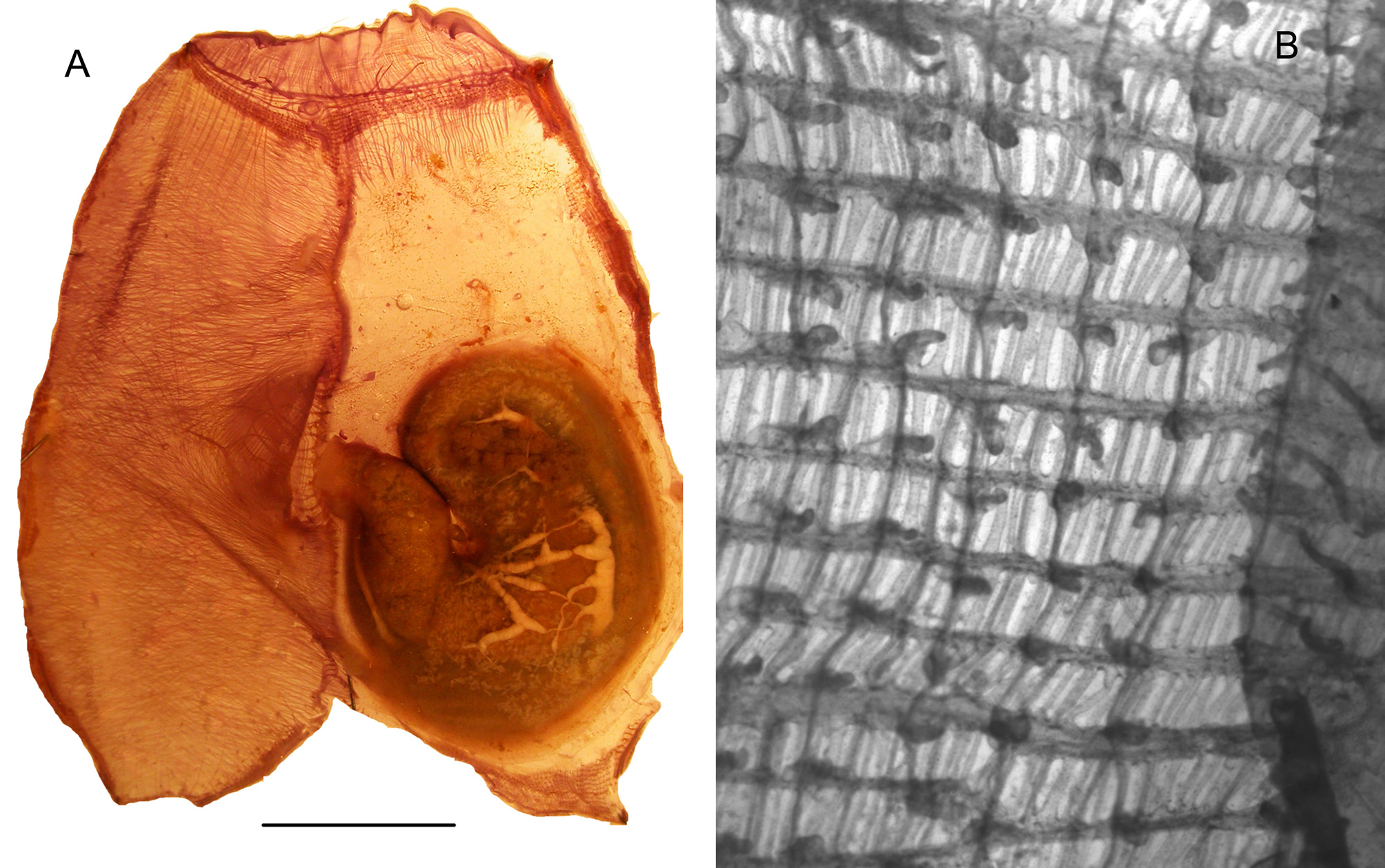
The single specimen 4.5 cm long has a clear cartilaginous tunic with a rough surface and some epibionts. The siphons are sessile the oral one apical end the atrial at half body length. There are 8 oral lobes with ocelli and on the undulated atrial rim 12 ocelli. The body wall is light brown in formalin ( Fig. 9A View FIGURE 9 ). The oral tentacles are spaced in 3 orders of size. The anterior lamina of the prepharyngeal groove has papillae but no papillae were seen on the prepharyngeal space. The dorsal tubercle has horns curved outwardly. The dorsal lamina begins with 2 blades and is prolonged in a high blade far behind the oesophageal aperture. The rapheal ribs end in small papillae ( Fig.9B View FIGURE 9 ). The branchial tissue is thin with meshes of irregular size containing 4 to 7 stigmata. The branchial papillae are round or somewhat elongate ( Fig.9B View FIGURE 9 ), most of them with 2 small protrusions at their base. There are no intermediate papillae. The branchial sac extends below the gut. The gut loop occupies half of the left side ( Fig. 9A View FIGURE 9 ). The stomach is oval with indistinct folds. The primary intestinal loop contains a massive ovary. The descending intestinal limb and rectum are inflated but do not form a protruding pouch. The rectum forms a closed secondary loop and ends in a smooth anus. The testis in small vesicles is spread on the whole external side of the gut, its sperm ducts are gathered in large channels with a scalariform design ( Fig.9 A View FIGURE 9 ) which join in a single duct along the rectum and the oviduct. With a single specimen it is not possible to determine if this peculiar design of the sperm duct is specific or is only an occasional development of the testis.
The musculature is almost absent on the left body side with longitudinal fibres issued from the oral siphon extending only a short distance behind the prepharyngeal area ( Fig. 9A View FIGURE 9 ). On the right side the longitudinal fibres issued from the oral part become thinner downwards and disappear exept for a few that extend almost to the posterior end of the body. The transverse musculature is very dense on the whole right body side and stop abruptly along the dorsal line ( Fig.9A View FIGURE 9 ). Some fibres of the atrial siphon complete this dense meshwork. This musculature design recalls that of Ascidia interrupta but is denser.
By its external aspect this species looks like Ascidia curvata but differs by a stronger musculature on the right side and the shape of the gonoducts
Many Ascidia species are present in the western tropical Atlantic ( Van Name 1945; Bonnet & Rocha 2011), but none have exactly the same set of characters. The variability in a single population in the genus Ascidia is too important to assign this Madibenthos specimen to a species with a single specimen collected.
No known copyright restrictions apply. See Agosti, D., Egloff, W., 2009. Taxonomic information exchange and copyright: the Plazi approach. BMC Research Notes 2009, 2:53 for further explanation.
|
Kingdom |
|
|
Phylum |
|
|
Class |
|
|
Order |
|
|
Family |
Ascidia
| Monniot, Françoise 2018 |
Ascidia
| Linnaeus 1767 |
Ascidia
| Linnaeus 1767 |