
Microlarnaca zhengi Lu & Bian, 2021
|
publication ID |
https://doi.org/ 10.11646/zootaxa.5061.2.7 |
|
publication LSID |
lsid:zoobank.org:pub:3E56554E-8165-4990-9FB7-D97A4167ADF4 |
|
DOI |
https://doi.org/10.5281/zenodo.5649658 |
|
persistent identifier |
https://treatment.plazi.org/id/03D9D94E-FFB1-BC18-88BB-71E0FCC8EACD |
|
treatment provided by |
Plazi |
|
scientific name |
Microlarnaca zhengi Lu & Bian |
| status |
sp. nov. |
Microlarnaca zhengi Lu & Bian View in CoL sp. nov.
Ẹ氏小Dzō
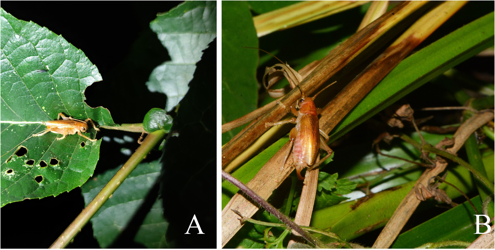
Figs 1–5 View FIGURE 1 View FIGURE 2 View FIGURE 3 View FIGURE 4 View FIGURE 5 , 10B View FIGURE 10
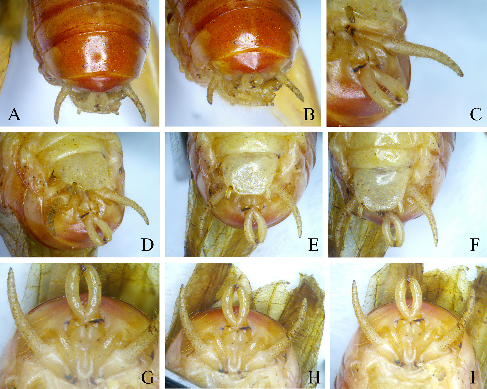
Diagnosis. The new species differs from other congeneric species in: processes of male ninth abdominal tergite with its apices directing forward ( Fig. 2I View FIGURE 2 ), processes of male tenth abdominal tergite shorter and stouter than styli ( Fig. 2G View FIGURE 2 ), its apices compressed and slighlty curved inward with apical margin truncate ( Fig. 2E View FIGURE 2 ); process of female subgenital plate stout, longer than subgenital plate ( Fig. 4E View FIGURE 4 ).
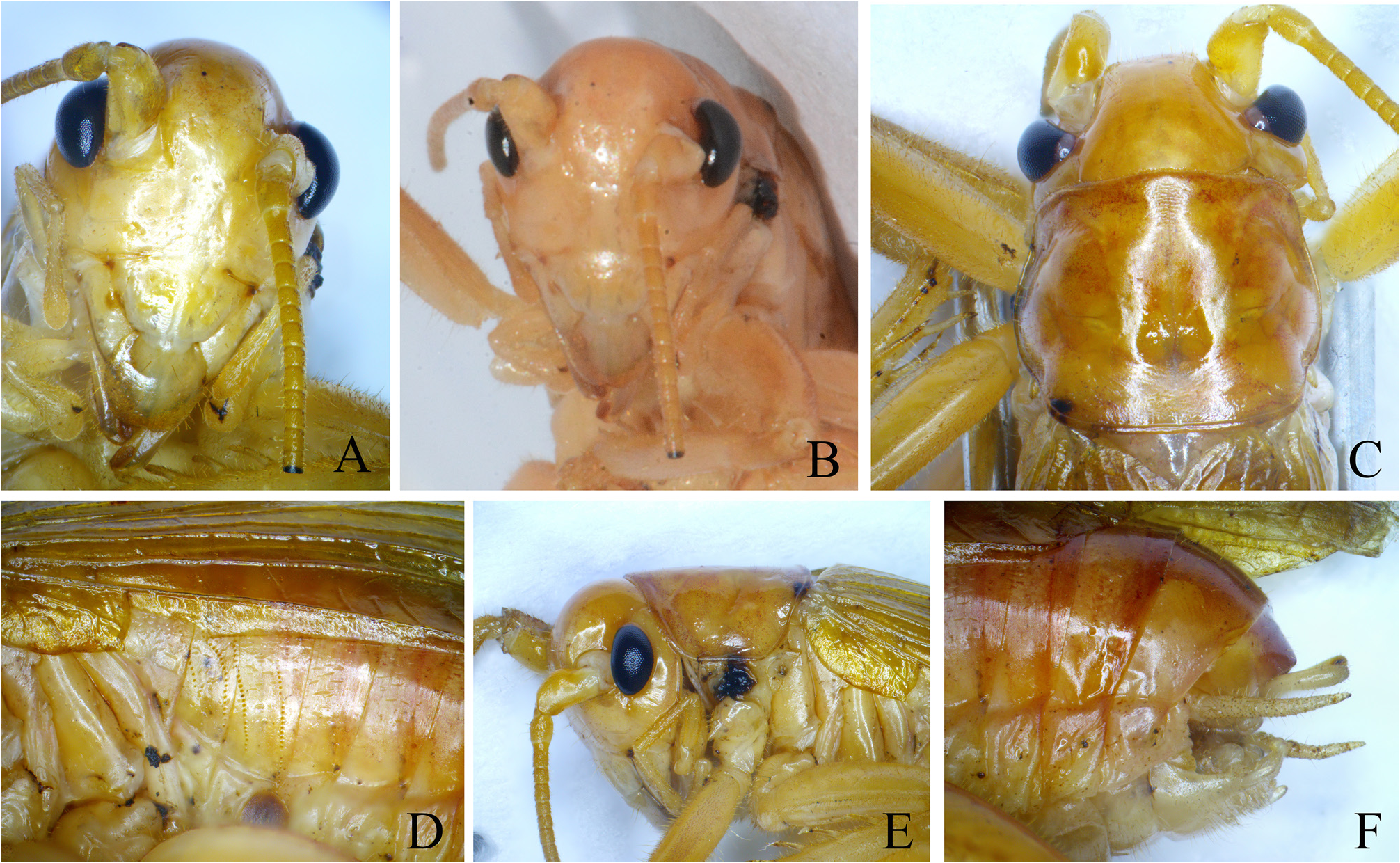
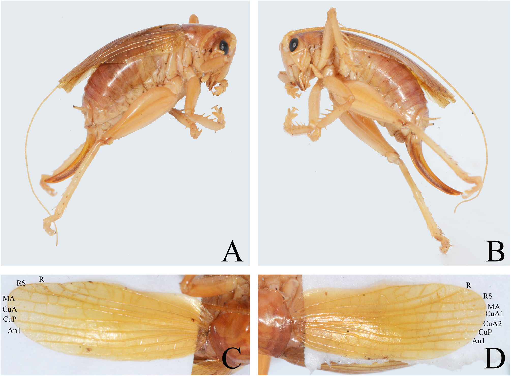
Male. Small species. Face ovoid; fastigium verticis wider as scape; face with scattered impressed spots ( Fig. 1A View FIGURE 1 ). Ocelli indistinct.
Fore coxae with 1 small spine; fore and middle femora unarmed, tibiae ventrally with 4 pairs of spurs and 1 pair of apical spurs; dorsal surface of middle tibiae with 1 interno-apical spine. Hind femora with 2 – 3 internal and 4 – 6 external spines on ventral surface; dorsal surface of tibiae with 4 – 6 internal and 6 external spines and 1 pair of apical spurs, vetral surface with 1 pair of subapical spurs and 2 pairs of apical spurs.
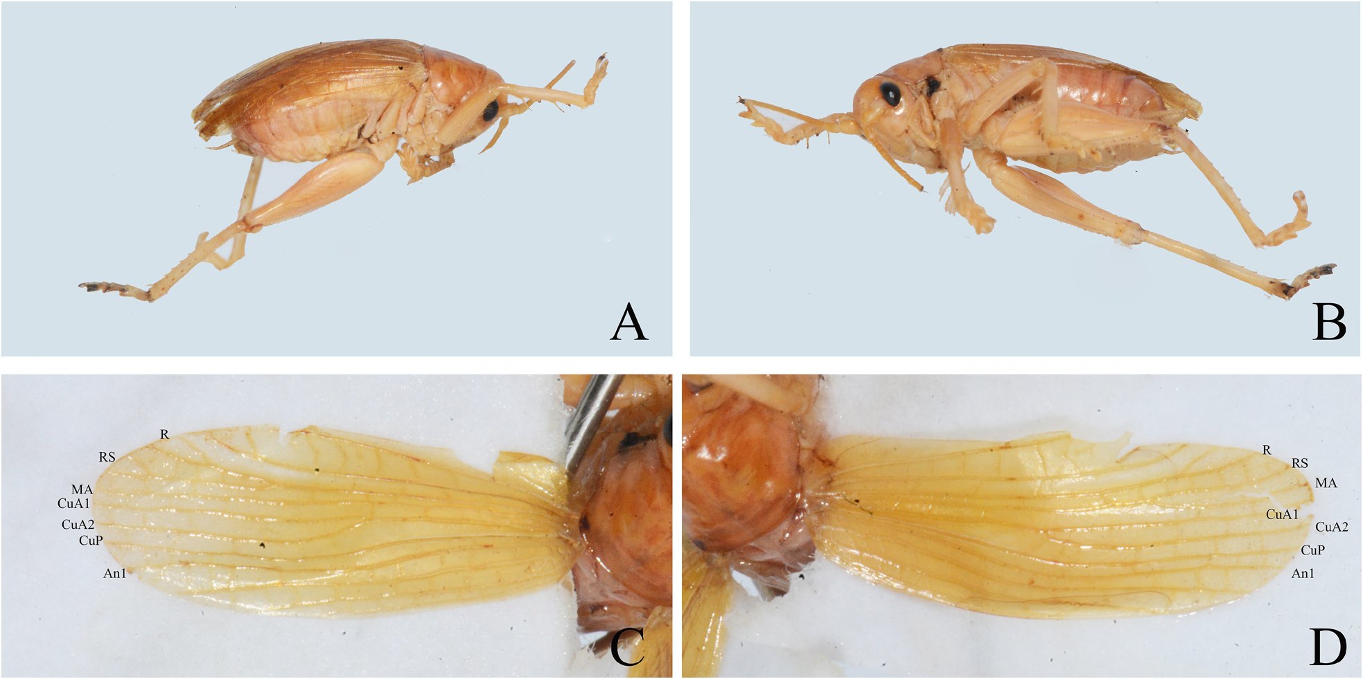
Wings slightly surpassing the apex of abdomen ( Fig. 3A–B View FIGURE 3 ). Tegmina: Radius with two branches, both forked near tip. Media anterior of left tegmen unbranched leaning on radius in basal area ( Fig. 3C View FIGURE 3 ). On the right tegmen ( Fig. 3D View FIGURE 3 ), media anterior fused in basal area with radius, then receives a short, oblique connection branch from radius behind middle of tegmen thus that MA and RS become fused for a short distance and divided again in apical third. Both tegmina: media posterior absent; cubitus anterior forks before mid-length of tegmina into two veins, CuA1 and CuA2; cubitus posterior undivided, free throughout; with 4 anal veins.
Second and third abdominal tergites with two rows of parallel stridulatory pegs separately ( Fig. 1D View FIGURE 1 ). Eighth abdominal tergite prolonged ( Fig. 1F View FIGURE 1 ). Apical area of ninth abdominal tergite projecting posteriorly ( Fig. 1F View FIGURE 1 ), with one process at each side pointing ventrad and forward ( Fig. 2G–I View FIGURE 2 ); basal area of the process stout, terminating to apex, apical area contorted as Fig. 2I View FIGURE 2 . Middle area of tenth abdominal tergite with a pair of swellings ( Fig. 2G View FIGURE 2 ), upper margins these swellings with a pair of short, cylindrical processes ( Fig. 2G–I View FIGURE 2 ); the apical area of processes connected each other ( Fig. 2G–I View FIGURE 2 ), apices compressed with truncate margin ( Fig. 2E, G View FIGURE 2 ). Subgenital plate trapezoidal, apical area projected with a shallowly median concavity on posterior margin ( Fig. 2F View FIGURE 2 ). Styli inserted at latero-apical angles of subgenital plate ( Fig. 2F View FIGURE 2 ).
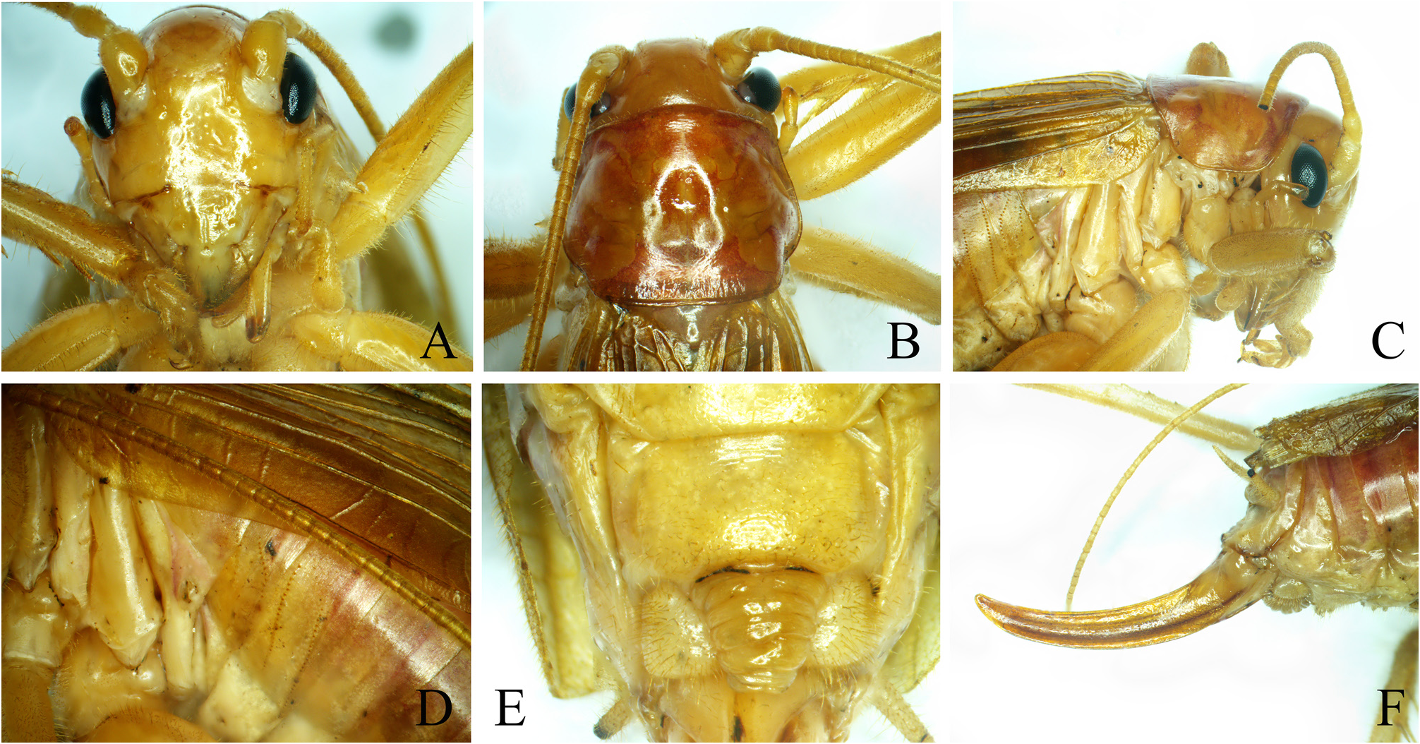
Female. Tegmina just reaching the apex of abdomen ( Fig. 5A–B View FIGURE 5 ). Tegmina ( Fig. 5C–D View FIGURE 5 ): Radius releases RS in subapical area; media leaning on radius in basal area but does not fuse with it; cubitus anterior forks behind midlength of right tegmen into two veins, CuA1 and CuA2, while the media on left tegmen remains single-branched; cubitus posterior undivided, free throughout; with 4 anal veins. Between posterior margin of seventh abdominal sternite and basal area of subgenital plate with one conical process ( Fig. 4E View FIGURE 4 ), which surpassing the posterior margin of subgenital plate and covered the largely area of subgenital plate, ventral surface of the process with sclerotised transverse folds ( Fig. 4E View FIGURE 4 ). Subgenital plate about 3.3 times as wide as long, the lateral margins convex ( Fig. 4E View FIGURE 4 ). Ovipositor short, apical half moderately uncurved; with gradually approaching margins, apices obtuse ( Fig. 4F View FIGURE 4 ).
Measurements (mm). body: ♂ 13.1, ♀ 13.6; pronotum: ♂ 4.2, ♀ 3.9; tegmen: ♂ 12.8, ♀ 11.4; hind femur: ♂ 7.7, ♀ 7.8; ovipositor: 7.7.
Coloration. Body yellowish brown. Eyes black. Tegmina semi-transparent yellowish brown. Dorsal surface purplish red.
Material examined. Holotype: male, Huazhuping, Funing, Yunnan, 23 July, 2021, coll. by Wei Bin, Xiaoyu Peng & Xun Bian . Paratye: 1 female, other information as holotype.
Etymology. The new species is named in honor of Professor Zhemin Zhen (Ẹ哲民) for his great contibutions to the classification of Chinese locusts and grasshoppers.
No known copyright restrictions apply. See Agosti, D., Egloff, W., 2009. Taxonomic information exchange and copyright: the Plazi approach. BMC Research Notes 2009, 2:53 for further explanation.